Review
. 2016 Dec;16(12):775-788.
doi: 10.1038/nrc.2016.105. Epub 2016 Oct 21.

Gap junctions and cancer: communicating for 50 years

Affiliations
Review

Gap junctions and cancer: communicating for 50 years

Trond Aasen et al. Nat Rev Cancer. 2016 Dec.

Erratum in

Abstract

Fifty years ago, tumour cells were found to lack electrical coupling, leading to the hypothesis that loss of direct intercellular communication is commonly associated with cancer onset and progression. Subsequent studies linked this phenomenon to gap junctions composed of connexin proteins. Although many studies support the notion that connexins are tumour suppressors, recent evidence suggests that, in some tumour types, they may facilitate specific stages of tumour progression through both junctional and non-junctional signalling pathways. This Timeline article highlights the milestones connecting gap junctions to cancer, and underscores important unanswered questions, controversies and therapeutic opportunities in the field.


Figures

FIGURE 1
FIGURE 1. of key discoveries related to gap junctions and cancer
cGAMP, 2′3′-cyclic GMP-AMP; EMT, epithelial to mesenchymal transition; GJIC, gap junctional intercellular communication; miRNAs, microRNAs.
FIGURE 2
FIGURE 2. Assembly of connexins into gap junctions
Schematic depiction of typical connexins with the characteristic four-transmembrane topology consisting of four transmembrane domains, two extra cellular loop domains, a cytoplasmic amino terminal, one cytoplasmic loop and a highly variable cytoplasmic carboxy-terminal domain. Six connexins oligomerize into a connexon or hemichannel that docks in homotypic, heterotypic and combined heterotypic/heteromeric gap junction arrangements. The permeability properties depend on the connexin isoforms expressed, and since cells can coexpress and intermix different isoforms a huge number of possible combinations exist making functional evaluation highly complex. Exchange of possible types of cancer-associated signalling molecules between two cells or a cell and the extracellular environment is illustrated. For simplicity only a few examples for each class of signalling molecule are shown. cAMP, cyclic AMP; cGAMP, 2′3′-cyclic GMP-AMP; IP3, inositol-1,4,5-trisphosphate; miR-125b, microRNA-125b.
FIGURE 3
FIGURE 3. Connexin involvement during cancer progression
(a) At early stages, connexins appear to mainly act as tumour suppressors, whereby loss of connexin expression or gap junctional intercellular communication (GJIC) may promote growth, survival and possibly angiogenesis. Within this context, the role of connexins in cancer stem cells (CSCs) remains unclear, as isoform-specific effects of connexins may be opposing. (b) Likewise, different connexins play different roles during epithelial to mesenchymal transition (EMT) and invasion, although overall loss of connexins seems to promote this phenotype. (c) During later stages, when cancer cells metastasise, connexin expression generally seems to facilitate rather than block intravasation of tumour cells into blood vessels. (d) Increased connexin levels can also promote tumour cell survival and adherence within the circulation (e) Extravasation of tumour cells out of blood vessels is also supported by upregulation of connexin expression. (f) Once at the metastatic site, the roles of connexins are more unclear, with some evidence for connexins promoting tumour cell dormancy but also promoting survival within that context. (g) Some evidence suggests connexins reduce cell growth directly in metastases, but at the same time may stimulate local invasion and survival. (h) Therapy response, which includes chemoresistance, can be achieved via multicellular connections between cancer cells or between cancer cells and healthy cells and may be connexin isoform-specific. Moreover, this process will be highly influenced by the specific microenvironment and whether the overall exchange of signals promotes the “Kiss of Life” or “Kiss of Death” (BOX 2). Purple boxes indicate overall connexin effect and green boxes denote the specific stage in cancer progression where connexins function.
FIGURE 4
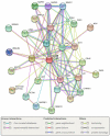
FIGURE 4. The Cx43 interactome
Example of STRING analysis of the most prevalent connexin Cx43 (encoded by GJA1) revealing a wide range of putative interactions, of which many are growth regulators or oncogenes such as SRC, AKT1, JUN, FOS and nephroblastoma overexpressed (NOV). The network was retrieved and constructed using the STRING database version 10.0 (http://string-db.org), using the most stringent confidence score prediction setting (>0.9), resulting in 28 interactions. Extended interaction-network (high confidence prediction, >0.7, 99 interactions) can be viewed in Supplementary Figure 1. Full names, score prediction and details of specific proteins are available in Supplementary Table 1.

References

    1. Loewenstein WR, Socolar SJ, Higashino S, Kanno Y, Davidson N. Intercellular Communication: Renal, Urinary Bladder, Sensory, and Salivary Gland Cells. Science. 1965;149:295–8. - PubMed
    1. Kanno Y, Loewenstein WR. Cell-to-cell passage of large molecules. Nature. 1966;212:629–30. - PubMed
    1. Loewenstein WR, Kanno Y. Studies on an Epithelial (Gland) Cell Junction. I. Modifications of Surface Membrane Permeability. J Cell Biol. 1964;22:565–86. - PMC - PubMed
    1. Loewenstein WR, Kanno Y. Intercellular communication and the control of tissue growth: lack of communication between cancer cells. Nature. 1966;209:1248–9. - PubMed
    1. Loewenstein WR, Kanno Y. Intercellular communication and tissue growth. I. Cancerous growth. J Cell Biol. 1967;33:225–34. - PMC - PubMed