Review
. 2016 Nov;274(1):33-58.
doi: 10.1111/imr.12500.

Complement component C3 - The "Swiss Army Knife" of innate immunity and host defense

Affiliations
Review

Complement component C3 - The "Swiss Army Knife" of innate immunity and host defense

Daniel Ricklin et al. Immunol Rev. 2016 Nov.

Abstract

As a preformed defense system, complement faces a delicate challenge in providing an immediate, forceful response to pathogens even at first encounter, while sparing host cells in the process. For this purpose, it engages a tightly regulated network of plasma proteins, cell surface receptors, and regulators. Complement component C3 plays a particularly versatile role in this process by keeping the cascade alert, acting as a point of convergence of activation pathways, fueling the amplification of the complement response, exerting direct effector functions, and helping to coordinate downstream immune responses. In recent years, it has become evident that nature engages the power of C3 not only to clear pathogens but also for a variety of homeostatic processes ranging from tissue regeneration and synapse pruning to clearing debris and controlling tumor cell progression. At the same time, its central position in immune surveillance makes C3 a target for microbial immune evasion and, if improperly engaged, a trigger point for various clinical conditions. In our review, we look at the versatile roles and evolutionary journey of C3, discuss new insights into the molecular basis for C3 function, provide examples of disease involvement, and summarize the emerging potential of C3 as a therapeutic target.

Keywords: compstatin; convertase; immune evasion; inflammation; therapeutics.


Conflict of interest statement

CONFLICT OF INTEREST STATEMENT

D. R. and J. D. L. are co-inventors of patents and/or patent applications describing complement inhibitors and their clinical use. J. D. L. is the founder of Amyndas Pharmaceuticals.

Figures

FIGURE 1
FIGURE 1
The various roles of C3 in complement activation and effector functions. (A) Schematic representation of the complement cascade. Initiation of classical and lectin pathway (CP, LP) via pattern recognition molecules, and of the alternative pathway via tick-over or adsorption, leads to the formation of initial C3 convertases, which activate C3. C3 can also be cleaved directly by extrinsic proteases. Independent of the activation route, the cleavage product C3b can form new convertases after engagement of factor B and D (FB, FD), thereby fueling an amplification loop that leads to rapid opsonization of the target surface with C3b. Increasing C3b densities facilitate the formation of C5 convertases that initiate the terminal pathway with generation of the lytic membrane attack complex (MAC) and the potent anaphylatoxin C5a. At the same time, C3 fragments exert direct effector functions. Although C3a binds to the anaphylatoxin receptor C3aR, C3b and its degradation products iC3b and C3dg interact with a variety of complement receptors (CR) to mediate immune adhesion, phagocytosis, and adaptive immune stimulation. Properdin (factor P, FP) enhances convertase stability, but regulators of complement activation (RCA) promote its decay and enable the degradation of C3b. (B) C3-induced complement initiation via hydrolysis in solution and physical adsorption on surfaces (i.e. “tick-over”) or via proposed capturing by bridging molecules such as properdin or P-selectin. (C) Amplification of the initial complement response, driven by the formation of C3bBb complexes via interaction of C3b with FB to form the pro-convertase C3bB and subsequent activation by FD. (D) Examples of major effector functions of C3 fragments, including chemotaxis and cell activation via C3a, phagocytosis of opsonized particles, immune adhesion and shuttling, and adaptive modulation via B-and T-cell stimulation. (E) Induction of the terminal pathway by increasing densities of C3b, leading to the generation of C5a and MAC. Ab, antibody; Ag, antigen; BCR, B-cell receptor; C3aR, C3a receptor; C5aR1, C5a receptor 1 (CD88); C5aR2, C5a receptor 2 (C5L2); CL-11, collectin 11; CRIg, CR of the immunoglobulin family; Fcn, ficolins; FI, factor I; MASP, MBL-associated serine protease; MBL, mannose-binding lectin; PAMP, pathogen-associated molecular patterns; RBC, red blood cell; TCR, T-cell receptor
FIGURE 2
FIGURE 2
Evolution of C3 in the context of innate (complement) and adaptive immunity. The phylogenetic tree shows the early occurrence of ancestral C3 in evolution, the development of thioester-containing proteins (TEP) in insects, and the formation of increasingly mature and versatile alternative and lectin pathway (LP) systems, the striking diversity of multiple C3 isoforms in teleost fish (i.e. osteichthyes), with development of the classical pathway alongside adaptive immunity in vertebrate species
FIGURE 3
FIGURE 3
Multifaceted connectivity of C3 fragments with endogenous and exogenous ligands. Although C3 engages in very few interactions with endogenous ligands, its activation to C3b and further degradation to iC3b and C3dg generate some of the most versatile ligands of host defense. This excerpt from the main map of the Complement Map database (CMAP; www.complement.us/cmap) shows protein-protein/ligand interactions based on experimental data from literature. Only the area of the main map that involves C3 and its fragments are shown (red box in insert); other elements have been removed
FIGURE 4
FIGURE 4
Structural anatomy of C3. (A) Crystal structure of native human C3 (PDB 2A73), with domains colored individually. (B, C) Schematic representation of domain arrangements, shown as linear bars divided into the β-chain and α-chain of C3 (B; amino acid numbering corresponding to mature protein without signal peptide) and as a scheme of their relative orientations in the crystal structure (C). (D) Positioning of the α-chain and β-chain of C3 as shown in a cartoon representation of the crystal structure. (E) Structural transformation of C3 to C3b upon complement activation. The major structural areas based on the analogy of a puppeteer with body, shoulder, head, and arm holding a puppet are shown in surface representations of the crystal structure of C3 and C3b (PDB 2I07). The location of the exposed thioester bond in C3b is highlighted in yellow
FIGURE 5
FIGURE 5
Molecular mechanisms driving C3 convertase formation, activity, and regulation. (A) Convertase formation and C3 activation. Upon activation of C3 and deposition of C3b, factor B (FB) binds to the newly exposed sites on C3b and undergoes a conformational transition between a closed “loading” and an open “activation” state. Factor D (FD) binds to the open form of the pro-convertase (C3bB) and cleaves Ba to leave the final AP C3 convertase C3bBb. The C3 substrate binds to C3bBb via a C3:C3b dimerization site and, upon presumed movement of the C3b-bound Bb, becomes activated to C3b, which can undergo new convertase formation. (B) Regulation of convertase activity. Although the C3bBb complex dissociates within a few minutes, with the Bb segment not being able to re-associate, regulators of complement activation (RCA) accelerate convertase decay by competing with the FB/Bb binding site on C3b. Bound RCA proteins also form a joint binding site for factor I (FI) that enables the cleavage of C3b to iC3b and, in case of CR1/CD35, to C3dg. To generate this figure, the structures of C3 (PDB 2A73), C3b (2I07), C3a (4HW5), C3d (1C3D), C3b2Bb2SCIN2 (2WIN; only C3bBb part shown), C3bB (2XWJ), C3bBD* (2XWB), C3b-FH[1–4] (2WII), FB (2OK5), Bb (1RRK), FD (1DSU), and FI (2XRC) were used in PyMOL. For visualization purposes, the closed form of C3bB was generated by using FB from CVF-FB (3HS0), Ba was extracted from the FB structure, full-length FH was composed of five copies of FH[1–4], and a hypothetical iC3b model was prepared using the structures of C3d and C3c (2A74). The model of C3 bound to C3bBb was prepared as described in, and the model of C3b-FH[1–4]-bound FI was created according to
FIGURE 6
FIGURE 6
Structural transformations guide the differential ligand binding of C3-derived opsonins. During the activation of C3 to C3b and the subsequent degradation of C3b to iC3b and C3dg, the ligand binding and signaling profile of the opsonins change to allow for a differential immune response. The extension of the TED-CUB interface in C3b enables binding of regulators and receptors of the RCA family (i.e. CR1/CD35, CD55, CD46, FH, FHL-1) and, in the presence of a cofactor, factor I (FI). The FI-mediated cleavage in CUB to form iC3b presumably leads to a degradation of the domain, thereby removing the RCA-binding site but providing access to sites for complement receptors CR2 and CR3 that were sterically restricted in C3b. These sites are also present in C3dg, although the release of the C3c segment eliminates the MG key ring that harbors a binding site for CRIg in C3b and iC3b. The structures of C3 (PDB 2A73), C3b (2I07) and C3d (1C3D) were used for this figure; the hypothetical structure of iC3b was generated using the structures of C3c (2A74) and C3d connected by a freeform line. The color scheme defined in Figure 4 was used to color individual domains
FIGURE 7
FIGURE 7
Examples of immune evasion strategies employed by human pathogens. (A) Capturing of host complement regulators to provide protection from complement attack. Factor H (FH) is able to recognize polyanionic host surface markers such as glycosaminoglycans (GAG) to direct complement regulation to self-cells (left). Although microbial surfaces typically lack such structures, many pathogens express specialized proteins that capture FH from circulation to protect their surfaces. This successful microbial evasion strategy can also be exploited for therapeutic purposes, for example by coating biomaterials with FH-binding peptides. (B) Convertase inhibition by staphylococcal evasion proteins. Bacterial proteins of the Efb and SCIN families both impair complement activity but use distinct mechanisms. Efb binds to the TED and induces a conformational change in C3b that largely reduces binding of FB and generation of AP C3 convertases (i.e., C3bBb). SCIN, on the other hand, stabilizes the assembled AP C3 convertase in an inactive (dimeric) form that prevents binding of the C3 substrate and renders the convertase unresponsive to decay acceleration
FIGURE 8
FIGURE 8
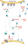
Therapeutic intervention strategies at the level of C3. Inhibition of complement activation at the level of C3 is primarily achieved through three major routes. Although FB and FD inhibitors (small molecules or antibodies) prevent the formation of the AP C3 convertase, engineered versions of complement regulators such as mini-FH or TT30 block convertases by accelerating their decay and enabling the proteolytic degradation of C3b by factor I; both approaches are specific for the AP. C3 inhibitors of the compstatin family (e.g. Cp40) bind to C3 and protect the substrate from being activated by any of the C3 convertases. Complement-targeted drugs available in the clinic, i.e. the anti-C5 mAb eculizumab and C1-inhibitor preparations, which all act at peripheral steps of the cascade, are also shown in the scheme

References

    1. Ricklin D, Lambris JD. Preformed mediators of defense— Gatekeepersenter the spotlight. Immunol Rev. 2016;274:5–8. - PubMed
    1. Chaplin H., Jr Review: The burgeoning history of the complement system 1888–2005. Immunohematology. 2005;21:85–93. - PubMed
    1. Ricklin D, Hajishengallis G, Yang K, Lambris JD. Complement: A key system for immune surveillance and homeostasis. Nat Immunol. 2010;11:785–797. - PMC - PubMed
    1. Ricklin D, Reis ES, Lambris JD. Complement in disease: A defence system turning offensive. Nat Rev Nephrol. 2016;12:383–401. - PMC - PubMed
    1. Merle NS, Noe R, Halbwachs-Mecarelli L, Fremeaux-Bacchi V, Roumenina LT. Complement system Part II: Role in immunity. Front Immunol. 2015;6:257. - PMC - PubMed

Publication types