Review
. 2016 Nov;274(1):270-280.
doi: 10.1111/imr.12470.

Injury site-specific targeting of complement inhibitors for treating stroke

Affiliations
Review

Injury site-specific targeting of complement inhibitors for treating stroke

Ali Alawieh et al. Immunol Rev. 2016 Nov.

Abstract

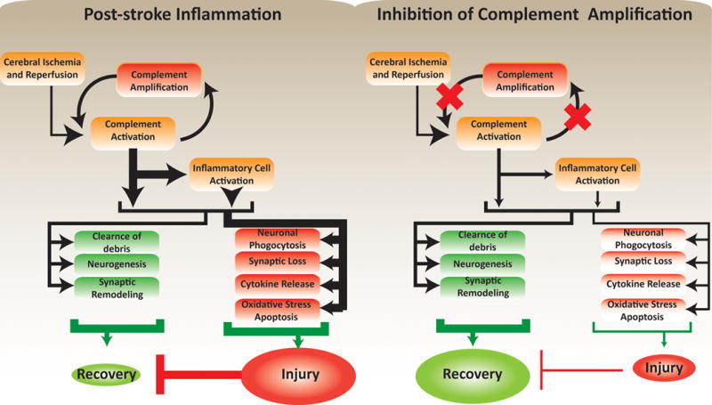
Cumulative evidence indicates a role for the complement system in both pathology and recovery after ischemic stroke. Here, we review the current understanding of the dual role of complement in poststroke injury and recovery, and discuss the challenges of anti-complement therapies. Most complement directed therapeutics currently under investigation or development systemically inhibit the complement system, but since complement is important for immune surveillance and is involved in various homeostatic activities, there are potential risks associated with systemic inhibition. Depending on the target within the complement pathway, other concerns are high concentrations of inhibitor required, low efficacy and poor bioavailability. To overcome these limitations, approaches to target complement inhibitors to specific sites have been investigated. Here, we discuss targeting strategies, with a focus on strategies developed in our lab, to specifically localize complement inhibition to sites of tissue injury and complement activation, and in particular to the postischemic brain. We discuss various injury site-specific targeted complement inhibitors as potential therapeutic agents for the treatment of ischemic stroke treatment, as well as their use as investigative tools for probing complement-dependent pathophysiological processes.

Keywords: complement system; inflammation; stroke; stroke therapeutics; tissue targeting.


Conflict of interest statement

The authors have no conflicts of interest

Figures

Figure 1
Figure 1. Overview of the complement system and complement regulators
In addition to extrinsic pathways that may contribute to complement activation through serum proteases (plasmin and thrombin), complement is activated by one of three pathways: 1. The classical pathway, via C1q binding to Fc-domains of antigen-bound antibodies or directly to apoptotic or stressed cells, 2. The lectin pathway, via binding of MBL to sugar moieties on pathogenic surfaces and glycosylated proteins, and 3. The alternative pathway, via spontaneous hydrolysis of serum complement C3 (“tick-over”) with the involvement of factors B and D. Both classical and lectin pathway form a C3 convertase by cleaving C4 and C2 to form C4b2a. The alternative pathway C3 convertase is formed by association of C3b with factor B and subsequent cleavage of factor B by factor D forming C3bBb. The initial products of C3 cleavage are C3b and C3a. Deposited C3b can further amplify C3 cleavage by recruitment of the alternative pathway, and additionally serves as an opsonin that is subsequently processed into iC3b and C3d. C3b can also associate with C3 convertase (C4b2a or C3bBb), forming a C5 convertase that cleaves C5 into C5b and C5a. C3a and C5a are termed anaphylatoxins and signal through G-protein coupled receptors. C5b deposits on cell surfaces and recruits downstream complement proteins C6-C9 leading to the formation of membrane attack complex (MAC). Several complement regulators that function at different points in the complement pathway serve as checkpoints that prevent uncontrolled complement activation (brown circles in the figure).
Figure 2
Figure 2. Triggers and consequences of complement activation after stroke
Following stroke or cerebral ischemia-reperfusion injury, complement can be activated by the direct binding of C1q and MBL to stressed cells, or via natural antibodies that recognize danger-associated molecular patterns (DAMPs). Reperfusion and activation of serum proteases like thrombin and plasmin can also provide an extrinsic mechanism to activate complement after stroke. Following initial activation, complement activity is amplified by the alternative pathway leading to deposition of opsonins, MAC and release of anaphylatoxins, all of which are involved in injury and recovery after stroke. Signaling through C3aR and C5aR1 has been implicated in promoting expression of adhesion molecules on endothelial surfaces, and to directly stimulate activation and migration of neutrophils and monocytes/microglia. Complement anaphylatoxins have also been shown to play a role in promoting neuronal stem cell proliferation and migration after stroke. C3d and other complement opsonins can also serve as targets and activators for phagocytic cells, but can also contribute to exacerbated loss of function after stroke by promoting synaptic and neuronal uptake by microglia and other phagocytic cells. Through a similar mechanism, complement opsonins can also beneficially assist with the clearance of apoptotic and dead cells, the resolution of inflammation, and synaptic remodeling after stroke. Finally, although several studies have shown MAC deposition in the brain after stroke, the role of MAC in post-stroke injury and recovery is still not well understood.
Figure 3
Figure 3. Brain localization of targeted versus untargeted fH to ischemic brain
Adult male C57BL/6 mice were subjected to 60 min right MCAO followed by reperfusion, and fluorescently labeled fH or CR2-fH were administered 30 min after reperfusion. In-vivo fluorescence tomography was performed daily and the average signal per unit area in the head was calculated. Figure shows localization of CR2-fH in brains of mice after ischemic stroke with a calculated half-life of 48.5 hrs. The right panel shows ex-vivo imaging of isolated brains at 5 days after injury, revealing the presence of CR2-fH specifically in the ipsilateral hemisphere.
Figure 4
Figure 4
Mechanism of action of different site-targeted complement inhibitors in the context of ischemic stroke
Figure 5
Figure 5. Pathophysiological responses after ischemic stroke and its modulation by targeted inhibition of complement amplification
Inhibition of complement amplification reduces inflammation-associated neuropathology. The reduced injury lessens the inhibitory effects of injury on regeneration, leading to optimal recovery.

References

    1. Mozaffarian Dariush, et al. Executive Summary: Heart Disease and Stroke Statistics—2016 Update A Report From the American Heart Association. Circulation. 2016;133.4:447–454. APA. - PubMed
    1. Kochanek K, Murphy S, Xu J, Arias E. Mortality in the United States, 2013. Hyattsville, MD: National Center for Health Statistics; 2014. (NCHS data brief, no. 178). 2014. - PubMed
    1. Murray CJ, et al. Disability-adjusted life years (DALYs) for 291 diseases and injuries in 21 regions, 1990–2010: a systematic analysis for the Global Burden of Disease Study 2010. Lancet. 2012;380:2197–2223. - PubMed
    1. Towfighi A, Saver JL. Stroke declines from third to fourth leading cause of death in the United States: historical perspective and challenges ahead. Stroke. 2011;42:2351–2355. - PubMed
    1. Tissue plasminogen activator for acute ischemic stroke. The National Institute of Neurological Disorders and Stroke rt-PA Stroke Study Group. N Engl J Med. 1995;333:1581–1587. - PubMed

Publication types

Substances