. 2016 Dec;5(23):3046-3055.
doi: 10.1002/adhm.201600699. Epub 2016 Oct 26.

Pericyte Seeded Dual Peptide Scaffold with Improved Endothelialization for Vascular Graft Tissue Engineering

Affiliations

Pericyte Seeded Dual Peptide Scaffold with Improved Endothelialization for Vascular Graft Tissue Engineering

Paola Campagnolo et al. Adv Healthc Mater. 2016 Dec.

Abstract

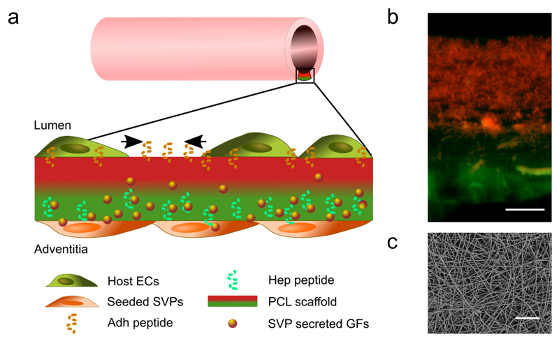
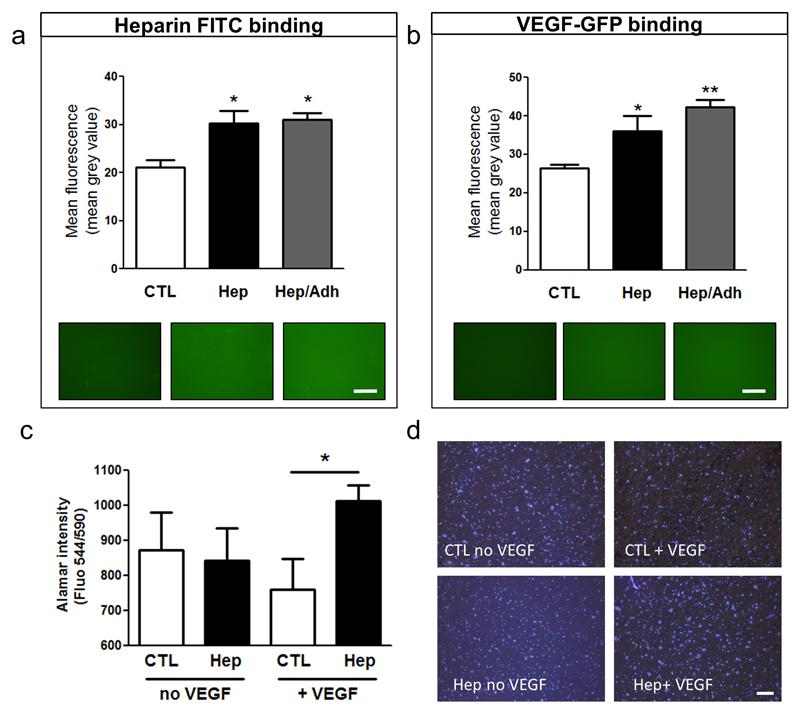
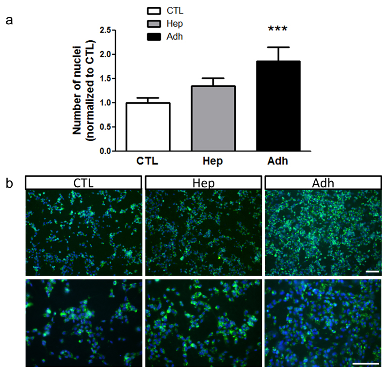
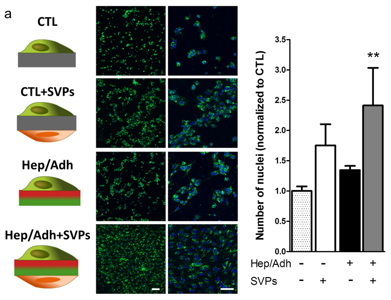
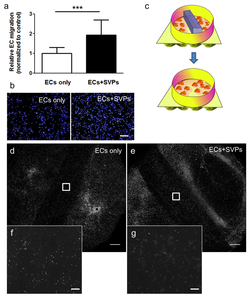
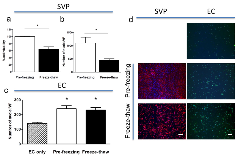
The development of synthetic vascular grafts for coronary artery bypass is challenged by insufficient endothelialization, which increases the risk of thrombosis, and the lack of native cellular constituents, which favors pathological remodeling. Here, a bifunctional electrospun poly(ε-caprolactone) (PCL) scaffold with potential for synthetic vascular graft applications is presented. This scaffold incorporates two tethered peptides: the osteopontin-derived peptide (Adh) on the "luminal" side and a heparin-binding peptide (Hep) on the "abluminal" side. Additionally, the "abluminal" side of the scaffold is seeded with saphenous vein-derived pericytes (SVPs) as a source of proangiogenic growth factors. The Adh peptide significantly increases endothelial cell adhesion, while the Hep peptide promotes accumulation of vascular endothelial growth factor secreted by SVPs. SVPs increase endothelial migration both in a transwell assay and a modified scratch assay performed on the PCL scaffold. Seeding of SVPs on the "abluminal"/Hep side of the scaffold further increases endothelial cell density, indicating a combinatory effect of the peptides and pericytes. Finally, SVP-seeded scaffolds are preserved by freezing in a xeno-free medium, maintaining good cell viability and function. In conclusion, this engineered scaffold combines patient-derived pericytes and spatially organized functionalities, which synergistically increase endothelial cell density and growth factor retention.

Keywords: biofunctionalization; electrospinning; endothelialization; pericytes; tissue engineered vascular graft.


Figures

Figure 1
Figure 1. Characterization of the engineered scaffold for blood vessel graft applications.
The bifunctional scaffold is composed of electrospun peptide-conjugated polycaprolactone (PCL) fibres seeded on the abluminal side with patient-derived pro-angiogenic saphenous vein pericytes (SVPs). As shown in the schematic (a) and the overlaid fluorescence microscopy image (b), the luminal side of the scaffold is mainly decorated with the Adh peptide (in red), to increase host endothelial cell (ECs) adhesion, migration and spreading. The outer layer presents mainly the Hep peptide (in green), which binds and present the SVP-produced growth factors (GFs). SEM image showing the size and distribution of the fibres in the scaffold (top view, c). Scale bars: 50 μm.
Figure 2
Figure 2. Hep peptide binds and coordinates SVP-produced growth factors.
Mean fluorescence (mean grey value) measuring the binding of heparin-FITC (a) and SVP-secreted VEGF-GFP (b) to unconjugated (CTL), Hep-conjugated (Hep) and dual peptide (Hep/Adh) scaffolds. Alamar blue (c) and representative images (d) show the increased HUVEC growth on VEGF loaded Hep mats. *P<0.05, **P<0.01. Scale bars: 500 μm. N = 3; n=3.
Figure 3
Figure 3. Adh peptide specifically increases endothelial cell adhesion and growth.
HUVECs were seeded on electrospun PCL scaffolds either unconjugated (CTL) or functionalised with Hep or Adh peptides. Cell density was measured at 48 hours post-seeding and is expressed as fold change over CTL (a, N = 4; n = 2). Representative images showing endothelial coverage on the different scaffolds (green: WGA-488, blue: DAPI, b). ***P<0.001. Scale bars: 100 μm.
Figure 4
Figure 4. The combination of bifunctional scaffold and SVP seeding increases endothelial coverage.
Fluorescently labelled HUVECs were seeded on plain PCL (CTL) or dual peptide scaffolds (Hep/Adh), in presence or absence of pericytes (SVPs). Representative confocal micrographs showing the resulting coverage in each condition (a; green: WGA-488; blue: DAPI). Number of nuclei quantified at 48 hours is shown as a ratio over the control (b; plain PCL, no SVPs; N = 3; n = 2). **P<0.01 vs. CTL. Scale bar: 100 μm.
Figure 5
Figure 5. Seeding of SVPs induces EC migration and gap closure.
In a transwell™ migration assay, the seeding of SVPs (ECs+SVPs) in the lower chamber induces EC migration, as compared to EC spontaneous migration (ECs only). Relative migration quantification (a) and representative pictures are shown (b, blue: DAPI; N = 7; n = 4). The Hep/Adh scaffold was secured in a CellCulture™ crown, after seeding with SVPs on the lower side (ECs+SVPs) and a barrier was placed in the middle to prevent HUVEC adhesion and create a gap. HUVECs were plated on the top chamber and the barrier was removed to allow their migration (c). Gap closure was monitored at 48 hours, showing that seeding of the SVPs on the lower side of the scaffold promotes HUVEC gap invasion (d-g; grey scale: WGA-488; N=3). ***P<0.001. Scale bars: 400 μm (b, f and g) and 2 mm (d and e).
Figure 6
Figure 6. Freezing of SVP-seeded PCL preserves cell viability and function.
SVP seeded grafts were frozen and recovered, preserving over 65% viability (a) and 50% cell confluency (b). Freeze-thawed SVP were equally able to promote EC density, as compared to freshly plated SVP (c). SVP after the freeze and thaw process remain adherent to the scaffold (d, red: WGA-568) and promoted EC density (d, green: WGA-488). *P<0.05. Scale bars: 200 μm. N = 3.

References

    1. Ratcliffe A. On the occasion of the XVII Meeting of the FECTS. 2000;19:353.
    1. Rocco KA, Maxfield MW, Best CA, Dean EW, Breuer CK. Tissue Eng Part B Rev. 2014;20:628. - PubMed
    1. Hasan A, Memic A, Annabi N, Hossain M, Paul A, Dokmeci MR, Dehghani F, Khademhosseini A. Acta Biomater. 2014;10:11. - PMC - PubMed
    1. Cleary MA, Geiger E, Grady C, Best C, Naito Y, Breuer C. Trends Mol Med. 2012;18:394. - PubMed
    1. Kaushal S, Amiel GE, Guleserian KJ, Shapira OM, Perry T, Sutherland FW, Rabkin E, Moran AM, Schoen FJ, Atala A, Soker S, et al. Nat Med. 2001;7:1035. - PMC - PubMed

Publication types

MeSH terms