. 2016 Oct 26:5:e18790.
doi: 10.7554/eLife.18790.

NPAS1-ARNT and NPAS3-ARNT crystal structures implicate the bHLH-PAS family as multi-ligand binding transcription factors

Affiliations

NPAS1-ARNT and NPAS3-ARNT crystal structures implicate the bHLH-PAS family as multi-ligand binding transcription factors

Dalei Wu et al. Elife. .

Abstract

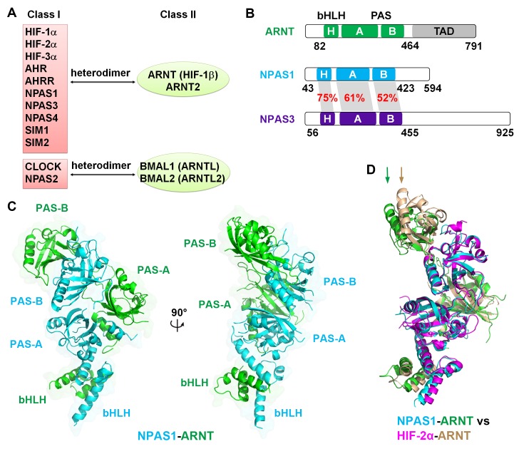
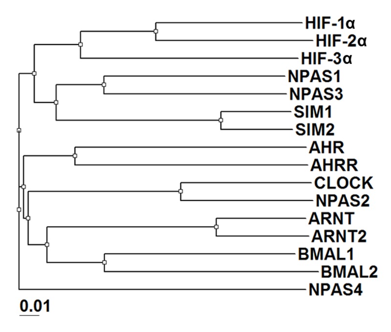
The neuronal PAS domain proteins NPAS1 and NPAS3 are members of the basic helix-loop-helix-PER-ARNT-SIM (bHLH-PAS) family, and their genetic deficiencies are linked to a variety of human psychiatric disorders including schizophrenia, autism spectrum disorders and bipolar disease. NPAS1 and NPAS3 must each heterodimerize with the aryl hydrocarbon receptor nuclear translocator (ARNT), to form functional transcription complexes capable of DNA binding and gene regulation. Here we examined the crystal structures of multi-domain NPAS1-ARNT and NPAS3-ARNT-DNA complexes, discovering each to contain four putative ligand-binding pockets. Through expanded architectural comparisons between these complexes and HIF-1α-ARNT, HIF-2α-ARNT and CLOCK-BMAL1, we show the wider mammalian bHLH-PAS family is capable of multi-ligand-binding and presents as an ideal class of transcription factors for direct targeting by small-molecule drugs.

Keywords: Hypoxia-Inducible Factor; Neuronal PAS Proteins; PAS domains; bHLH-PAS proteins; biophysics; crystal structure; drug discovery; human; ligand binding pockets; mouse; psychiatric disorders; structural biology; transcription factor.


Conflict of interest statement

The authors declare that no competing interests exist.

Figures

Figure 1.
Figure 1.. Comparison of bHLH-PAS proteins.
(A) Heterodimerization patterns between bHLH-PAS proteins. (B) Protein domain arrangements of ARNT, NPAS1 and NPAS3. Percent amino-acid identities between corresponding domains of NPAS1 and NPAS3 are in red. (C) Overall crystal structure of the NPAS1-ARNT complex shown in two views. (D) Superposition of NPAS1-ARNT and HIF-2α-ARNT heterodimers. The arrows on top show the shift in position for the PAS-B domain of ARNT. DOI: http://dx.doi.org/10.7554/eLife.18790.003
Figure 1—figure supplement 1.
Figure 1—figure supplement 1.. Phylogenetic tree of all mouse bHLH-PAS family members based on protein sequences at their bHLH-PAS-A-PAS-B regions.
DOI: http://dx.doi.org/10.7554/eLife.18790.004
Figure 1—figure supplement 2.
Figure 1—figure supplement 2.. Comparative protein sequence analysis of mouse bHLH-PAS proteins.
The bHLH, PAS-A and PAS-B segments, and the secondary structure components of NPAS1 are shown above the alignment. The NPAS1 residues interacting directly with ARNT (at interface 1–4) are marked using cyan round dots, while those at intra-molecular interfaces (5 and 6) are marked with brown round dots. Triangles show pocket amino-acids in NPAS1, HIF-2α, CLOCK, ARNT and BMAL1 (in cyan, magenta, brown, green and lime colors, respectively). DOI: http://dx.doi.org/10.7554/eLife.18790.005
Figure 1—figure supplement 3.
Figure 1—figure supplement 3.. Comparison of the overall structures of NPAS1-ARNT and CLOCK-BMAL1 complexes.
The two complexes are superimposed by aligning the bHLH domains (A) or PAS-B domains (B) of ARNT with BMAL1, respectively. DOI: http://dx.doi.org/10.7554/eLife.18790.006
Figure 2.
Figure 2.. Comparison of NPAS1-ARNT and NPAS3-ARNT complexes.
(A) DNA-binding affinities measured using fluorescence anisotropy. The Kd values of NPAS1-ARNT and NPAS3-ARNT binding to the same HRE element were 16.3 ± 1.1 nM and 17.5 ± 1.1 nM, as calculated from three technical replicates respectively. (B) Overall structures of the NPAS3-ARNT-DNA, with the hexameric HRE site colored in cyan and blue. (C) Structure comparison of NPAS3-ARNT-DNA and HIF-2α-ARNT-DNA complexes aligned on their DNA. (D) Superposition of NPAS3-ARNT and NPAS1-ARNT structures. DNA was omitted from the NPAS3-ARNT complex. Arrows show the extension of α1 helices likely associated with its DNA binding. DOI: http://dx.doi.org/10.7554/eLife.18790.008
Figure 3.
Figure 3.. Testing of domain interfaces in NPAS1-ARNT and other ARNT heterodimers using mutagenesis.
(A) Detailed interactions at each of six interfaces in NPAS1-ARNT. Each junction is circled in the context of the overall structure of NPAS1-ARNT on the left, and each interface is further listed in the table. (B) Co-IP experiments showing the effects of ARNT mutations at interfaces 1–4 on the cellular stabilities of heterodimers formed with NPAS1, NPAS3, SIM1, NPAS4 and AHR, respectively. (C) Co-IP experiments showing the effects of NPAS1 mutations at interfaces 5 and 6 on the stabilities of NPAS1-ARNT heterodimer. (D) Luciferase reporter assay testing the effects of NPAS1 (wide-type and three mutants) on HRE-driven (left) and tyrosine hydroxylase (TH) promoter-driven (right) transactivation. Each sample represents the average reading of cells from three wells. (E) Co-IP experiments showing the effects of ARNT2 mutations at interfaces 1–4 (corresponding to ARNT) on the stabilities of heterodimers formed with SIM1 and NPAS4. The residues and mutants of ARNT, NPAS1 and ARNT2 are labelled in green, cyan and brown, respectively. DOI: http://dx.doi.org/10.7554/eLife.18790.009
Figure 3—figure supplement 1.
Figure 3—figure supplement 1.. Amino-acid sequence alignment of full-length mouse ARNT and ARNT2 proteins.
Secondary structures and positions of the bHLH, PAS-A and PAS-B domains of ARNT are indicated on top. The ARNT residues participating at dimerization interfaces are indicated with green dots. DOI: http://dx.doi.org/10.7554/eLife.18790.010
Figure 4.
Figure 4.. Comparison of ligand-binding pockets among multiple bHLH-PAS proteins.
(A and E) Relative positions of the four pockets (red circles) within the structures of NPAS1-ARNT (A) and CLOCK-BMAL1 (E) complexes. (B–D and F–H) Empty pockets in each of PAS-A and PAS-B domains of NPAS1 (B), ARNT (C), HIF-2α (D), CLOCK (F), BMAL1 (G) and SIM1 (H). The accessible cavities are shown in red meshes, together with the amino-acid residues lining each pocket. For NPAS1, the surrounding residues are further covered with 2FO – FC map contoured at 1.0σ (B). The NPAS1 residues with identical counterparts in NPAS3 are in cyan and labelled in black, while those with non-conserved NAPS3 counterparts (indicated in parentheses) are in magenta and labelled in red (B). ARNT pocket residues from apo HIF-2α-ARNT complex (PDB: 4ZP4) (in wheat) and those from NPAS1-ARNT complex (in green) are superposed for comparison (C). The HIF-2α residues with identical counterparts in HIF-1α are in magenta, while those with non-conserved HIF-1α counterparts (indicated in parentheses) are in green (D). Structures of CLOCK (F) and BMAL1 (G) are from the CLOCK-BMAL1 complex (PDB: 4F3L) in brown and lime, respectively. The SIM1 structure in orange was modelled from NPAS1 and HIF-2α; and the pocket residues not conserved in SIM2 are in green with their SIM2 counterparts indicated in parentheses (H). DOI: http://dx.doi.org/10.7554/eLife.18790.011

References

    1. Adams PD, Afonine PV, Bunkóczi G, Chen VB, Davis IW, Echols N, Headd JJ, Hung LW, Kapral GJ, Grosse-Kunstleve RW, McCoy AJ, Moriarty NW, Oeffner R, Read RJ, Richardson DC, Richardson JS, Terwilliger TC, Zwart PH. PHENIX: a comprehensive Python-based system for macromolecular structure solution. Acta Crystallographica Section D Biological Crystallography. 2010;66:213–221. doi: 10.1107/S0907444909052925. - DOI - PMC - PubMed
    1. Ao A, Wang H, Kamarajugadda S, Lu J. Involvement of estrogen-related receptors in transcriptional response to hypoxia and growth of solid tumors. PNAS. 2008;105:7821–7826. doi: 10.1073/pnas.0711677105. - DOI - PMC - PubMed
    1. Bersten DC, Bruning JB, Peet DJ, Whitelaw ML. Human variants in the neuronal basic helix-loop-helix/Per-Arnt-Sim (bHLH/PAS) transcription factor complex NPAS4/ARNT2 disrupt function. PLoS One. 2014;9:e85768. doi: 10.1371/journal.pone.0085768. - DOI - PMC - PubMed
    1. Bersten DC, Sullivan AE, Peet DJ, Whitelaw ML. Bhlh-pas proteins in cancer. Nature Reviews Cancer. 2013;13:827–841. doi: 10.1038/nrc3621. - DOI - PubMed
    1. Biasini M, Bienert S, Waterhouse A, Arnold K, Studer G, Schmidt T, Kiefer F, Gallo Cassarino T, Bertoni M, Bordoli L, Schwede T. SWISS-MODEL: modelling protein tertiary and quaternary structure using evolutionary information. Nucleic Acids Research. 2014;42:W252–W258. doi: 10.1093/nar/gku340. - DOI - PMC - PubMed

Publication types

MeSH terms

Substances