. 2016 Oct 26:5:e20934.
doi: 10.7554/eLife.20934.

Eukaryotic translation initiation factor 3 plays distinct roles at the mRNA entry and exit channels of the ribosomal preinitiation complex

Affiliations

Eukaryotic translation initiation factor 3 plays distinct roles at the mRNA entry and exit channels of the ribosomal preinitiation complex

Colin Echeverría Aitken et al. Elife. .

Abstract

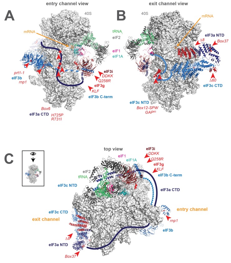
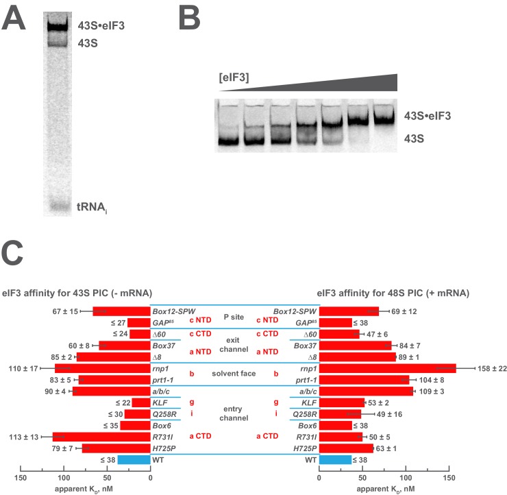
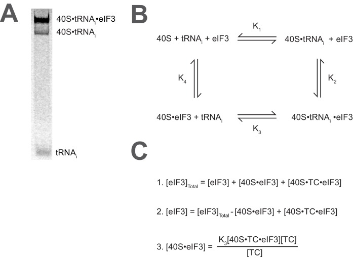
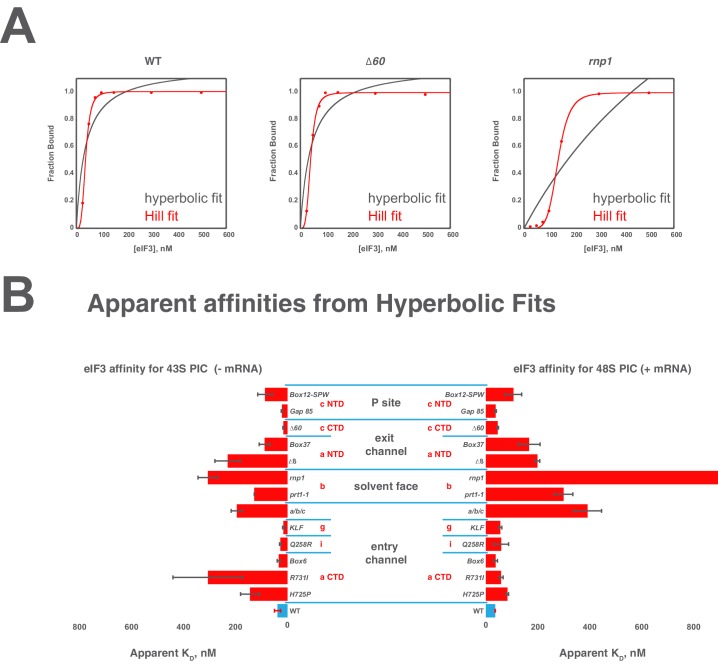
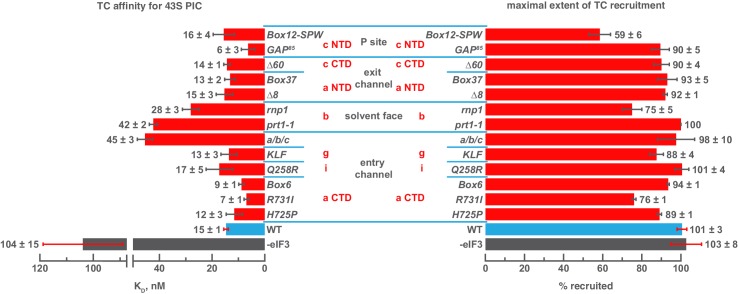
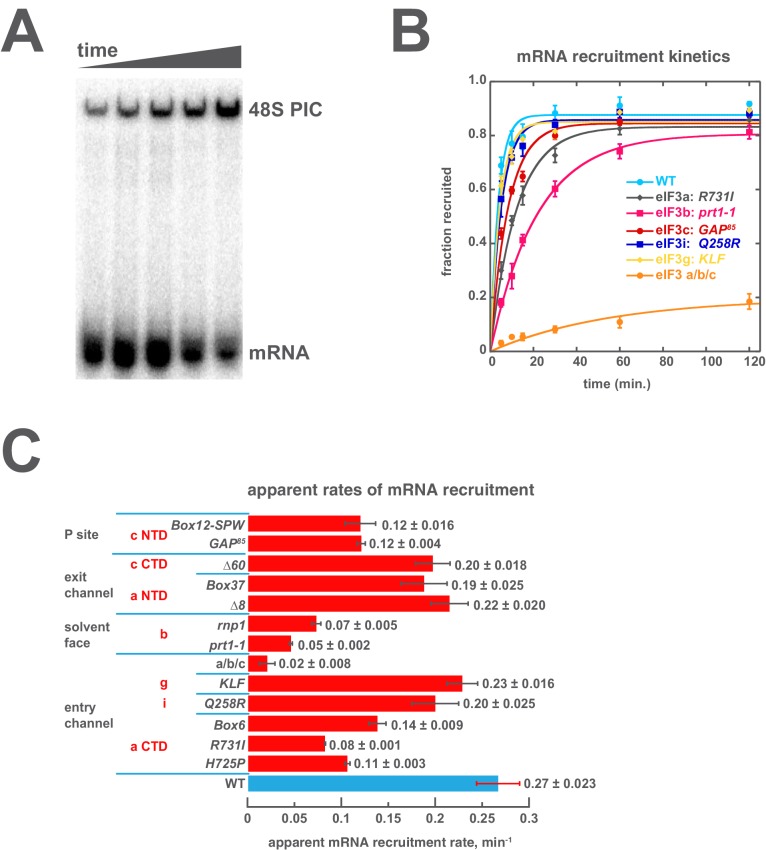
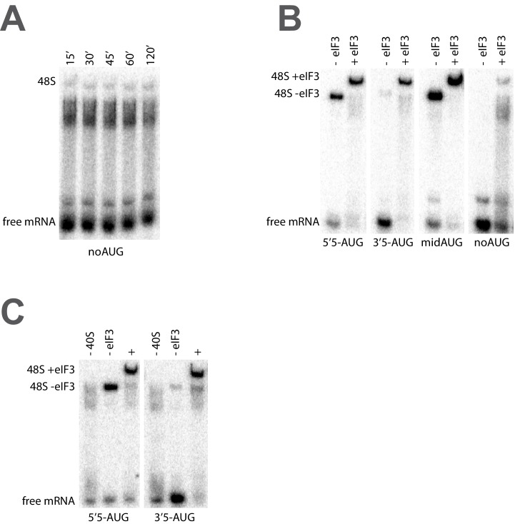
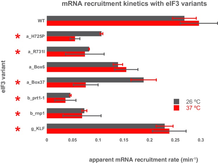
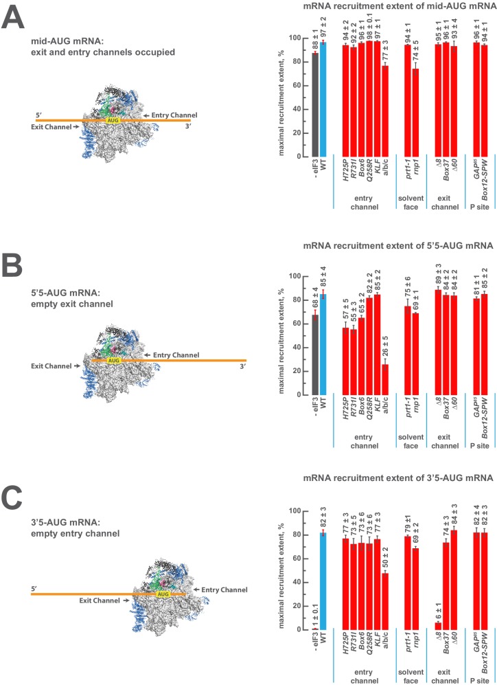
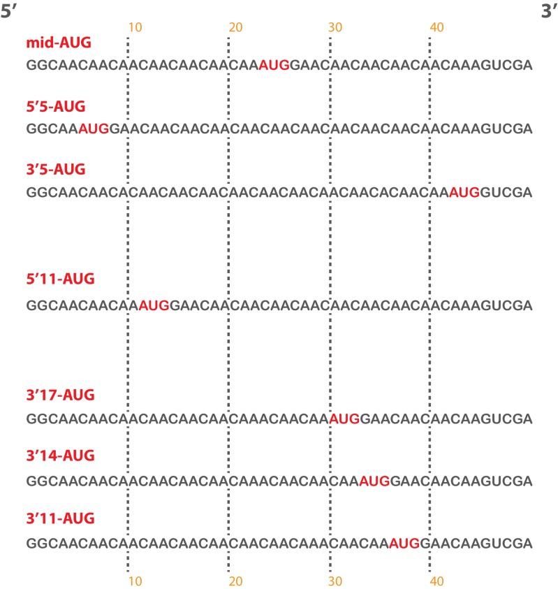
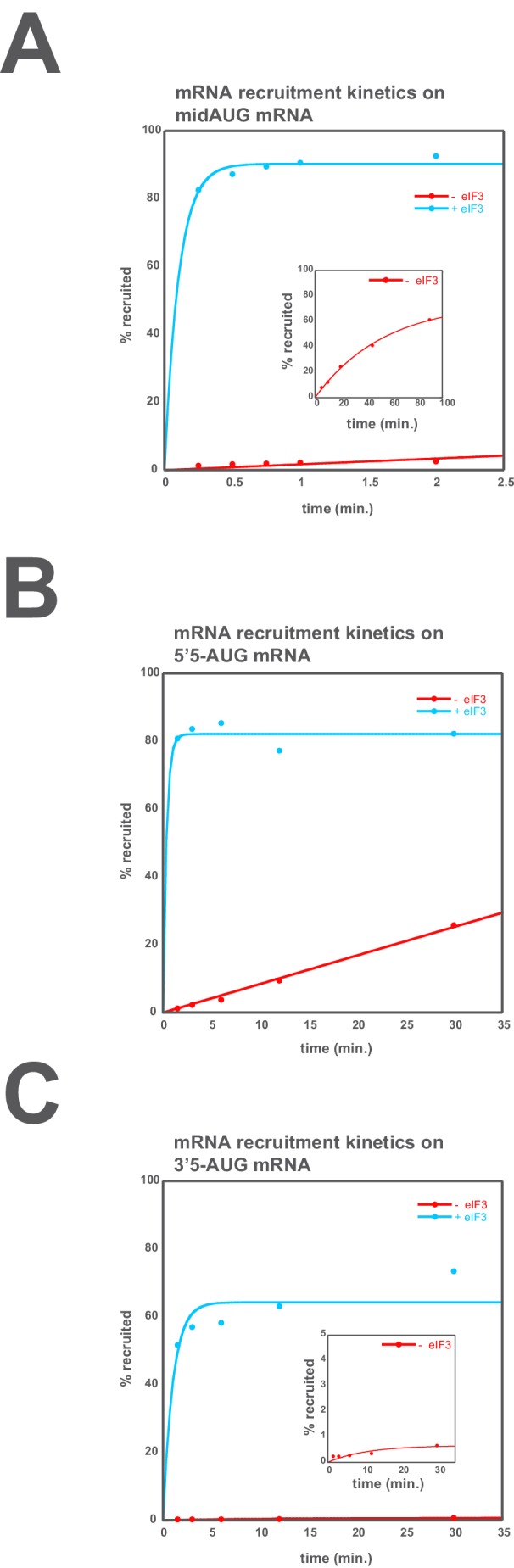
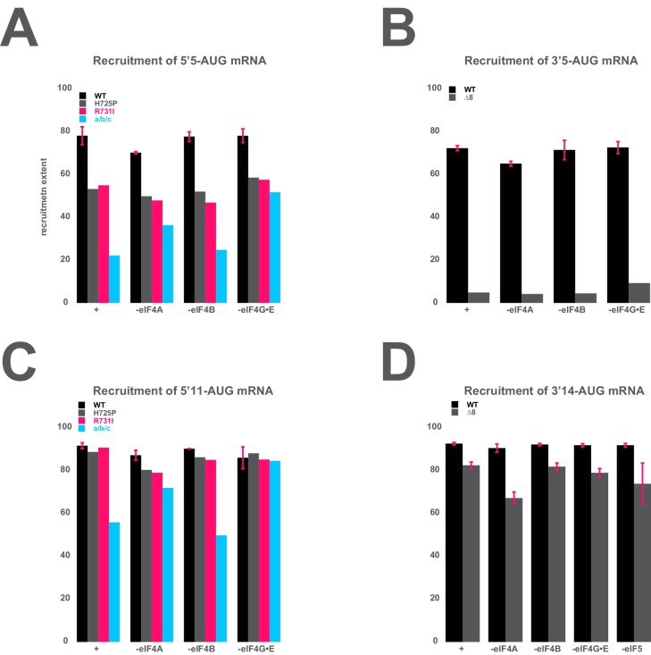
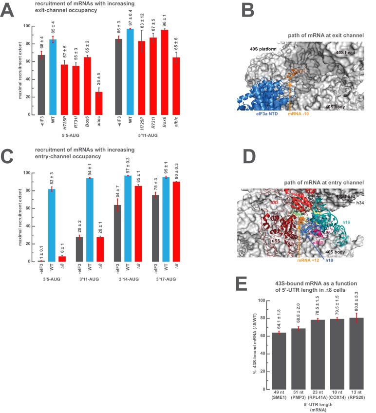
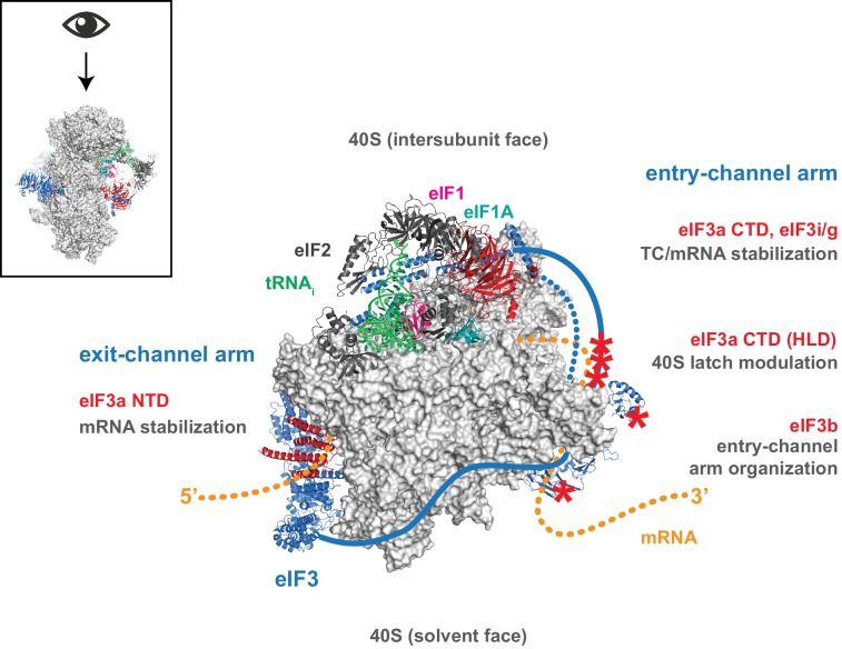
Eukaryotic translation initiation factor 3 (eIF3) is a central player in recruitment of the pre-initiation complex (PIC) to mRNA. We probed the effects on mRNA recruitment of a library of S. cerevisiae eIF3 functional variants spanning its 5 essential subunits using an in vitro-reconstituted system. Mutations throughout eIF3 disrupt its interaction with the PIC and diminish its ability to accelerate recruitment to a native yeast mRNA. Alterations to the eIF3a CTD and eIF3b/i/g significantly slow mRNA recruitment, and mutations within eIF3b/i/g destabilize eIF2•GTP•Met-tRNAi binding to the PIC. Using model mRNAs lacking contacts with the 40S entry or exit channels, we uncovered a critical role for eIF3 requiring the eIF3a NTD, in stabilizing mRNA interactions at the exit channel, and an ancillary role at the entry channel requiring residues of the eIF3a CTD. These functions are redundant: defects at each channel can be rescued by filling the other channel with mRNA.

Keywords: S. cerevisiae; biochemistry; biophysics; eIF3; initiation; mRNA recruitment; ribosome; structural biology; translation; yeast.


Conflict of interest statement

AGH: Reviewing editor, eLife. The other authors declare that no competing interests exist.

Figures

Figure 1.
Figure 1.. A library of S. cerevisiae eIF3 functional variants affected by mutations spanning all five essential subunits.
Three views of the cryo-EM structure of eIF3 bound to the partial yeast 48S PIC in the closed conformation (py48S-closed), as reported by Llacer, et al. The 40S subunit is shown in light grey, with resolved domains of eIF3 shown as ribbons and unresolved regions cartooned as solid or dotted lines: eIF3a is shown in dark blue, eIF3c and eIF3b in light blue, eIF3i in maroon, and eIF3g in red. The location of mutations throughout eIF3 are shown colored in red and indicated by red arrowheads; the eIF3i and eIF3g subunits are also depicted in red hues owing to their absence in the a/b/c subcomplex resulting from the DDKK mutation in eIF3i. The mRNA (orange), Met-tRNAi (green), eIF2 (charcoal), eIF1 (pink), and eIF1A (teal) are also visible in this structure. (A) View looking towards the mRNA entry channel, with the 40S intersubunit face on the right, and the solvent face on the left. (B) Opposite view looking towards the mRNA exit channel, with the 40S intersubunit face on the left, and the solvent face on the right. (C) View of the PIC looking down at the 40S head, showing the relative orientation of the entry- and exit-channel arms of eIF3 and its contacts on the 40S solvent face. The PIC is oriented such that the intersubunit face appears at the top, while the solvent face appears at the bottom. DOI: http://dx.doi.org/10.7554/eLife.20934.003
Figure 2.
Figure 2.. Mutations in the mRNA entry- or exit-channel arms of eIF3 or in eIF3b destabilize eIF3 binding to the PIC.
(A) Binding of ternary complex containing [35S]-Met-tRNAiMet to 40S ribosomal subunits in the presence of eIF1, eIF1A, and WT or mutant eIF3 was measured using a native gel-shift assay, which separates free [35S]-Met-tRNAiMet from that bound to 43S PICs alone or 43S PICs containing eIF3 (43S·eIF3). (B) The titration of eIF3 into reactions containing 43S PICs produces a well-resolved gel-shift that monitors the binding of eIF3 to the PIC. The amounts of [35S]-Met-tRNAiMet free, bound to 43S PICs, and bound to 43S∙eIF3 complexes were quantified and analyzed as described in Figure 2—figure supplement 1. (C) The apparent equilibrium dissociation constant (KD) of WT (blue bars) and eIF3 variants (red bars) for the PIC, both in the absence (left) and presence (right) of mRNA, obtained by fitting the data with the Hill equation (see Figure 2—figure supplement 2B). Bars and errors represent mean and SEM, respectively (as determined by individual fitting of each experiment), of ≥2 experiments. Owing to the conditions of these assays, apparent affinities ≤ 30 nM likely represent upper limits. DOI: http://dx.doi.org/10.7554/eLife.20934.005
Figure 2—figure supplement 1.
Figure 2—figure supplement 1.. Calculating the affinity of eIF3 for PICs under conditions where free 40S subunits compete for binding.
(A) Native gel electrophoresis of PICs formed in the presence of [35S]-Met-tRNAiMet resolves free tRNAi and all other TC-bound species. (B) Thermodynamic cycle for the binding of both eIF3 and tRNAi (as TC) to 40S (as 40S•eIF1•eIF1A). At 40S concentrations near or below the dissociation constant for tRNAi binding to 40S (K1), free 40S subunits compete with 40S•tRNAi for eIF3 such that the concentration of total eIF3 is not equivalent to the concentration of free eIF3 necessary to determine its affinity for 40S•tRNAi (K2) (C) The concentration of free eIF3 ([eIF3]) can be determined from the total concentration of eIF3 ([eIF3]Total) and the concentration of all other eIF3-containing species. The concentration of eIF3 bound to PICs ([40S•TC•eIF3]) can be determined empirically, as this species contains radiometrically-labeled tRNAi and can thus be measured. The concentration of eIF3 bound to 40S subunits alone ([40S•eIF3]) cannot be measured directly, but can be calculated from the measured concentrations of eIF3 bound to PICs ([40S•TC•eIF3]), the concentration of free tRNAi ([TC]) (both of which also contain radiometrically-labeled tRNAi and can thus also be measured empirically), and the measured dissociation constant for the binding of tRNAi to the 40S•eIF3 complex (K3). This value can then be used, together with the measured concentrations of other eIF3-bound species, to determine the concentration of free eIF3 and model the data with a Langmuir isotherm to determine K2. DOI: http://dx.doi.org/10.7554/eLife.20934.007
Figure 2—figure supplement 2.
Figure 2—figure supplement 2.. Comparison of hyperbolic and Hill equations for modeling eIF3 binding to the PIC.
(B) Comparison of representative hyperbolic (grey) and Hill (red) fits obtained upon modeling the binding of either WT, ∆60, or rnp1 eIF3 to 43S PICs. In all cases, the Hill fit more accurately models the binding data, and in the case of more severe binding defects, such as that observed for rnp1 eIF3, the hyperbolic (Langmuir) fit is unable to model the data. (A) Apparent affinities of WT (blue bars) and eIF3 variants (red bars) for 43S (left) and 48S (right) PICs, as determined by modeling binding data with a standard hyperbolic (Langmuir) binding isotherm. Bars and errors represent mean values and SEM, respectively (as determined by individual fitting of each experiment), of ≥2 experiments, with the exception of rnp1 eIF3 binding to 48S PICs (right), which could not be accurately modeled with a hypergolic (Langmuir) binding isotherm. DOI: http://dx.doi.org/10.7554/eLife.20934.008
Figure 3.
Figure 3.. Mutations in the mRNA entry-channel arm of eIF3 or in eIF3b destabilize binding of TC to the PIC.
The KD of TC (left) or maximal extent of TC recruitment (right) observed for PICs assembled either in the absence of eIF3 (grey bar), or the presence of either WT (blue bar) or variant eIF3 (red bars). Bars and errors represent mean and SEM, respectively (as determined by individual fitting of each experiment), of ≥2 experiments. DOI: http://dx.doi.org/10.7554/eLife.20934.009
Figure 4.
Figure 4.. Mutations throughout the eIF3 complex compromise its ability to accelerate recruitment of capped native RPL41A mRNA to the PIC.
(A) mRNA recruitment reactions were quenched at appropriate time points on a running native gel to separate free [32P]-capped RPL41A mRNA from mRNA recruited to form 48S PICs. The bands were quantified to determine the fraction of total mRNA bound at each time point. (B) Individual time courses were fit with single-exponential rate equations to determine the observed rate constant for each experiment. (C) The observed rates of mRNA recruitment of capped RPL41A mRNA measured in the presence of WT (blue bar) and variants of eIF3 (red bars). Bars and errors represent mean values and SEM, respectively (as determined by individual fitting of each experiment), of ≥2 experiments. DOI: http://dx.doi.org/10.7554/eLife.20934.011
Figure 4—figure supplement 1.
Figure 4—figure supplement 1.. The recruitment of unstructured model mRNAs to the PIC depends on the presence of an AUG codon and 40S subunits.
(A) Representative mRNA recruitment gel showing recruitment time course for the noAUG unstructured model mRNA, which lacks a start codon. (B) Comparison of mRNA recruitment gels at 120’, both in the presence and absence of eIF3, for the 5’5-AUG, 3’5-AUG, and midAUG mRNAs, all of which contain an AUG codon, and the noAUG mRNA, which lacks a start codon. (C) mRNA recruitment gels for the 5’5-AUG (left panel) and 3’5-AUG mRNAs (right panel), showing lanes containing reactions lacking either 40S subunits (- 40S, left lane) or eIF3 (- eIF3, middle lane), or in the presence of both (+, right lane). DOI: http://dx.doi.org/10.7554/eLife.20934.013
Figure 4—figure supplement 2.
Figure 4—figure supplement 2.. Comparison of the kinetics of mRNA recruitment for WT and variant eIF3 at 26°C and 37°C.
The apparent rates of mRNA recruitment of capped RPL41A mRNA observed in the presence of WT and eIF3 variants that confer Ts- phenotypes in vivo (denoted with asterisks) as well as the box6 variant, which does not. Bars represent apparent recruitment rates at either 26°C (grey bars) or 37°C (red bars). Bars and errors represent mean values and SEM, respectively (as determined by individual fitting of each experiment), of ≥2 experiments. DOI: http://dx.doi.org/10.7554/eLife.20934.014
Figure 5.
Figure 5.. eIF3 strongly stabilizes binding of mRNA at the exit channel of the PIC in a manner dependent on the eIF3a NTD.
(A) The maximal extent of mid-AUG mRNA recruited in the absence of eIF3 (grey bar), or in the presence of either WT (blue bar) or variant eIF3 (red bars). The locations of sequences 5’ or 3’ of the AUG in the PIC, with AUG in the P site, are shown schematically on the left, indicating that both the mRNA entry and exit channels of the PIC are fully occupied. Bars and errors represent mean and SEM, respectively, of ≥2 experiments. (B) The maximal extent of 5’5-AUG mRNA recruited in the absence of eIF3, or in the presence of either WT or variant eIF3. As shown on the left, 5’5 AUG mRNA programs a recruited complex in which only the mRNA entry channel is fully occupied, and thus is sensitized to changes in that channel. (C) The maximal extent of 3’5-AUG mRNA recruited in the absence of eIF3, or in the presence of either WT or variant eIF3. The 3’5-AUG programs a recruited complex in which only the mRNA exit channel is occupied (left), and thus is sensitized to changes in that channel. DOI: http://dx.doi.org/10.7554/eLife.20934.015
Figure 5—figure supplement 1.
Figure 5—figure supplement 1.. Short, unstructured model mRNAs with distinct start codon positions.
Specific sequences of the unstructured model mRNAs used in this study, with the AUG start codon highlighted in red. All model mRNAs are 50 nucleotides in length, and primarily contain unstructured CAA repeats. Each mRNA was in-vitro transcribed and capped, as described in the materials and methods. DOI: http://dx.doi.org/10.7554/eLife.20934.017
Figure 5—figure supplement 2.
Figure 5—figure supplement 2.. eIF3 accelerates the recruitment of unstructured model mRNAs.
Apparent mRNA recruitment kinetics observed in both the presence (blue) and absence (red) of eIF3, for the mid-AUG (A), 5’5-AUG (B), and 3’5-AUG (C) mRNAs. Extended time courses are shown (as insets) for both the midAUG and 3’5-AUG mRNAs. DOI: http://dx.doi.org/10.7554/eLife.20934.018
Figure 5—figure supplement 3.
Figure 5—figure supplement 3.. The mRNA recruitment extent defects observe for entry- and exit-channel variants of eIF3 are not exacerbated in the absence of the eIF4 factors.
(A) The extent of 5’5-AUG mRNA recruited to the PIC in the presence of WT eIF3 (black bars) and eIF3 entry-channel variants (grey, pink, and blue bars), in either the presence all factors (+), or the absence of either eIF4A, eIF4B, or eIF4G•E. (B) The extent of 3’5-AUG mRNA recruited to the PIC in the presence of WT (black bars) and ∆8 (grey bars) eIF3, in either the presence of all factors (+), or the absence of either eIF4A, eIF4B, or eIF4G•E. (C) The extent of 5’11-AUG mRNA recruited to the PIC in the presence of WT eIF3 (black bars) and eIF3 entry-channel variants (grey, pink, and blue bars), in either the presence of all factors (+), or the absence of either eIF4A, eIF4B, or eIF4G•E. (D) The extent of 3’14-AUG mRNA recruited to the PIC in the presence of WT (black bars) and ∆8 (grey bars) eIF3, in either the presence of all factors (+), or the absence of either eIF4A, eIF4B, or eIF4G•E. DOI: http://dx.doi.org/10.7554/eLife.20934.019
Figure 6.
Figure 6.. The presence of sufficient mRNA in the opposite channel rescues the destabilization of mRNA binding caused by mutations in the entry- or exit-channel arms of eIF3.
(A) Bars represent the maximal extent of recruitment of mRNAs that program complexes with either 5 (5’5-AUG) or 11 (5’11-AUG) nucleotides in the mRNA exit channel, in the absence of eIF3 (grey bars) or in the presence of either WT eIF3 (blue bars) or variants with mutations near the mRNA entry channel (red bars). Bars and errors represent mean and SEM, respectively, of ≥2 experiments. (B) The path of mRNA (orange) at the exit channel of the 40S subunit (grey) in the py48S-closed structure (Llácer et al., 2015). mRNA is visualized up to the −10 nucleotide, where it emerges from the exit channel pore and is located approximately 15 Å from the eIF3a NTD (blue). (C) Bars represent the maximal extent of recruitment of mRNAs with 5 (3’5-AUG), 11 (3’11-AUG), 14 (3’14-AUG), or 17 (3’17-AUG) nucleotides in the entry channel in the absence of eIF3 (grey bars) or in the presence of either WT eIF3 (blue bars) or the ∆8 variant (red bars). Bars and errors represent mean and SEM, respectively, of ≥2 experiments. (D) The path of mRNA (orange) at the entry channel of the 40S subunit (grey) in the py48S PIC lacking eIF3 (Hussain et al., 2014). mRNA is visualized up to the +12 nucleotide, where it emerges from the 40S latch composed of h34 (charcoal), h18 (blue), and uS3 (red), and projects towards a constriction formed by uS5 (burgundy), uS3 (red), and eS30 (pink), near h16 (teal). (E) The amount of mRNA cross-linked to PICs in ∆8 cells, as a percentage of mRNA cross-linked to PICs in WT cells, determined for five mRNAs with decreasing 5’-UTR lengths. Bars and errors represent means and SD from two independent biological replicates. DOI: http://dx.doi.org/10.7554/eLife.20934.020
Figure 7.
Figure 7.. Distinct roles for the mRNA entry- and exit-channel arms of eIF3.
A proposed model for the roles of the eIF3 entry- and exit-channel arms during translation initiation. The py48S-closed complex is shown from above (boxed schematic), looking down at the head of the 40S (light grey), with the solvent face at the bottom, and the intersubunit face at the top with Met-tRNAi (green), eIF2 (dark grey), eIF1 (pink), and eIF1A (teal) bound to the decoding center. eIF3 is shown in blue, with areas of interest highlighted in red or denoted with red asterisks; resolved regions of eIF3 are depicted as ribbons, with unresolved regions cartooned as solid or dashed lines. The mRNA is cartooned in orange, entering the PIC though the entry channel at right, and exiting at the pore near the platform at left. Our results suggest that the eIF3 exit-channel arm, and specifically the eIF3a NTD, is critical for stabilizing mRNA binding to the PIC at the exit channel, whereas the eIF3a CTD (unresolved regions appear as solid blue line) and eIF3i/g enhance the stability of mRNA interactions at the entry-channel. The entry-channel arm and its attachment to the solvent face via eIF3b (unresolved regions appear as dashed blue line) is also important for stabilizing TC binding to the PIC and promoting steps that control the kinetics of mRNA recruitment, perhaps as a function of modulating the 40S latch. DOI: http://dx.doi.org/10.7554/eLife.20934.022

References

    1. Acker MG, Kolitz SE, Mitchell SF, Nanda JS, Lorsch JR. Reconstitution of yeast translation initiation. Methods in Enzymology. 2007;430:111–145. doi: 10.1016/S0076-6879(07)30006-2. - DOI - PubMed
    1. Aitken CE, Lorsch JR. A mechanistic overview of translation initiation in eukaryotes. Nature Structural & Molecular Biology. 2012;19:568–576. doi: 10.1038/nsmb.2303. - DOI - PubMed
    1. Algire MA, Maag D, Savio P, Acker MG, Tarun SZ, Sachs AB, Asano K, Nielsen KH, Olsen DS, Phan L, Hinnebusch AG, Lorsch JR. Development and characterization of a reconstituted yeast translation initiation system. RNA. 2002;8:382–397. doi: 10.1017/S1355838202029527. - DOI - PMC - PubMed
    1. Asano K, Clayton J, Shalev A, Hinnebusch AG. A multifactor complex of eukaryotic initiation factors, eIF1, eIF2, eIF3, eIF5, and initiator tRNA(Met) is an important translation initiation intermediate in vivo. Genes & Development. 2000;14:2534–2546. doi: 10.1101/gad.831800. - DOI - PMC - PubMed
    1. Asano K, Phan L, Anderson J, Hinnebusch AG. Complex formation by all five homologues of mammalian translation initiation factor 3 subunits from yeast Saccharomyces cerevisiae. Journal of Biological Chemistry. 1998;273:18573–18585. doi: 10.1074/jbc.273.29.18573. - DOI - PubMed

MeSH terms

LinkOut - more resources