. 2017 Mar;74(6):1117-1131.
doi: 10.1007/s00018-016-2398-4. Epub 2016 Oct 25.

NEAT1 modulates herpes simplex virus-1 replication by regulating viral gene transcription

Affiliations

NEAT1 modulates herpes simplex virus-1 replication by regulating viral gene transcription

Ziqiang Wang et al. Cell Mol Life Sci. 2017 Mar.

Abstract

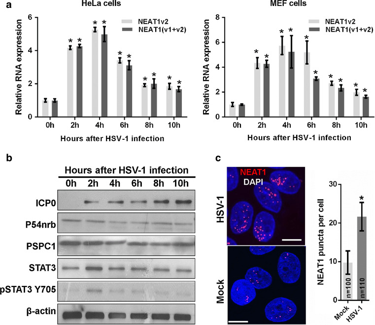
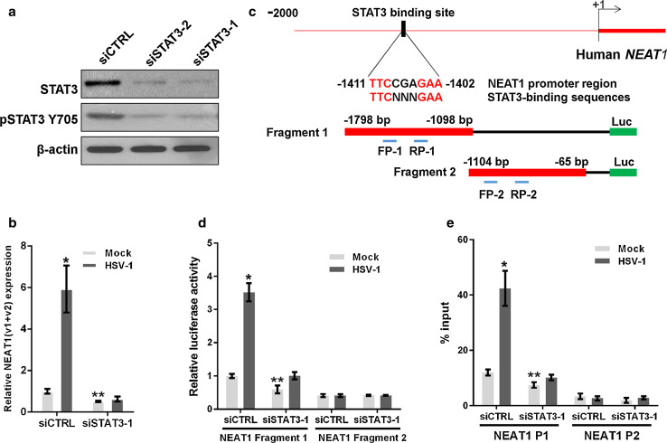
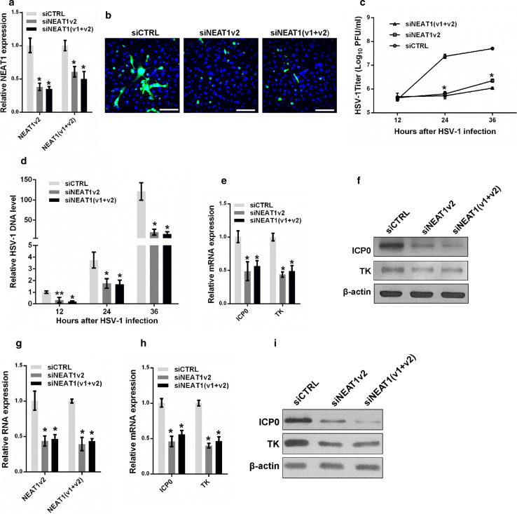
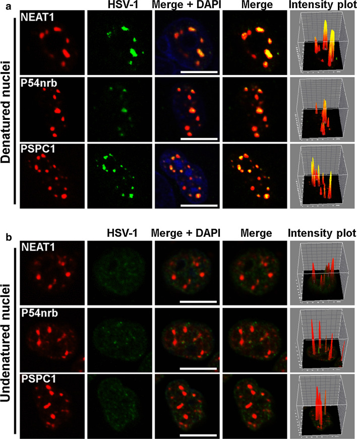
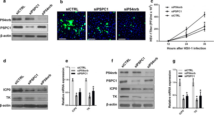
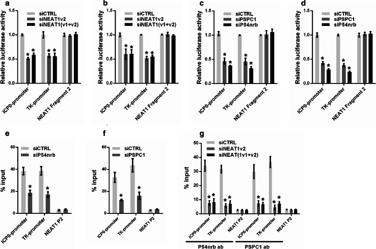
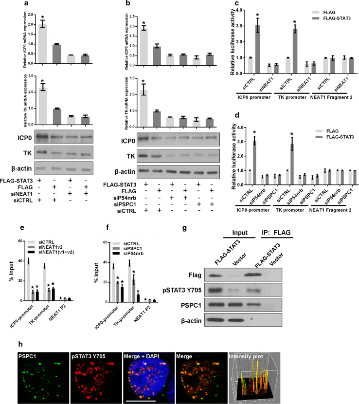
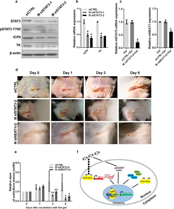
Nuclear paraspeckle assembly transcript 1 (NEAT1) is the crucial structural platform of paraspeckles, which is one type of nuclear bodies. As a stress-induced lncRNA, the expression of NEAT1 increases in response to viral infection, but little is known about the role of NEAT1 or paraspeckles in the replication of herpes simplex virus-1 (HSV-1). Here, we demonstrate that HSV-1 infection increases NEAT1 expression and paraspeckle formation in a STAT3-dependent manner. NEAT1 and other paraspeckle protein components, P54nrb and PSPC1, can associate with HSV-1 genomic DNA. By binding with STAT3, PSPC1 is required for the recruitment of STAT3 to paraspeckles and facilitates the interaction between STAT3 and viral gene promoters, finally increasing viral gene expression and viral replication. Furthermore, thermosensitive gel containing NEAT1 siRNA or STAT3 siRNA effectively healed the skin lesions caused by HSV-1 infection in mice. Our results provide insight into the roles of lncRNAs in the epigenetic control of viral genes and into the function of paraspeckles.

Keywords: Gene regulation; HSV-1; NEAT1; Paraspeckle; STAT3; Viral replication.


Figures

Fig. 1
Fig. 1
HSV-1 infection increases NEAT1 expression and paraspeckle formation. a HeLa and MEF cells were infected with HSV-1 and harvested at the indicated time points. The expression levels of NEAT1 relative to those of β-actin mRNA were determined with real-time PCR. The data were normalized to the NEAT1 level at 0 h after HSV-1 infection. b HeLa cells were infected with HSV-1 and collected at the indicated time points for western blot to analyze the expression of ICP0, P54nrb, PSPC1, STAT3, pSTAT3 Y705 and β-actin. c HeLa cells were fixed 4 h after HSV-1 infection and incubated with the NEAT1 probe (red). Fluorescence images were captured with a confocal microscope. Scale bars 10 μm. The number of NEAT1 puncta per cell was analyzed. *p < 0.01
Fig. 2
Fig. 2
STAT3 is involved in NEAT1-expression-mediated HSV-1 infection. a HeLa cells were transfected with STAT3 siRNA or the negative control siRNA for 36 h. The levels of STAT3 and pSTAT3 Y705 were determined by western blotting. b HeLa cells transfected with STAT3 or the control siRNA were mock infected or infected with HSV-1 for 4 h. Relative NEAT1(v1 + v2) levels (compared with β-actin mRNA) were analyzed with real-time PCR. The data were normalized to the control level in mock-infected cells. c Schematic representation of the STAT3-binding site in the human NEAT1 gene. The black box shows the potential binding site, and the red characters indicate matching sequences. Two luciferase promoter reporter constructs are shown, designated Fragment 1 and Fragment 2. FP: forward primer; RP reverse primer. d Luciferase activity assay in HeLa cells transfected with STAT3 siRNA and the luciferase reporter containing either Fragment 1 or Fragment 2 of the NEAT1 gene. The data were normalized to the control level in mock-infected cells. e ChIP assays were performed with an anti-pSTAT3 Y705 antibody to determine the fold enrichment of the NEAT1 fragment by pSTAT3 Y705. NEAT1 P1 and NEAT1 P2 refer to the region that was amplified by paired primers (FP1/RP1 and FP2/RP2, respectively). NEAT1 P2 was used as a negative control. *p < 0.01, **p < 0.05
Fig. 3
Fig. 3
NEAT1 modulates HSV-1 replication and viral gene expression. a Analysis of NEAT1 expression in HeLa cells transfected with NEAT1 siRNA or the negative control for 36 h. b HeLa cells transfected with NEAT siRNA were infected with HSV-1 and immuno-stained with an anti-HSV-1 glycoprotein antibody (green). Images were captured with a confocal microscope. Nuclei were stained with DAPI (blue). Scale bars 80 μm. c Plaque-forming assay in HeLa cells transfected with NEAT siRNA and infected with HSV-1 at the indicated time points. d Quantification of viral DNA levels in HeLa cells transfected with NEAT1 siRNA and infected with HSV-1 at indicated time points. e The relative mRNA expression levels of ICP0 and TK relative to that of ACTB were determined in HeLa cells transfected with NEAT1 siRNA and infected with HSV-1 for 4 h. f The samples in e were subjected to western blot to analyze the expression of ICP0, TK and β-actin. g Analysis of NEAT1 expression in MEF cells transfected with NEAT1 siRNA or the negative control for 36 h. h The relative mRNA expression levels of ICP0 and TK were determined in MEF cells transfected with NEAT1 siRNA and infected with HSV-1 for 4 h. i Western blot analysis of ICP0, TK and β-actin in MEF cells transfected with NEAT1 siRNA and infected with HSV-1 for 4 h. *p < 0.01, **p < 0.05
Fig. 4
Fig. 4
NEAT1, P54nrb, and PSPC1 co-localize with HSV-1 genomic DNA. The biotin-labeled HSV-1 genomic DNA probe (green) incubated with denatured (a) or undenatured (b) HeLa cells nuclei infected with HSV-1(blue) and then incubated with the NEAT1 probe (red), the anti-P54nrb antibody (red) or anti-PSPC1 antibodies (red). The images were captured with a confocal microscope. The intensity plots for the red and green channels were analyzed with ImageJ software. DAPI (blue) was used to stain the nuclei. Scale bars 10 μm
Fig. 5
Fig. 5
P54nrb and PSPC1 modulate HSV-1 replication and gene expression. a HeLa cells transfected with P54nrb, PSPC1 or the control siRNA were infected with HSV-1 for 4 h. The samples were collected for western blot to analyze the expression of P54nrb, PSPC1 and β-actin. b The samples in a. were fixed and immuno-stained with an anti-HSV-1 glycoprotein antibody (green). Images were captured with a confocal microscope. Nuclei were stained with DAPI (blue). Scale bars 80 μm. c Plaque-forming assay in HeLa cells transfected with P54nrb, PSPC1 or the control siRNA and infected with HSV-1 for the indicated time points. d Western blot analysis of ICP0, TK and β-actin in HeLa cells transfected with P54nrb or PSPC1 siRNA and infected with HSV-1. e HeLa cells transfected with the indicated siRNAs were infected with HSV-1 for 4 h. The relative expression of ICP0 and TK relative to that of ACTB was measured with real-time PCR. f MEF cells transfected with the indicated siRNAs were infected with HSV-1 for 4 h. Samples were collected for western blot to analyze the expression of P54nrb, PSPC1, ICP0, and TK. g The samples in f were collected to analyze the mRNA levels of ICP0 and TK relative to that of ACTB. *p < 0.01
Fig. 6
Fig. 6
NEAT1 and paraspeckle protein components regulate viral gene transcription. Luciferase activity assay in HeLa (a) or MEF cells (b) co-transfected with NEAT siRNA and the luciferase reporter plasmid containing either the ICP0 promoter or the TK promoter. NEAT1 Fragment 2 reporter was used as the control. Luciferase activity assay in HeLa (c) or MEF cells (d) co-transfected with P54nrb or PSPC1 siRNA and a luciferase reporter plasmid containing either the ICP0 promoter or the TK promoter. The NEAT1 Fragment 2 reporter was used as the control. HeLa cells transfected with P54nrb siRNA (e) or PSPC1 siRNA (f) were infected with HSV-1 for 4 h. A ChIP assay was performed with anti-P54nrb or anti-PSPC1 antibody, and the fold enrichment of the ICP0 or TK promoter by P54nrb or PSPC1 relative to the input level was determined with real-time PCR. NEAT1 P2 refers to the region of NEAT1 Fragment 2 that was amplified by paired primers (FP2 and RP2, in Fig. 2b) and was used as a negative control. g HeLa cells transfected with NEAT1 siRNA were infected with HSV-1 for 4 h. ChIP assays were performed with anti-P54nrb or anti-PSPC1 antibody, and the fold enrichment of the ICP0 and TK promoters by P54nrb or PSPC1 relative to the input level was examined with real-time PCR. NEAT1 P2 refers to the region of NEAT1 Fragment 2 that was amplified by paired primers (FP2 and RP2, in Fig. 2b) and was used as a negative control. *p < 0.01
Fig. 7
Fig. 7
NEAT1 and paraspeckle protein components regulate STAT3-mediated viral gene expression. a HeLa cells co-transfected with FLAG-STAT3 and NEAT siRNA were infected with HSV-1. The levels of ICP0 and TK were measured with real-time PCR and western blotting. b HeLa cells co-transfected with FLAG–STAT3 and the indicated siRNAs were subjected to HSV-1 infection. The levels of ICP0 and TK were analyzed with real-time PCR and western blotting. HeLa cells were co-transfected with FLAG–STAT3 and NEAT1 siRNA (c) or FLAG–STAT3 and P54nrb, PSPC1 siRNAs (d) and luciferase reporter plasmids. Cells were infected with HSV-1 for 4 h and collected for the luciferase activity assay. The NEAT1 Fragment 2 reporter was used as the control. HeLa cells infected with NEAT1 siRNA (e) or PSPC1 and P54nrb siRNAs (f) were subjected to HSV-1 infection. Samples were collected for ChIP assays to analyze the fold enrichment of the ICP0 and TK promoters by pSTAT3 Y705 relative to the input level. NEAT1 P2 refers to the region of NEAT1 Fragment 2 that was amplified by paired primers (FP2 and RP2, in Fig. 2b) and was used as a negative control. g HeLa cells transfected with FLAG-STAT3 or the empty vector were infected with HSV-1 for 4 h. The cell lysates were collected and immuno-precipitated with an anti-FLAG antibody. The immuno-precipitates were subjected to western blotting analysis of FLAG, pSTAT3 Y705, PSPC1 and β-actin. h HeLa cells infected with HSV-1 were immuno-stained with PSPC1 (green) and pSTAT3 Y705 (red) and subjected to confocal analysis. The intensity plots for the red and green channels were analyzed with ImageJ software. Scale bars 10 μm. *p < 0.01
Fig. 8
Fig. 8
Depletion of STAT3 reduces the development of zosteriform lesions. a MEF cells transfected with STAT3 siRNA or negative control siRNA were infected with HSV-1, and the expression levels of STAT3, pSTAT3 Y705, ICP0, and TK were analyzed with western blot. b Quantification of the mRNA levels of ICP0 and TK in MEF cells transfected with STAT3 siRNA and infected with HSV-1 with real-time PCR. c Thermosensitive gel (100 μL) containing M siSTAT3-2, M siNEAT1v2, negative control, or mock was placed on the skin of C57BL/6 mice. Two days later, the skin was cut off, and the cell lysates were harvested. The relative expression of STAT3 or NEAT1 was determined with real-time PCR. d The mice that developed zosteriform lesions were treated with thermosensitive gel containing M siSTAT3-2, M siNEAT1v2 or the negative control siRNA on their skin. The zosteriform lesions were observed on days 0, 1, 3, and 6 after incubation with the gel. The relative sizes of the zosteriform lesions were quantified by measuring the widths of the zosteriform lesions at indicated time points (e). **p < 0.05. f. Schematic model of the roles of NEAT1, P54nrb and PSPC1 in viral gene expression

References

    1. Chen LL, Carmichael GG. Altered nuclear retention of mRNAs containing inverted repeats in human embryonic stem cells, functional role of a nuclear noncoding RNA. Mol Cell. 2009;35:467–478. doi: 10.1016/j.molcel.2009.06.027. - DOI - PMC - PubMed
    1. West JA, Davis CP, Sunwoo H, Simon MD, Sadreyev RI, Wang PI, et al. The long noncoding RNAs NEAT1 and MALAT1 bind active chromatin sites. Mol Cell. 2014;55:791–802. doi: 10.1016/j.molcel.2014.07.012. - DOI - PMC - PubMed
    1. Mao YS, Sunwoo H, Zhang B, Spector DL. Direct visualization of the co-transcriptional assembly of a nuclear body by noncoding RNAs. Nat Cell Biol. 2011;13:95–101. doi: 10.1038/ncb2140. - DOI - PMC - PubMed
    1. Clemson CM, Hutchinson JN, Sara SA, Ensminger AW, Fox AH, Chess A, et al. An architectural role for a nuclear noncoding RNA, NEAT1 RNA is essential for the structure of paraspeckles. Mol Cell. 2009;33:717–726. doi: 10.1016/j.molcel.2009.01.026. - DOI - PMC - PubMed
    1. Yamazaki T, Hirose T. The building process of the functional paraspeckle with long non-coding RNAs. Front Biosci (Elite Ed) 2015;7:1–41. doi: 10.2741/s420. - DOI - PubMed

MeSH terms

LinkOut - more resources