Review
. 2017 Apr;22(2-3):381-394.
doi: 10.1007/s00775-016-1399-y. Epub 2016 Oct 25.

Go it alone: four-electron oxidations by mononuclear non-heme iron enzymes

Affiliations
Review

Go it alone: four-electron oxidations by mononuclear non-heme iron enzymes

Spencer C Peck et al. J Biol Inorg Chem. 2017 Apr.

Abstract

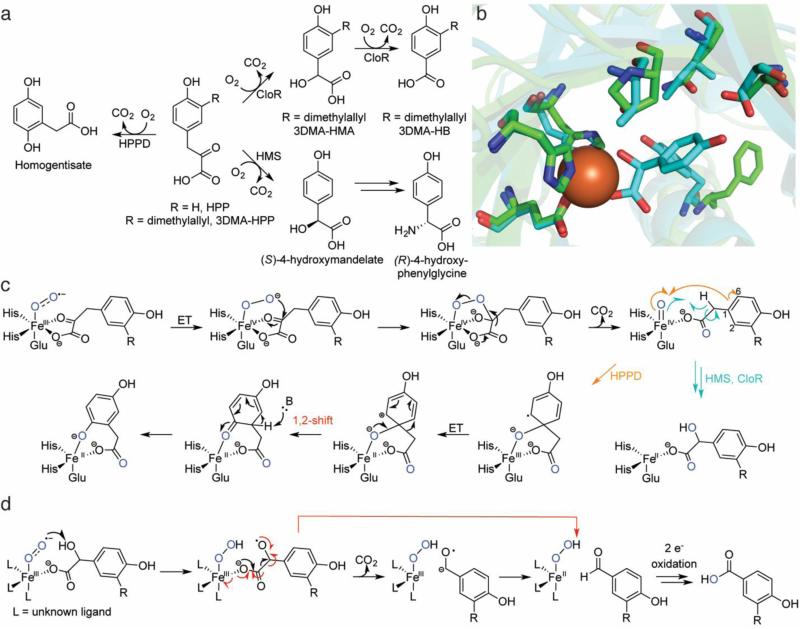
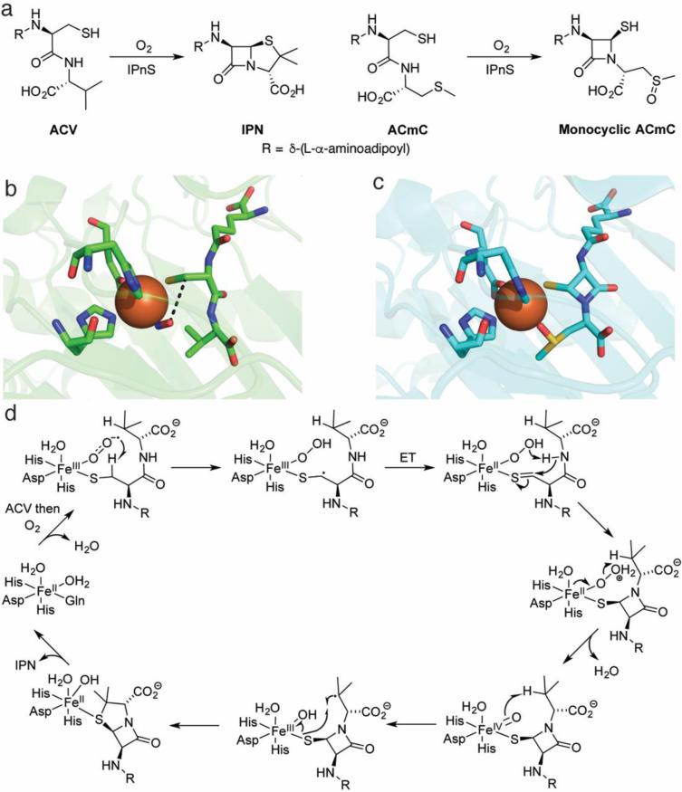
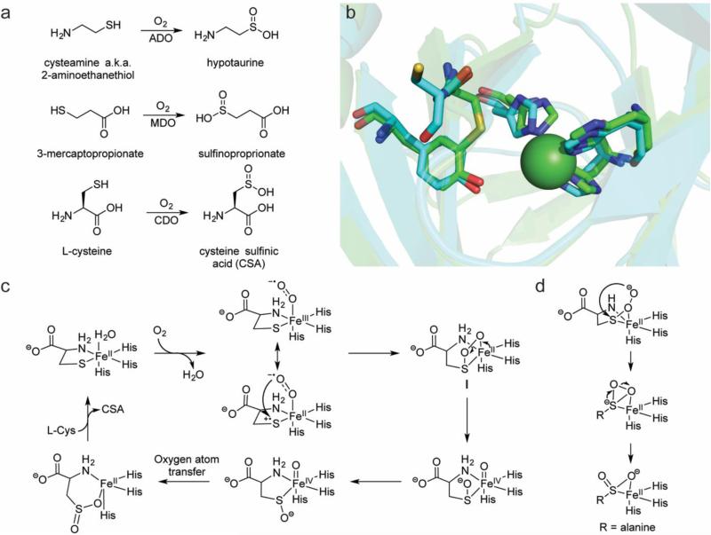
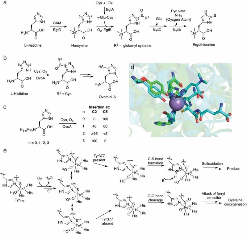
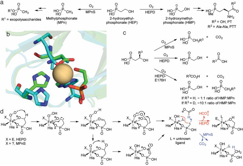
This review discusses the current mechanistic understanding of a group of mononuclear non-heme iron-dependent enzymes that catalyze four-electron oxidation of their organic substrates without the use of any cofactors or cosubstrates. One set of enzymes acts on α-ketoacid-containing substrates, coupling decarboxylation to oxygen activation. This group includes 4-hydroxyphenylpyruvate dioxygenase, 4-hydroxymandelate synthase, and CloR involved in clorobiocin biosynthesis. A second set of enzymes acts on substrates containing a thiol group that coordinates to the iron. This group is comprised of isopenicillin N synthase, thiol dioxygenases, and enzymes involved in the biosynthesis of ergothioneine and ovothiol. The final group of enzymes includes HEPD and MPnS that both carry out the oxidative cleavage of the carbon-carbon bond of 2-hydroxyethylphosphonate but generate different products. Commonalities amongst many of these enzymes are discussed and include the initial substrate oxidation by a ferric-superoxo-intermediate and a second oxidation by a ferryl species.

Keywords: Ferryl; Iron oxo; Non-heme iron; Oxidase; Oxygenase; Superoxo.


Figures

Fig. 1
Fig. 1
Overview of HPPD, HMS, and CloR. a The reactions catalyzed by HPPD, HMS, and CloR. The stereochemistry of the initial CloR hydroxylation has not been defined. b Overlays of the crystal structures of Fe-HPPD (carbons in green, PDB ID: 1CJX) aligned with Co-HMS bound to 4-hydroxymandelate (carbons in aqua, PDB ID: 2R5V). Given the sequence divergence of CloR from structurally characterized proteins, CloR did not yield high-confidence homology models from I-TASSER [125] and is therefore omitted. c A proposed mechanism for catalysis by HPPD, HMS, and the first oxidative decarboxylation by CloR. For details and discussion, see the text. d A mechanistic proposal for the second oxidative decarboxylation by CloR. A concerted β-scission mechanism is shown in red. ET = electron transfer
Fig. 2
Fig. 2
Overview of the chemistry effected by IPNS. a The native substrate of IPNS catalysis (left) and a substrate analog that does not undergo a second cyclization (right). b The crystal structure of the ternary complex of Fe(II)-IPNS-ACV-NO (PDB ID: 1BLZ). The distance between the oxygen atom of NO to the β carbon of cysteine in ACV is 3.3 Å (black dashes). c The structure of the product of the in crystallo reaction of Fe(II)-IPNS with ACmC reveals formation of the thiazolidine ring and sulfoxidation of the methylcysteine moiety (PDB ID: 1QJF). d A proposed mechanism for catalysis by IPNS. Instead of a formal ET in the second step after substrate binding, the two structures could also be considered resonance forms. For details, see the text
Fig. 3
Fig. 3
Overview of the current knowledge of the thiol dioxygenases. a Reactions catalyzed by ADO, MDO, and CDO. b The crystal structure of a eukaryotic Ni-CDO (carbons in green, PDB ID: 2ATF) overlaid with a prokaryotic Fe-CDO (carbons in aqua, PDB ID: 3EQE) demonstrates the preservation of the 3-His coordination sphere in each active site. The Cys-Tyr crosslink is observed only in the eukaryotic version; in the prokaryotic version, the position of the Cys is occupied by a Gly and the Cys is instead one residue removed and unable to form a crosslink. c Mechanistic proposal for catalysis by CDO. The majority of mechanistic research on the TDOs has focused on CDO, but a similar mechanism may be operative in ADO and MDO catalysis. The attack on the sulfur by the ferric-superoxo may be enhanced by a Fe(II)-thiyl radical cation resonance form in which the sulfur has donated an electron to the metal center. d The persulfenate intermediate that was observed crystallographically [80]
Fig. 4
Fig. 4
Overview of catalysis by the sulfoxide-inserting enzymes EgtB and OvoA. a The biosynthetic pathway to ergothioneine. b The ovothiol A biosynthetic pathway. c The site-selectivity of OvoA catalysis is dependent on the number of methyl groups on Nα of L-His. d An overlay of the Mn-EgtB-dimethylhistidine-γ-glutamyl cysteine complex (carbons in aqua, PDB ID: 4X8D) with the apo OvoA homology model (carbons in green), generated for this review using I-TASSER [125]. Key active site residues are preserved in the OvoA homology model, including the three histidines that would ligate the active site metal (one is partially obscured by the substrates in the EgtB structure). The homology model lacks an active site metal, explaining why the histidine side chains do not overlay with the histidine side chains in the EgtB structure. e A mechanistic proposal for catalysis by EgtB and OvoA. As with CDO, a minor resonance form with a Fe(II)-thiyl radical cation may activate O2 for attack on the sulfur atom. The ferrous-peroxysulfur species initially implicated based on DFT calculations (bottom) has instead been proposed to lead to non-productive dioxygenation to generate CSA as in CDO (Fig. 3c)
Fig. 5
Fig. 5
Overview of catalysis by HEPD and MPnS. a The biosynthetic pathways in which HEPD and MPnS are operative. b An overlay of the Cd-HEPD-HEP crystal structure (carbons in green, PDB ID: 3GBF) and an apo MPnS homology model (carbons in aqua) generated by I-TASSER [125]. An isoleucine is predicted to occupy the space of the carboxylate ligand in HEPD (E176), but a Gln residue in MPnS that aligns with the Fe-binding Glu of hydroxypropylphosphonate epoxidase is also predicted to be nearby. The identity of the third iron-binding ligand in MPnS remains unclear. c Scheme detailing the results of labeling experiments with MPnS, HEPD, and HEPD-E176H. d A mechanistic proposal invoking a consensus mechanism for HEPD and MPnS. Product identity is governed by whether the MPn radical combines with the ferric-hydroxide (HEPD) or attacks formate (MPnS). For details, see the text

References

    1. Kovaleva EG, Lipscomb JD. Nat Chem Biol. 2008;4:186–193. - PMC - PubMed
    1. Loenarz C, Schofield CJ. Nat Chem Biol. 2008;4:152–156. - PubMed
    1. Price JC, Barr EW, Glass TE, Krebs C, Bollinger JM., Jr. J Am Chem Soc. 2003;125:13008–13009. - PubMed
    1. Price JC, Barr EW, Tirupati B, Bollinger JM, Jr., Krebs C. Biochemistry. 2003;42:7497–7508. - PubMed
    1. Hoffart LM, Barr EW, Guyer RB, Bollinger JM, Jr., Krebs C. Proc Natl Acad Sci USA. 2006;103:14738–14743. - PMC - PubMed

Publication types

LinkOut - more resources