. 2017 Mar;187(3):418-427.
doi: 10.1111/cei.12892. Epub 2016 Dec 12.

Characterization of resident lymphocytes in human pancreatic islets

Affiliations

Characterization of resident lymphocytes in human pancreatic islets

M Radenkovic et al. Clin Exp Immunol. 2017 Mar.

Abstract

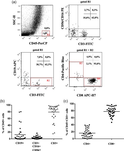
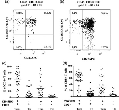
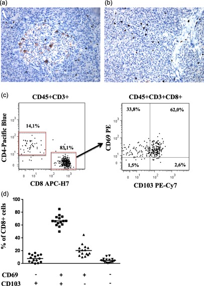
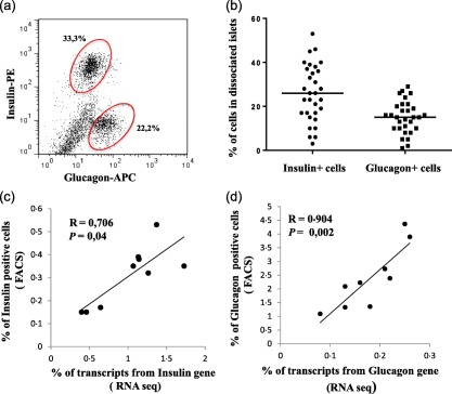
The current view of type 1 diabetes (T1D) is that it is an immune-mediated disease where lymphocytes infiltrate the pancreatic islets, promote killing of beta cells and cause overt diabetes. Although tissue resident immune cells have been demonstrated in several organs, the composition of lymphocytes in human healthy pancreatic islets have been scarcely studied. Here we aimed to investigate the phenotype of immune cells associated with human islets of non-diabetic organ donors. A flow cytometry analysis of isolated islets from perfused pancreases (n = 38) was employed to identify alpha, beta, T, natural killer (NK) and B cells. Moreover, the expression of insulin and glucagon transcripts was evaluated by RNA sequencing. Up to 80% of the lymphocytes were CD3+ T cells with a remarkable bias towards CD8+ cells. Central memory and effector memory phenotypes dominated within the CD8+ and CD4+ T cells and most CD8+ T cells were positive for CD69 and up to 50-70% for CD103, both markers of resident memory cells. The frequency of B and NK cells was low in most islet preparations (12 and 3% of CD45+ cells, respectively), and the frequency of alpha and beta cells varied between donors and correlated clearly with insulin and glucagon mRNA expression. In conclusion, we demonstrated the predominance of canonical tissue resident memory CD8+ T cells associated with human islets. We believe that these results are important to understand more clearly the immunobiology of human islets and the disease-related phenotypes observed in diabetes.

Keywords: T cells; diabetes; human; memory; pancreas.


Figures

Figure 1
Figure 1
Ex‐vivo flow cytometry analysis of B, natural killer (NK) and T lymphocytes in dissociated pancreatic islets of non‐diabetic donors. (a) Representative flow cytometry dot plots showing the gating strategy used for the identification of total lymphocyte (CD45+), total T cells (CD3+), NK cells (CD3CD56+CD16+), B cells (CD19+) as well as CD4+ and CD8+ T cells in dissociated human pancreatic islets (b) Cumulative analysis of CD4+ and (c) CD8+ T cells within the T cell compartment in the dissociated pancreatic islets from 38 organ donors. The data are shown as the mean percentage and standard deviation. [Colour figure can be viewed at wileyonlinelibrary.com]
Figure 2
Figure 2
Naive and memory CD4+ and CD8+ T cell subpopulations in dissociated pancreatic islets of non‐diabetic donors. Representative flow cytometry dot‐plots showing naive and memory subpopulations of (a) CD4+ and (b) CD8+ T cells from one representative organ donor. (c) Cumulative analysis of naive and memory subpopulation of CD4+ (n = 29) and D) CD8+ T cells (n = 35) cells from non‐diabetic donors. The data are shown as the mean percentage and standard deviation. Tcm = central memory T cells, Tem = effector memory T cells, Tte = terminal effector T cells, Tn = naive T cells.
Figure 3
Figure 3
Characterization of resident CD8+ T cells in dissociated pancreatic islets of non‐diabetic donors. (a) Immunohistochemical analysis of CD8+ T cells in pancreatic biopsy of a representative donor depicting the presence of CD8+ T cells within and in close proximity of one islet and in the surrounding exocrine tissue and (b) several CD8+ T cells spread within the exocrine tissue. Both images were taken with ×20 magnification. (c) Representative flow cytometric dot‐plots showing the expression of CD69 and CD103 on CD8+ T cells in islets cell suspensions of one representative organ donor. (d) Cumulative analysis of CD4+ and CD8+ T cells expressing the CD69 and CD103 from 15 organ donors. The data are shown as the mean percentage and standard deviation. [Colour figure can be viewed at wileyonlinelibrary.com]
Figure 4
Figure 4
Percentages of pancreatic insulin and glucagon positive cell populations of non‐diabetic donors and their correlation with RNA expression for insulin and glucagon. (a) Representative flow cytometry analysis of islets cell suspension depicting the percentage of insulin and glucagon positive cells and (b) cumulative analysis of 31 non‐diabetic donors are shown as the mean percentage and standard deviation. Spearman's correlation test between (c) insulin and (d) glucagon gene expression level determined by RNA sequencing in nine non‐diabetic donors and the percentage of insulin and glucagon cells assessed by flow cytometry. [Colour figure can be viewed at wileyonlinelibrary.com]

References

    1. Atkinson MA, von Herrath M, Powers AC, Clare‐Salzler M. Current concepts on the pathogenesis of type 1 diabetes–considerations for attempts to prevent and reverse the disease. Diabetes Care 2015; 38:979–88. - PMC - PubMed
    1. Campbell‐Thompson ML, Atkinson MA, Butler AE et al The diagnosis of insulitis in human type 1 diabetes. Diabetologia 2013; 56:2541–3. - PubMed
    1. Gianani R, Campbell‐Thompson M, Sarkar SA et al Dimorphic histopathology of long‐standing childhood‐onset diabetes. Diabetologia 2010; 53:690–8. - PubMed
    1. In't Veld P. Insulitis in human type 1 diabetes: the quest for an elusive lesion. Islets 2011; 3:131–8. - PMC - PubMed
    1. Krogvold L, Wiberg A, Edwin B et al Insulitis and characterisation of infiltrating T cells in surgical pancreatic tail resections from patients at onset of type 1 diabetes. Diabetologia 2016; 59:492–501. - PubMed

Publication types