. 2017 Jul;26(7):615-618.
doi: 10.1111/exd.13250. Epub 2016 Dec 19.

Digital imaging biomarkers feed machine learning for melanoma screening

Affiliations

Digital imaging biomarkers feed machine learning for melanoma screening

Daniel S Gareau et al. Exp Dermatol. 2017 Jul.

Abstract

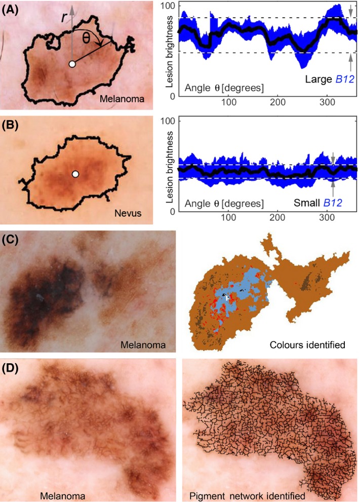
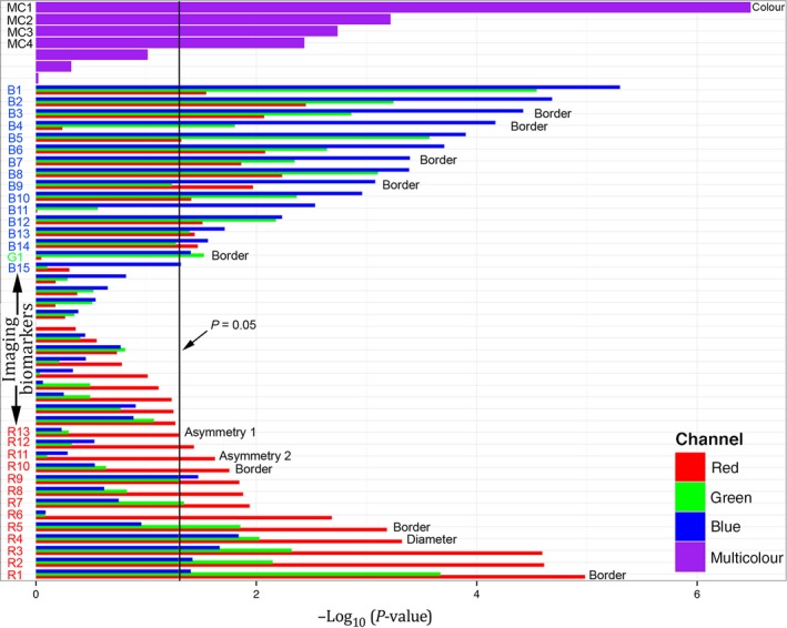
We developed an automated approach for generating quantitative image analysis metrics (imaging biomarkers) that are then analysed with a set of 13 machine learning algorithms to generate an overall risk score that is called a Q-score. These methods were applied to a set of 120 "difficult" dermoscopy images of dysplastic nevi and melanomas that were subsequently excised/classified. This approach yielded 98% sensitivity and 36% specificity for melanoma detection, approaching sensitivity/specificity of expert lesion evaluation. Importantly, we found strong spectral dependence of many imaging biomarkers in blue or red colour channels, suggesting the need to optimize spectral evaluation of pigmented lesions.

Keywords: dermoscopy; imaging biomarkers; machine learning; machine vision; melanoma; pigmented lesion; screening; skin optics.


Figures

Figure 1
Figure 1
Example melanoma imaging biomarkers. (A) and (B) show a melanoma and a nevus, respectively, where lesion centre (white circle) and peripheral border (black line) between lesion and normal skin are illustrated. The mean lesion brightness along the sweeping arm as a function of angle θ is plotted (black line) to the right with the standard deviation shown in blue. The melanoma imaging biomarker (MIB) B12 is graphically shown to be the brightness range over an angular sweep of the mean lesion pixel brightness. The range is divided by the mean to achieve the final B12 MIB. The images shown are of a melanoma that yields a large B12 value and a nevus that yields a small B12 value. A melanoma with multiple colours (C) is shown in colour map illustrating MIB MC1. A melanoma with an atypical reticular pigmented network (D) is shown with an overlay of the pigmented network branches. Each black line segment terminates on each end in either a branch point or an endpoint. Statistical analysis of these branches yielded MIBs B8, B11, B15, R3, R7 and R8
Figure 2
Figure 2
The length of the horizontal bar, for each image feature extracted, is negative the base 10 logarithm of the P‐value, where the P‐value is the standard statistical significance metric, calculated using univariate, two‐tailed, unpaired t‐tests (for continuous variables) and Fisher's exact test (for categorical variables). For single colour channel metrics, three adjoined bars, colour‐coated red, green and blue show the importance when evaluated in the respective colour channels of the image. The melanoma imaging biomarkers (MIBs) with statistical significance for melanoma discrimination (P<.05, vertical black line) are labelled on the vertical axis describing the colour channel they were used in: B1‐B14 from the blue channel, G1 from the green channel and R1‐R13 from the red channel. MC1‐MC4 denote MIBs that used multiple colour channel information. The text to the right of the bars indicates MIBs that contain information based on the dermoscopic ABCD criteria. The most significant MIB was the number of colours identified in the lesion while the diameter of the lesion had intermediate significance and the asymmetry of the lesion silhouette (Asymmetry 1, illustrated in Figure S10) had borderline significance. The lesion border features (see Figure S4) pertain to the edge demarcation.

References

    1. Hansen C., Wilkinson D., Hansen M., Argenziano G., J. Am. Acad. Dermatol. 2009, 61, 599. - PubMed
    1. Salerni G., Teran T., Puig S., Malvehy J., Zalaudek I., Argenziano G., Kittler H., J. Eur. Acad. Dermatol. Venereol. 2013, 27, 805. - PubMed
    1. Henning J. S., Dusza S. W., Wang S. Q., Marghoob A. A., Rabinovitz H. S., Polsky D., Kopf A. W., J. Am. Acad. Dermatol. 2007, 56, 45. - PubMed
    1. Monheit G., Cognetta A. B., Ferris L., Rabinovitz H., Gross K., Martini M., Grichnik J. M., Mihm M., Prieto V. G., Googe P., King R., Toledano A., Kabelev N., Wojton M., Gutkowicz‐Krusin D., Arch. Dermatol. 2011, 147, 188. - PubMed
    1. Doyle‐Lindrud S., Clin. J. Oncol. Nurs. 2015, 19, 31. - PubMed

Publication types

MeSH terms

Substances