. 2016 Oct 26;12(10):e1005982.
doi: 10.1371/journal.ppat.1005982. eCollection 2016 Oct.

Middle East Respiratory Coronavirus Accessory Protein 4a Inhibits PKR-Mediated Antiviral Stress Responses

Affiliations

Middle East Respiratory Coronavirus Accessory Protein 4a Inhibits PKR-Mediated Antiviral Stress Responses

Huib H Rabouw et al. PLoS Pathog. .

Abstract

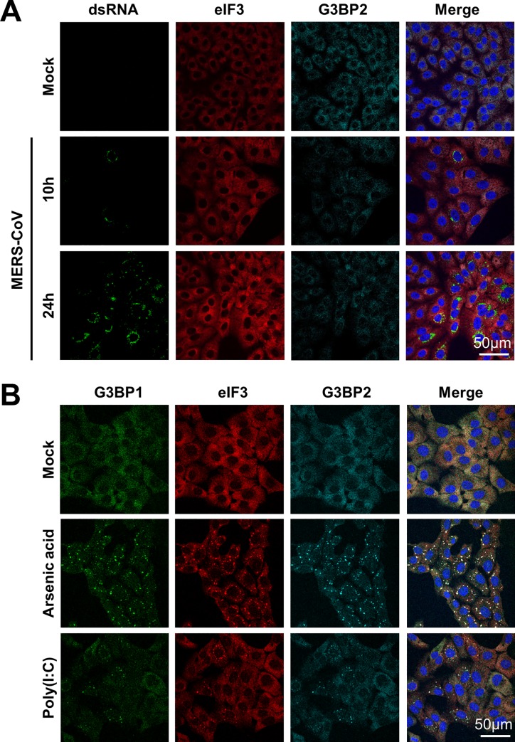
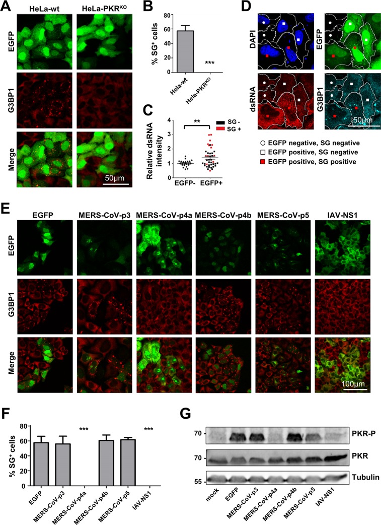
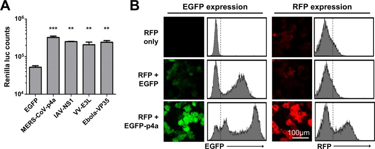
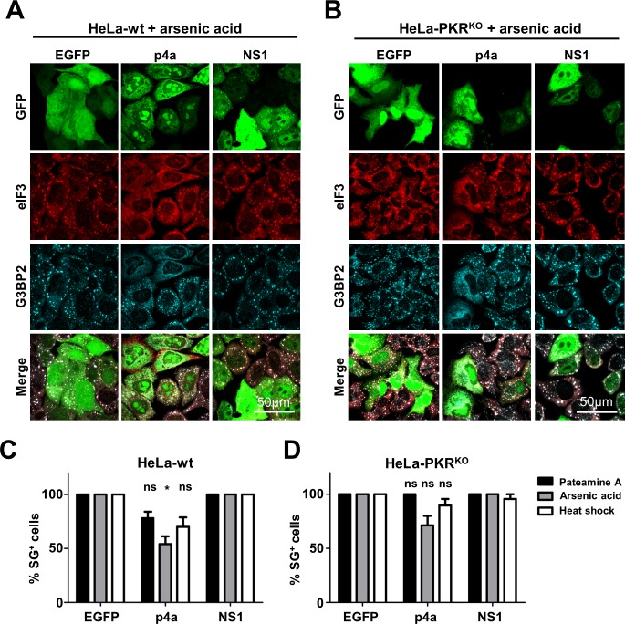
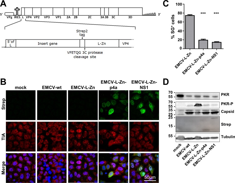
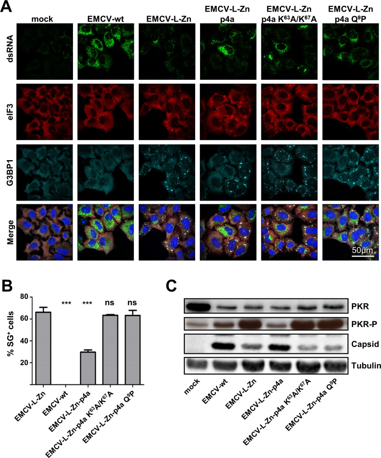
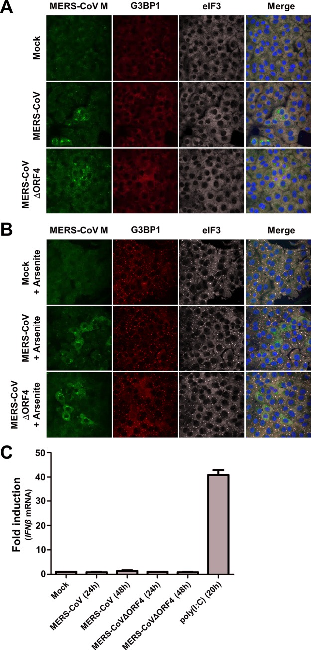
Middle East respiratory syndrome coronavirus (MERS-CoV) causes severe respiratory infections that can be life-threatening. To establish an infection and spread, MERS-CoV, like most other viruses, must navigate through an intricate network of antiviral host responses. Besides the well-known type I interferon (IFN-α/β) response, the protein kinase R (PKR)-mediated stress response is being recognized as an important innate response pathway. Upon detecting viral dsRNA, PKR phosphorylates eIF2α, leading to the inhibition of cellular and viral translation and the formation of stress granules (SGs), which are increasingly recognized as platforms for antiviral signaling pathways. It is unknown whether cellular infection by MERS-CoV activates the stress response pathway or whether the virus has evolved strategies to suppress this infection-limiting pathway. Here, we show that cellular infection with MERS-CoV does not lead to the formation of SGs. By transiently expressing the MERS-CoV accessory proteins individually, we identified a role of protein 4a (p4a) in preventing activation of the stress response pathway. Expression of MERS-CoV p4a impeded dsRNA-mediated PKR activation, thereby rescuing translation inhibition and preventing SG formation. In contrast, p4a failed to suppress stress response pathway activation that is independent of PKR and dsRNA. MERS-CoV p4a is a dsRNA binding protein. Mutation of the dsRNA binding motif in p4a disrupted its PKR antagonistic activity. By inserting p4a in a picornavirus lacking its natural PKR antagonist, we showed that p4a exerts PKR antagonistic activity also under infection conditions. However, a recombinant MERS-CoV deficient in p4a expression still suppressed SG formation, indicating the expression of at least one other stress response antagonist. This virus also suppressed the dsRNA-independent stress response pathway. Thus, MERS-CoV interferes with antiviral stress responses using at least two different mechanisms, with p4a suppressing the PKR-dependent stress response pathway, probably by sequestering dsRNA. MERS-CoV p4a represents the first coronavirus stress response antagonist described.


Conflict of interest statement

The authors have declared that no competing interests exist.

Figures

Fig 1
Fig 1. MERS-CoV infection fails to activate the stress response pathway.
(A) Immune fluorescence images of mock-treated an MERS-CoV infected Vero cells. Cells were infected with an MOI of 1 and fixed using 3% paraformaldehyde in PBS at 10h or 24h post infection. Cells were stained for dsRNA, and stress granule markers eIF3 and G3BP2. (B) Immune fluorescence images of cells treated with arsenic acid (0.5 mM for 60 min) or transfected with poly(I:C) and stained for eIF3, G3BP1 and G3BP2.
Fig 2
Fig 2. MERS-CoV p4a suppresses dsRNA-dependent and PKR-mediated stress in transfected cells.
(A) Immune fluorescence images of HeLa-wt or HeLa-PKRKO cells transfected with pEGFP-N3 plasmid (500 ng/well). Cells were fixed at 24h post transfection using paraformaldehyde and stained for G3BP1 (shown in red). EGFP expression is shown in green. (B) Quantification of SG-positive cells. SG-positive cells were quantified from three randomly selected images. Shown are means with standard deviations, analyzed using an unpaired t-test (***, p<0.001). (C) Quantification of the average dsRNA staining intensity in individual cells using imageJ software. Intensity levels are plotted relative to that of the non-transfected cells from the same images. Cells were classified as non-transfected or transfected based on EGFP expression, and as SG-positive or SG-negative based on presence of G3BP1 aggregates. Differences in relative dsRNA intensity levels were analyzed using an unpaired t-test (**, p<0.01). (D) Typical example of the IFA images used for quantification in C. Borders of two cells of each phenotype (EGFP-; EGFP+SG-; EGFP+SG+) are indicated in white. (E) Immune fluorescence images of HeLa cells transfected with pEGFP expression plasmids. Cells were fixed at 24h post transfection and stained for G3BP1 (shown in red). EGFP expression is shown in green. (F) Quantification of SG-positive cells. Analysis was performed as described in panel B (***, p<0.001). (G) Western blot analysis of PKR and phospho-PKR in HeLa cell lysates at 24h post pEGFP plasmid transfection. Tubulin expression was detected as loading control.
Fig 3
Fig 3. MERS-CoV p4a rescues protein translation upon plasmid DNA transfection-mediated stress.
(A) Bar-graph showing Renilla luciferase counts measured at 16h post co-transfection of pTK-RLuc and pEGFP expression plasmids. Means and standard deviations are shown of triplicate measurements. Data was analyzed using an unpaired t-test (***, p<0.001; **, p<0.01). (B) Flow cytometry analysis of HeLa cells expressing RFP, RFP and EGFP, or RFP and EGFP-p4a. The dashed lines in the histograms divide non-RFP/EGFP expressing cells from RFP/EGFP-expressing cells.
Fig 4
Fig 4. MERS-CoV p4a does not inhibit PKR-independent SG formation.
(A, B) Immune fluorescence images of HeLa-wt cells (A) and HeLa-PKRKO cells (B) transfected with the indicated pEGFP-expression plasmids. Next day, SG formation was triggered using arsenic acid (0.5 mM for 30 min). Cells were fixed and stained for eIF3 (shown in red) or G3BP2 (shown in cyan). EGFP expression is shown in green. (C, D) Quantification of SG-positive HeLa-wt cells (C) and HeLa-PKRKO cells (D) treated with Pateamine A (100 nM for 2h), arsenic acid (0.5 mM for 30 min), or heat shock (50°C for 30 min). SG-positive cells were quantified from three randomly selected images. Shown are means with standard deviations, which were analyzed using an unpaired t-test. (*, p<0.05; ns, not significant).
Fig 5
Fig 5. MERS-CoV p4a inhibits PKR activation during mengovirus infection.
(A) Schematic overview of the recombinant mengovirus system. The upper panel shows the wt mengovirus genome. The lower panel highlights the 5’-region showing the gene insertion upstream of the inactivated L. (B) Immune fluorescence images of HeLa-wt cells that were mock-infected or infected with wt mengovirus or the indicated recombinant mengoviruses (MOI = 10). Cells were fixed at 6h post infection and stained for TIA1 (shown in red) and Strep-tagged p4a or NS1 (shown in green). Nuclei were stained using Hoechst-33258 (shown in blue). (C) SG-positive cells were quantified from three randomly selected images. Shown are means with standard deviations, which were analyzed using an unpaired t-test (***, p<0.001). (D) Western blot analysis of PKR and phospho-PKR in cells infected with indicated viruses. Capsid staining was used as a control for virus replication efficiency, tubulin staining was used as loading control and Strep-tag staining showed expression of the MERS-CoV p4a and IAV NS1.
Fig 6
Fig 6. The dsRNA binding motif in MERS-CoV p4a is crucial for suppressing SG formation.
(A) Immune fluorescence images of HeLa-wt cells that were mock-treated or infected with wt mengovirus or the indicated recombinant mengoviruses (MOI = 10). Cells were fixed at 6h post infection and stained for dsRNA (shown in green), eIF3 (shown in red), and G3BP1 (shown in cyan). Nuclei were stained using Hoechst-33258 (shown in blue). (B) SG-positive cells were quantified from three randomly selected images. Shown are means with standard deviations, analyzed using an unpaired t-test (***, p<0.001; ns, not significant). (C) Western blot analysis of PKR and phospho-PKR in cells infected with indicated viruses. Capsid staining was used as a control for virus replication efficiency and tubulin staining was used as loading control.
Fig 7
Fig 7. MERS-CoV p4a is a type I IFN antagonist.
(A, B) Relative IFNβ mRNA levels induced by transfection of poly(I:C) (A) or 6.5 kb viral dsRNA (sequence derived from the Coxsackie virus B3 genome) (B) in HeLa-wt cells expressing EGFP or EGFP-p4a fusion proteins. To obtain a cell pool in which all cells express the protein of interest, plasmids encoding EGFP fusion proteins were co-transfected with a plasmid conferring puromycin resistance. Subsequent puromycin selection for two days eliminated non-transfected cells. RT-qPCR was used to quantify relative IFNβ mRNA levels 8h post RNA ligand transfection. Shown are means and standard deviations of the relative IFNβ mRNA levels compared to EGFP-expressing cells. Analysis was performed by unpaired t-test (***, p<0.001; **, p<0.01; ns, not significant). (C) Bar-graph showing IFNβ mRNA levels induced by recombinant mengovirus infection (MOI = 10) of HeLa cells. RT-qPCR was used to quantify relative IFNβ mRNA levels at 8h post infection. Means and standard deviations of the relative IFNβ mRNA levels of triplicates are shown and analyzed using an unpaired t-test (***, p<0.001; **, p<0.01; ns, not significant).
Fig 8
Fig 8. MERS-CoV p4a increases mengovirus fitness.
(A) Immune fluorescence images of HeLa-wt and HeLa-PKRKO cells infected with EMCV-L-Zn (MOI = 10). Cells were fixed at 6h post infection and SG formation was visualized using antibodies directed against G3BP1 (shown in green) and eIF3 (shown in red). Nuclei were stained using Hoechst-33258 (shown in blue). (B) In parallel with A, RNA was isolated at 8h post infection and relative IFNβ mRNA levels were quantified by RT-PCR. Means and standard deviations of triplicate measurements are shown. (C) Virus production after wt and recombinant mengovirus infection (MOI = 0.01) in HeLa and HeLa-PKRKO cells. Supernatant was collected 24h post infection and virus progeny was titrated by end-point dilution with 3-fold dilution steps. (D) Schematic representation of the virus competition assay. Briefly, two viruses are mixed 1:1 and used to infection HeLa-wt or HeLa-PKRKO cells. Progeny virus was collected 48h post infection and viral RNA was isolated. RT-PCR was used to amplify the MERS-CoV 4a insert, which was analyzed using agarose gel electrophoresis. (E, F, G) Agarose gel analysis of the 4a insert region from virus competition assays with the indicated viruses. To distinguish between wild-type and mutant 4a genes, 4a-wt specific HindIII digestion was used.
Fig 9
Fig 9. MERS-CoV encodes another suppressor of innate antiviral responses.
(A, B) Vero cells were infected (MOI = 1) with MERS-CoV wt or MERS-CoVΔORF4. At 16h p.i., cells were (A) mock treated, or (B) treated with 0.5 mM arsenic acid for 1h. Subsequently, MERS-CoV infection and SG formation were visualized by IFA using antibodies directed against MERS-CoV M, G3BP1, and eIF3, respectively. (C) Huh7 cells were transfected with poly(I:C), or infected (MOI = 1) with the indicated viruses. RT-qPCR was used to quantify relative IFNβ mRNA levels at the indicated time points. Shown are means and standard deviations of the relative IFNβ mRNA levels compared to mock treated cells.

Similar articles

Cited by

References

    1. Onomoto K, Jogi M, Yoo J-SS, Narita R, Morimoto S, Takemura A, et al. Critical Role of an Antiviral Stress Granule Containing RIG-I and PKR in Viral Detection and Innate Immunity. PLoS One. 2012;7: e43031 10.1371/journal.pone.0043031 - DOI - PMC - PubMed
    1. White JP, Lloyd RE. Regulation of stress granules in virus systems. Trends Microbiol. 2012;20: 175–83. 10.1016/j.tim.2012.02.001 - DOI - PMC - PubMed
    1. Wippich F, Bodenmiller B, Trajkovska MG, Wanka S, Aebersold R, Pelkmans L. Dual specificity kinase DYRK3 couples stress granule condensation/ dissolution to mTORC1 signaling. Cell. 2013;152: 791–805. 10.1016/j.cell.2013.01.033 - DOI - PubMed
    1. Arimoto K, Fukuda H, Imajoh-Ohmi S, Saito H, Takekawa M. Formation of stress granules inhibits apoptosis by suppressing stress-responsive MAPK pathways. Nat Cell Biol. 2008;10: 1324–1332. 10.1038/ncb1791 - DOI - PubMed
    1. Kim WJ, Back SH, Kim V, Ryu I, Jang SK. Sequestration of TRAF2 into stress granules interrupts tumor necrosis factor signaling under stress conditions. Mol Cell Biol. 2005;25: 2450–2462. 10.1128/MCB.25.6.2450-2462.2005 - DOI - PMC - PubMed

MeSH terms

Substances