Review
. 2016 Sep 15;6(4):1801-1850.
doi: 10.1002/cphy.c160002.

Neural Control of the Upper Airway: Respiratory and State-Dependent Mechanisms

Affiliations
Review

Neural Control of the Upper Airway: Respiratory and State-Dependent Mechanisms

Leszek Kubin. Compr Physiol. .

Abstract

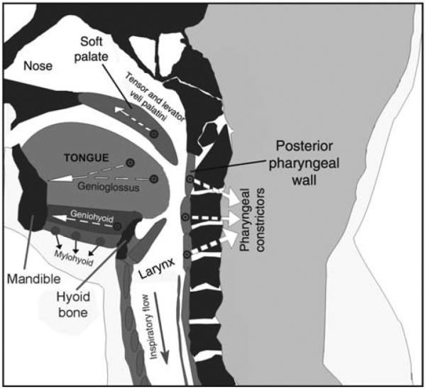
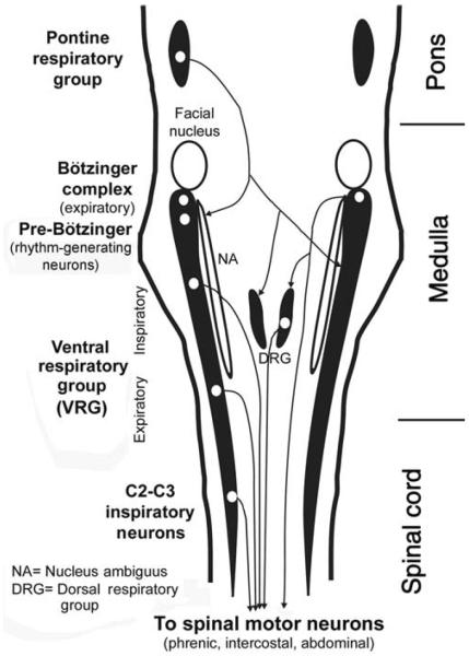
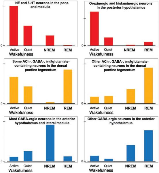
Upper airway muscles subserve many essential for survival orofacial behaviors, including their important role as accessory respiratory muscles. In the face of certain predisposition of craniofacial anatomy, both tonic and phasic inspiratory activation of upper airway muscles is necessary to protect the upper airway against collapse. This protective action is adequate during wakefulness, but fails during sleep which results in recurrent episodes of hypopneas and apneas, a condition known as the obstructive sleep apnea syndrome (OSA). Although OSA is almost exclusively a human disorder, animal models help unveil the basic principles governing the impact of sleep on breathing and upper airway muscle activity. This article discusses the neuroanatomy, neurochemistry, and neurophysiology of the different neuronal systems whose activity changes with sleep-wake states, such as the noradrenergic, serotonergic, cholinergic, orexinergic, histaminergic, GABAergic and glycinergic, and their impact on central respiratory neurons and upper airway motoneurons. Observations of the interactions between sleep-wake states and upper airway muscles in healthy humans and OSA patients are related to findings from animal models with normal upper airway, and various animal models of OSA, including the chronic-intermittent hypoxia model. Using a framework of upper airway motoneurons being under concurrent influence of central respiratory, reflex and state-dependent inputs, different neurotransmitters, and neuropeptides are considered as either causing a sleep-dependent withdrawal of excitation from motoneurons or mediating an active, sleep-related inhibition of motoneurons. Information about the neurochemistry of state-dependent control of upper airway muscles accumulated to date reveals fundamental principles and may help understand and treat OSA. © 2016 American Physiological Society. Compr Physiol 6:1801-1850, 2016.


Figures

Figure 1
Figure 1
Schematic representation of a sagittal cross-section through the upper airway. During inspiration, negative intraluminar pressure pulls three soft tissue elements, the tongue, posterior pharyngeal walls, and soft palate, toward each, thereby reducing the airway lumen in the pharyngeal region. This airway-collapsing action is opposed by pharyngeal dilator muscles, including the genioglossus, geniohyoid, and tensor and levator veli palatini. Additionally, activation of the pharyngeal constrictors stiffens the airway walls. Arrows show approximate directions of the forces exerted during contraction of these major pharyngeal muscles. Image based on a scan of the upper airway in an OSA patient—courtesy of Dr. Richard J. Schwab at the University of Pennsylvania. (Modified from Fig. 1 in Ref. 270 and republished with permission from Informa Healthcare, a member of the Taylor and Francis Group, obtained via the Copyright Clearance Center, Inc.)
Figure 2
Figure 2
Distribution of the major groups of respiratory neurons in the brainstem and upper spinal cord, as seen in a dorsal view. Brainstem respiratory neurons form longitudinal columns, of which the most prominent one is the VRG located in the ventrolateral medullary reticular formation. The rostralmost part of the VRG, the Bötzinger complex, contains mainly late expiratory neurons. Next region caudally, the pre-Bötzinger complex, contains pacemaker neurons that, at least in neonatal animals, are capable of producing basic respiratory rhythm under in vitro conditions. Farther caudal is a large group of mainly inspiratory-modulated neurons, of which many send axons to spinal motoneurons that innervate the diaphragm and external intercostal muscles. Nucleus ambiguus runs parallel to this part of VRG and contains cell bodies of laryngeal motoneurons. Caudal to the inspiratory part of the VRG is an expiratory region whose neurons send axons to spinal expiratory motoneurons that control internal intercostal and abdominal muscles. A spinal extension of the VRG, the C2 and C3 group, again contains inspiratory neurons whose function may be to reinforce the actions of the VRG. The dorsal respiratory group is located in the viscerosensory nucleus of the solitary tract. It contains mostly inspiratory-modulated neurons, of which some have connections with spinal motoneurons and some receive input from pulmonary and laryngeal receptors. The pontine respiratory group located in the dorsolateral pons comprises cells with different patterns of respiratory modulation. These neurons integrate peripheral and central respiratory and nonrespiratory inputs and have descending projections to medullary respiratory neurons. (Modified from Fig. 2 in Ref. 266 and republished with permission from the American Academy of Sleep Medicine.)
Figure 3
Figure 3
Major sources of pontomedullary afferent projections to the orofacial motor nuclei. (A) Schematic dorsal view of the most prominent afferent pathways common to all orofacial motor nuclei. The medullary IRt is the main source of bilateral projections to the trigeminal (V), facial (VII), hypoglossal (XII), and ambiguus (nA) motor nuclei (red arrows). The projections to these motor nuclei tend to exhibit a distinct rostrocaudal pattern. The projections to orofacial motor nuclei that originate in the pontine reticular region surrounding the trigeminal motor nucleus (peri-V), and the ventrolateral medullary gigantocellular region (GCv) tend to be unilateral (blue arrows). (Fig. 12 from Ref. 551 colorized and republished with permission from John Wiley and Sons obtained via the Copyright Clearance Center, Inc.) (B) Major medullary sources of afferent projections to the XII nucleus in relation to the descending spinal collaterals of selected XII premotor neurons, as revealed by large retrograde tracer injections into the XII motor nucleus and ventral horns of the lumbar (L2) spinal cord. The two medullary levels illustrated are located rostral to the XII nucleus. Consistent with data in A, the IRt region contains the largest number of neurons retrogradely labeled with Diamidino Yellow (DY) tracer from the XII nucleus (red dots). Notably, no cells retrogradely labeled with Fast Blue (FB) tracer from the spinal cord (black dots) are located in this region. Additional medullary XII premotor neurons are located along the midline (enlarged images in the middle), the gigantocellular region pars α (GiA), and the lateral paragigantocellular (LPGi) region. A small fraction of cells in these medial and ventral locations has divergent projections to the XII nucleus and lumbar spinal cord (blue triangles). The key to anatomical regions is shown on the right. (Modified from Fig. 3B in Ref. 323 and republished with permission from John Wiley and Sons obtained via the Copyright Clearance Center, Inc.) Additional abbreviations in A and B: Gi, gigantocellular reticular region; mlf, medial longitudinal fasiculus; MVe, medial vestibular nucleus; PCRtA, parvicellular reticular area; PrH, nucleus prepositus hypoglossi; py, pyramidal tract; RMg, Rob, Rpa, raphé magnus, pallidus, and obscurus nuclei; Sp5, spinal trigeminal sensory nucleus; SpVe, spinal vestibular nucleus.
Figure 4
Figure 4
Typical activity patterns during wakefulness and sleep states of selected neurochemically distinct groups of central neurons. Some groups have the highest activity during wakefulness (top/red); others have peaks during both wakefulness and REM sleep or during REM sleep only (middle/yellow), and still others during NREM or REM sleep (bottom/blue). These neuronal groups have direct and indirect connections with central respiratory neurons and upper airway motoneurons through which they impart state-dependent changes onto the respiratory system.
Figure 5
Figure 5
Schematic representation of the four key neuroanatomically and neurochemically distinct inputs to the XII nucleus. NE afferents originate mainly in the pontine A7 and A5 groups and the SubC region, whereas the largest group of NE neurons, the LC, has negligible projections to the XII nucleus. For clarity, NE projections are shown on one side only; they are bilateral with a minor ipsilateral predominance. 5-HT afferents come from the medullary raphé pallidus and obscurus nuclei, as well as the lateral wings of the medullary raphé. The medullary IRt contains glutamatergic and cholinergic XII premotor cells; the former provide inspiratory drive to XII motoneurons, and the latter may mediate pre- or postsynaptic modulatory influences that are either respiratory or state dependent. The LPGi and the adjacent areas contain cells that have been hypothesized to mediate active inhibitory effects of REM sleep to motoneurons. GABAergic, REM sleep-active neurons with divergent projections ascending to the pons and descending to the spinal cord have been located in this area (579). It is not known whether the same cells also have axonal projections to the XII nucleus. Abbreviations: A, nucleus ambiguus; IO, inferior olive, Po, nucleus pontis oralis; K-F, Kölliker-Fuse nucleus; NTS, nucleus of the solitary tract; py, pyramidal tract.
Figure 6
Figure 6
NE- and 5-HT-containing axon terminals are present throughout the XII nucleus. (A and B) Microscopic images of coronal cross-sections through the XII nucleus on one side. The section in A was immunostained for dopamine-ß-hydroxylase (DBH), which in the XII nucleus labels noradrenergic fibers and terminals (black). The section in B was immunostained for 5-HT fibers and terminals. In both panels, selected XII motoneurons that innervate the tongue also were labeled by retrograde transport from the base of the tongue (brown). Typical of motoneurons innervating the genioglossus, most labeled motoneurons are located in the ventromedial quadrant of the XII nucleus. Panels A1 and B1 show enlarged details framed in the corresponding panels on the left. The small dark-brown and black particles in the enlarged images represent fine axonal ramifications and terminals containing NE (in A1) and 5-HT (in B1). NE terminals are especially numerous in the ventromedial portion of the XII nucleus. CE, central canal. (Unpublished data from the study described in Ref. 443.)
Figure 7
Figure 7
Different firing rate outcomes resulting from different patterns of convergence of three functionally distinct excitatory inputs onto an upper airway motoneuron. The scheme shows three scenarios of how different combinations of inputs from functionally different sources may shape state-dependent changes in motoneuronal activity. (A) The three distinct excitatory inputs considered—respiratory, state-dependent, and reflex—are shown converging on an upper airway motoneuron. In B to D, the depolarizing actions contributed by each of these three inputs summate above the “baseline membrane potential” (black line at the bottom of each panel), which may result in the membrane potential crossing the firing threshold. Of the three distinct drives, the respiratory input is distinctly rhythmic (phasic), the central state-dependent input is tonic, and the reflex input includes both a phasic respiratory and a tonic component. The respiratory input is assumed to increase during sleep (e.g., to make up for reduced ventilation during NREM sleep, or as a result of central activation during REM sleep). In the three scenarios shown in B to D, different relative magnitudes of the three drives during wakefulness and their different changes at sleep onset result in quantitatively and qualitatively different firing rate outcomes at the motoneuronal level. (B) A case with a moderate phasic respiratory, moderate reflex, and strong central tonic input during wakefulness. A large drop of the tonic input at the onset of sleep is compensated for by an increase in the respiratory input and the associated phasic component of the reflex input. As a result, the motoneuron's activity minimally changes on the transition from wakefulness to sleep. (C) A case with a strong phasic respiratory input and weak central and reflex inputs during wakefulness. The increase in respiratory input during sleep more than makes up for the loss of the reflex and tonic drives. As a result, the motoneuron is more active during sleep than during wakefulness. (D) A case with all three inputs having similar magnitudes during wakefulness and the state-dependent input being profoundly reduced during sleep. In spite of a prominent increase in the respiratory input and a moderate increase in the reflex input during sleep, the motoneuron becomes silent. (Modified from Fig. 5 in Ref. 270 and republished with permission from Informa Healthcare, a member of the Taylor and Francis Group, obtained via the Copyright Clearance Center, Inc.)
Figure 8
Figure 8
Time course of lingual muscle activity during transition from wakefulness (W) to NREM sleep in healthy humans and rats. (A) In healthy humans, peak firing rate of inspiratory phasic motor units recorded at the base of the tongue significantly declined in association with a transition from quiet wakefulness to NREM sleep (squares). Transitions were defined based on EEG power shift from α-frequency to Θ-frequency. Mean data from 29 inspiratory phasic motor units. In contrast, inspiratory tonic motor units (defined as those firing continuously throughout the respiratory cycle with firing rate increases during inspiration) either had minimal declines of firing rate in association with the onset of NREM sleep or were entirely silenced around the time of the transition. Filled circles show data for all 58 inspiratory tonic motor units studied, including those that became silent at NREM sleep onset. Open circles show data for the subset of inspiratory tonic motor units that were not silenced at the onset of NREM sleep; for this group (n = 29), there was only a very small and transient decrease of firing rate. Asterisks indicate significantly lower firing rate after NREM sleep onset when compared to the mean before the transition. (Fig. 2 in Ref. 592 republished with permission from the American Academy of Sleep Medicine obtained via the Copyright Clearance Center, Inc.) (B) Lingual and nuchal EMGs averaged over multiple transitions from W to NREM sleep and from NREM sleep to W in rats. The top graphs show average levels of lingual EMG determined during successive 10 s intervals over 2-min periods before and after the state transitions, as indicated above the panels. Root mean squares of muscle activity were normalized within each animal and recording session by its average level during W. The middle graphs show the corresponding changes in postural activity recorded from dorsal neck muscles. The bottom panels show the corresponding average changes in cortical delta power which characteristically increases during NREM sleep. Lingual activity is nearly abolished following entry into NREM sleep. Such a low level of activity is maintained throughout the duration of NREM sleep episodes and rapidly returns to the wakefulness level after arousal (right panels). (Modified from Fig. 5 in Ref. 316 and republished with permission from Elsevier.)
Figure 9
Figure 9
Upper airway muscle activity during REM sleep in subjects with fully patent upper airway is dominated by nonrespiratory, phasic bursts that emerge from an otherwise atonic state. (A) Activity of the arytenoid (laryngeal) muscle during REM sleep in a healthy human subject. (Fig. 9 in Ref. 278 republished with permission from the American Physiological Society.) (B) Average time course of lingual muscle activity during transitions from NREM to REM sleep in rats. Lingual and nuchal EMG levels were measured during successive 10 s intervals from 60 s before to 200 s after the state transition (time zero) and averaged over multiple transitions in multiple animals. Root mean squares of muscle activity were normalized within each animal by their average levels during wakefulness. The bottom graph shows the corresponding average time course of the ratio of EEG powers in β-2 to Δ-2 bands (the ratio characteristically increases during REM sleep). Lingual and nuchal EMGs follow a different time course during the transition. Whereas nuchal EMG declines prior to the onset of REM sleep and then maintains a low level (atonia), lingual EMG is nearly atonic prior to the onset of REM sleep (see Fig. 8B) and first gradually increases and then declines after REM sleep onset. The relatively smooth time course of the increase and then decline is a result of averaging of many short bursts of activity (twitches) across many NREM to REM sleep transitions. The resulting mean time course indicates that, after the onset of REM sleep, individual twitches first become gradually larger and more frequent and then gradually become smaller and less frequent when the duration of REM sleep episodes extends beyond its mean (90 s in rats). (Modified from Fig. 6 in Ref. 446 and republished with permission from Archives Italiennes de Biologie and the University of Pisa.)
Figure 10
Figure 10
REM sleep exerts a powerful excitatory effect on the activity of most brainstem respiratory neurons. In this example, cumulative activity of an augmenting medullary inspiratory neuron was recorded from a chronically instrumented cat over a period covering a transition from NREM to REM sleep and then from REM sleep to awakening. The period of REM sleep is marked by EEG desynchronization, appearance of PGO waves, and a slightly reduced and variable end-expiratory CO2. The bottom panel shows that the neuron has a distinctly increased activity during REM sleep when compared to either the preceding period of NREM sleep or the period after awakening, as indicated by the slope of cumulative activity. (Fig. 1 in Ref. 392 republished with permission from John Wiley and Sons obtained via the Copyright Clearance Center, Inc.)
Figure 11
Figure 11
REM sleep-like episodes of depression of XII nerve activity accompanied by the characteristic cortical and hippocampal activation can be elicited by microinjections of a cholinergic agonist, carbachol, into the dorsal mesopontine tegmentum in urethane-anesthetized, paralyzed, and artificially ventilated rats. (A) Typical REM sleep-like episode. The moving average of XII nerve activity (top) shows inspiratory bursts of activity. The amplitude of the bursts is reduced and the central respiratory rate declines within less than 2 min following pontine carbachol injection (at arrow). Parallel to the depression of XII nerve activity, there is cortical and hippocampal activation. The second trace from top shows raw signal recorded from the hippocampus. The third trace from top shows the power of hippocampal activity in the 3 to 5 Hz frequency band, which represents theta-like rhythm under urethane anesthesia. The increase in power of cortical EEG in the 6 to 9 Hz frequency band (bottom) indicates activation relative to the period prior to carbachol injection. The entire episode lasts about 5 min, after which all signals return to their precarbachol levels and patterns. The ability to repeatedly elicit such REM sleep-like episodes allows one to investigate cellular, neurochemical, and network mechanisms of the interaction between REM sleep and the regulation of breathing and upper airway muscle activity. (Unpublished record from the study described in Ref. 142.) B: The sites at which carbachol microinjections elicit REM sleep-like episodes in urethane-anesthetized rats superimposed onto two standard cross sections of the brainstem from levels 8.3 mm and 8.72 mm caudal to bregma. Abbreviations: LDN, laterodorsal tegmental nucleus; PPN, pedunculopontine tegmental nucleus; scp, superior cerebellar peduncle; VT, ventral tegmental nucleus. (Modified from Fig. 2 in Ref. 272 and republished with permission from Elsevier.)
Figure 12
Figure 12
Antagonism of glycinergic inhibition at the level of the XII motor nucleus did not prevent the REM sleep-like depression of XII nerve activity elicited in decerebrate, paralyzed, vagotomized, and artificially ventilated cats by pontine microinjections of a cholinergic agonist, carbachol. Strychnine, a blocker of glycinergic receptors, was repeatedly microinjected into the right XII nucleus 3 to 16 min prior to the beginning of the record. The traces show moving averages of activities recorded from both XII nerves (upward deflections represent inspiratory bursts) and a cervical nerve branch that innervates dorsal neck muscles (a marker of postural activity). Pontine carbachol injection made at the marker initiated the REM sleep-like suppression of activity in both XII nerves and the postural nerve. The activities of both XII nerves were similarly depressed despite the prior injection of strychnine into the right XII nucleus. (Modified from Fig. 2B in Ref. 273 and republished with permission from Elsevier.)
Figure 13
Figure 13
Results from different laboratories and different animal models consistently indicate that antagonism of glycinergic inhibition at the motoneuronal level by strychnine does not abolish the REM sleep-related depression of activity in orofacial motoneurons. (A) In chronically instrumented, behaving cats, microinjections of strychnine into the trigeminal motor nucleus only marginally diminished the REM sleep-related depression of reflexly elicited activation of the masseter muscle. (Modified from Fig. 3 in Ref. 505 and republished with permission from Elsevier.) (B) In unanesthetized, decerebrate cats, the REM sleep-like depression of spontaneous activity of the XII nerve elicited by microinjection of a cholinergic agonist, carbachol, into the dorsomedal pontine tegmentum was well maintained despite microinjections of strychnine into the XII nucleus. (Modified from Fig. 5 in Ref. 273 and republished with permission from Elsevier.) (C) REM sleep atonia of the masseter muscle was not reduced in naturally sleeping rats by continuous microperfusion of strychnine into the trigeminal motor nucleus. QW, quiet wakefulness. (Data extracted and replotted from Fig. 4D in Ref. 64 with the authors' permission.) (C) In naturally sleeping rats, depression of activity of the genioglossus muscle (GG) measured in arbitrary units (A.U.) during the period of REM sleep without muscle twitches (“TONIC”) occurred with a similar magnitude during continuous microperfusion of the XII nucleus region with strychnine and when the nucleus was perfused with a vehicle (Control). (Data extracted and re-plotted from Fig. 4 in Ref. 358 with the authors' permission.)
Figure 14
Figure 14
Evidence that the REM sleep-related depression of activity in XII motoneurons is caused by a concurrent withdrawal of endogenous activation mediated by NE and 5-HT. When a cocktail containing the α1-adrenergic receptor antagonist, prazosin, and a broad-spectrum 5-HT receptor antagonist, methysergide, is injected into the XII nucleus at multiple antero-posterior levels, spontaneous XII nerve activity is depressed to 20% to 30% of its control level and remains depressed for several hours (line graph at the top). This indicates that XII motoneurons are under a strong endogenous excitatory drive mediated by NE and 5-HT. The three sets of bar graphs at the bottom show XII nerve activity levels measured just before (B), during (D), and just after (A) REM sleep-like episodes elicited by pontine microinjections of carbachol. The first set of bars characterizes typical REM sleep-like depression elicited under the control conditions (before prazosin and methysergide injection into the XII nucleus; see Fig. 11A for a detailed time course and pattern of a typical REM sleep-like episode). Under these control conditions, XII nerve activity is depressed to 20% to 30% of the control. The second set of bars represents XII nerve activity during REM sleep-like episodes elicited 40 to 50 min after prazosin and methysergide microinjections, that is, when the antagonists exert maximal effect (cf. top graph). Under these conditions, there is no additional depression of XII nerve activity during the REM sleep-like episode beyond that already caused by the antagonists. The third set of bars shows XII nerve activity during the REM sleep-like episodes elicited when XII nerve activity partially recovered from the effect of the antagonists. At this time after the antagonist injections (about 170 min), XII nerve activity is again depressed during the REM sleep-like episodes. Thus, when endogenous activation of XII motoneurons mediated by NE and 5-HT is fully blocked (middle set of bars), no depression of XII nerve activity occurs during the REM sleep-like episodes. This indicates an occlusion between the depression caused by the antagonists and the effect of REM sleep-like state that takes place at the level of XII motoneurons. (Data from Ref. 142 visualized as Fig. 3 in Ref. 267 and republished with permission from Elsevier.)
Figure 15
Figure 15
Semiquantitative representation of afferent projections to the XII nucleus from distinct groups of pontomedulary NE neurons based on retrograde tracing studies. (A) When expressed relative to the total number of retrogradely labeled NE neurons found throughout the brainstem following tracer injections into the XII nucleus, projections from the A5 group are most prominent, followed by SubC region, and then A1/C1 and A7 groups. (B) When the numbers of NE cells retrogradely labeled from the XII nucleus found in each group are expressed relative to the total numbers of neurons present in this cell group, the A7 group contains the highest percentage of cells sending axons to the XII nucleus, followed by the A5 group and SubC. Regardless of the quantification method, projections from the LC are negligible. In both schemes, arrow thickness is proportional to the corresponding percentage of cells retrogradely labeled from the XII nucleus. (Data from Ref. 447 modified from Fig. 4 in Ref. 267 and republished with permission from Elsevier.)
Figure 16
Figure 16
Pathways transmitting activation from the hypothalamic wake-active orexin neurons to orofacial motoneurons. Anatomical studies indicate that ACh, 5-HT, and NE brainstem neurons as well as motoneurons, are among the major targets of axons descending from hypothalamic orexin-containing neurons. However, physiological and pharmacological studies reveal that nonorexin cells located in the posterior hypothalamic orexinergic region also can significantly influence the respiratory output, including upper airway motoneurons. In particular, neither the combined antagonism of 5-HT and NE-mediated excitation at the XII nucleus level (147), nor microinjections of a dual orexin receptor antagonist into the XII nucleus (518), could significantly attenuate activation of XII motoneurons from the hypothalamic region containing orexin neurons. On the other hand, microinjections of a dual orexin receptor antagonist into the hypothalamic orexin cell field significantly attenuated activation of XII motoneurons from this hypothalamic region (518). These results support the presence of an excitatory pathway that descends from the posterior hypothalamus parallel to the orexinergic pathway and targets different components of the respiratory system, including upper airway motoneurons. Additional connections may exist, such as nonorexinergic excitatory hypothalamo-brainstem pathways to ACh, 5-HT and NE neurons, and there may be additional orexinergic connections to central respiratory neurons, although specific details are not yet available. Cholinergic effects within the respiratory network may be excitatory or inhibitory (marked with yellow circles).
Figure 17
Figure 17
Effects of microdialysis perfusion of the XII nucleus region with different K+ channel blockers in chronically instrumented, behaving rats. (A) Perfusion with a solution containing barium ions, which primarily block the inwardly rectifying (Kir) channels, resulted in GG muscle activity being elevated in all sleep-wake states and attaining the highest levels during wakefulness and REM sleep with muscle twitches (REM(+)). (B) During perfusion with an antagonist of the tandem pore domain (TASK) channels, methanandamide, activation was limited to wakefulness. (C) Perfusion with 4-aminopyridine (4-AP, a blocker of voltage-dependent Kv4 channels) or TEA (a blocker of voltage-dependent Kv2 channels) increased GG activity across all sleep-wake states, with the strongest activation attained during REM sleep with twitches. Collectively, these experiments demonstrate that closure of certain K+ channels can elevate GG muscle activity during sleep to, or above, the levels seen during wakefulness. In all panels, integrated GG activity was measured during inspiration within different sleep-wake states using arbitrary units while the XII nucleus region was perfused with a vehicle [artificial cerebrospinal fluid (ACSF) with or without an emulsificant] or with one of the blockers. Symbols in the graphs indicate statistically significant elevation of activity with the blocker when compared to vehicle. (Panels A–C show Figs. 1E, 2B and 3C, respectively, from Ref. 171 and are republished with permission from the American Academy of Sleep Medicine obtained via the Copyright Clearance Center, Inc.)
Figure 18
Figure 18
Summary of neurochemically distinct pathways that converge on upper airway motoneurons and determine the respiratory and sleep-wake state-dependent patterns of motoneuronal activity. Among the many excitatory pathways (shown on the left) the glutamatergic one mediates the respiratory drive, whereas the effects of all the remaining ones are likely to be wakefulness (or wakefulness and REM sleep) related. Among the inhibitory pathways (shown on the right), GABA, and glycine are especially important for shaping the reflex responses and rhythmic activities of orofacial motoneurons, whereas ACh may mediate pre- and postsynaptic, state-dependent inhibitory effects through muscarinic (M) receptors [in addition to the excitatory effects of ACh mediated by nicotinic (N) receptors]. Additional state-dependent and state-independent presynaptic effects are exerted on the respiratory (glutamatergic) input to motoneurons by 5-HT and GABA.
Figure 19
Figure 19
Upper airway muscle activity changes across sleep-wake states have different patterns in healthy subjects and OSA subjects with anatomically compromised upper airway. The graph compares measurements obtained from recordings of sternohyoid muscle activity in English bulldogs, who present with OSA (especially during REM sleep), and in normal dogs (beagles). During wakefulness, sternohyoid EMG is higher in English bulldogs than in beagles. Furthermore, whereas in bulldogs sternohyoid EMG steadily declines from wakefulness to NREM sleep and then REM sleep, in beagles, there is a decline between wakefulness and NREM sleep and then an increase during REM sleep. The increase during REM sleep is similar to that in rats (315, 316, 340), other healthy dogs (455), cats (437), and healthy humans (91, 278). (Graphical representation of numerical data in Ref. 191 published as Fig. 3B in Ref. 270 and republished with permission from Informa Healthcare, a member of the Taylor and Francis Group, obtained via the Copyright Clearance Center, Inc.)
Figure 20
Figure 20
Rats exposed to CIH have increased density of NE terminals in the ventromedial quadrant of the XII nucleus (top section) and increased endogenous excitatory drive to XII motoneurons mediated by α1-adrenergic receptors (bottom section). (A1 and B1) Comparison of the appearance of NE-containing terminals in 100 × 100 μm images of the ventromedial quadrant of the XII nucleus in a rat exposed to CIH for 35 days (A1) and a sham-treated animal (B1). Also visible are several XII motoneurons retrogradely labeled from the base of the tongue (dark brown). (A2 and B2) Graphic renditions of all NE-containing terminals found in the images shown in A1 and B1. Red dots indicate terminals that were closely apposed to the somatic membrane of retrogradely labeled XII motoneurons (gray), whereas black dots represent the remaining NE terminals. (C) Average numbers of NE terminals (immunostained for dopamine-β-hydroxylase) counted in 24 matched for the anteroposterior level pairs of brain sections from eight pairs of CIH/sham-treated rats. (Panels A–C modified from Fig. 2 and 3 in Ref. 443 and republished with permission from the American Thoracic Society.) (D) Microinjections of the α1-adrenergic receptor antagonist, prazosin (PZ), into the XII nucleus caused larger decrements of spontaneous XII nerve activity in anesthetized, paralyzed, and artificially ventilated rats that were earlier exposed to CIH for 35 days than in sham-treated animals. (E) Prazosin injections did not cause any changes of the central respiratory rate. (Panels D and E modified from Fig. 3 in Ref. 517 and republished with permission from the American Physiological Society.)

References

    1. Adachi M, Nonaka S, Katada A, Arakawa T, Ota R, Harada H, Takakusaki K, Harabuchi Y. Carbachol injection into the pontine reticular formation depresses laryngeal muscle activities and airway reflexes in decerebrate cats. Neurosci Res. 2010;67:40–50. - PubMed
    1. Adachi T, Robinson DM, Miles GB, Funk GD. Noradrenergic modulation of XII motoneuron inspiratory activity does not involve α2-receptor inhibition of the Ih current or presynaptic glutamate release. J Appl Physiol. 2005;98:1297–1308. - PubMed
    1. Aghajanian GK, Sprouse JS, Sheldon P, Rasmussen K. Electrophysiology of the central serotonin system: Receptor subtypes and transducer mechanisms. Ann N Y Acad Sci. 1990;600:93–103. - PubMed
    1. Agnati LF, Zoli M, Strömberg I, Fuxe K. Intercellular communication in the brain wiring versus volume transmission. Neuroscience. 1995;69:711–726. - PubMed
    1. Ahmed WA, Tsutsumi M, Nakata S, Mori T, Nishimura Y, Fujisawa T, Kato I, Nakashima M, Kurahashi H, Suzuki K. A functional variation in the hypocretin neuropeptide precursor gene may be associated with obstructive sleep apnea syndrome in Japan. Laryngoscope. 2012;122:925–929. - PubMed

Publication types