. 2016 Oct 25;17(5):1369-1382.
doi: 10.1016/j.celrep.2016.09.087.

Roles of H3K27me2 and H3K27me3 Examined during Fate Specification of Embryonic Stem Cells

Affiliations

Roles of H3K27me2 and H3K27me3 Examined during Fate Specification of Embryonic Stem Cells

Aster H Juan et al. Cell Rep. .

Erratum in

Abstract

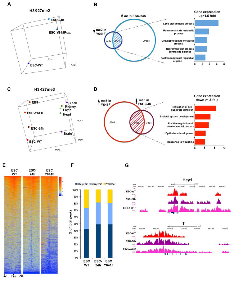
The polycomb repressive complex 2 (PRC2) methylates lysine 27 of histone H3 (H3K27) through its catalytic subunit Ezh2. PRC2-mediated di- and tri-methylation (H3K27me2/H3K27me3) have been interchangeably associated with gene repression. However, it remains unclear whether these two degrees of H3K27 methylation have different functions. In this study, we have generated isogenic mouse embryonic stem cells (ESCs) with a modified H3K27me2/H3K27me3 ratio. Our findings document dynamic developmental control in the genomic distribution of H3K27me2 and H3K27me3 at regulatory regions in ESCs. They also reveal that modifying the ratio of H3K27me2 and H3K27me3 is sufficient for the acquisition and repression of defined cell lineage transcriptional programs and phenotypes and influences induction of the ESC ground state.

Keywords: H3K27 methylation; embryonic stem cells; polycomb proteins.


Figures

Figure 1
Figure 1. Dynamic Distribution of H3K27 Methylation States during ESC Differentiation
(A) Average normalized tag density profiles depicting H3K27me3 enrichment in ESC-24h for prospective DHS regions within the top quartile of either H3K27me3+ (blue trace) or H3K27me2+ (red trace) in pluripotent ESCs. (B) Transition of H3K27me3 across bivalent vs. repressed promoters and poised enhancers in ESC to different H3K27 states during early ESC differentiation (ESC-24h). (C) GO-biological processes associated with genes within ±20Kb proximity from H3K27me3+ regions in ESCs that maintain their H3K27me3 status in ESC-24h (shadowed area). Expression cut-off ≥1.5-fold. (D) Average normalized tag density profiles depicting H3K27 acetylation enrichment in ESC-24h for prospective DHS regions within the top quartile of either H3K27me3+ (blue trace) or H3K27me2+ (red trace) in pluripotent ESCs. (E) Transition of H3K27me2 across H3K4me3+/− promoters and H3K4me1+ enhancers in ESCs to different H3K27 states during early ESC differentiation (ESC-24h). (F) GO-biological processes associated with genes within ±20Kb proximity from the regions that are H3K27me2+ regions in ESCs that acquire H3K27ac in ESC-24h (shadowed area). Expression cut-off ≥1.5-fold. (G) Box plot of H3K27me3 (left panel) and H3K27me2 (right panel) peak intensities for ±5Kb intergenic genomic regions surrounding the summit of each peak at two different ESCs developmental stages and in skeletal myoblasts (MB). P-values were determined by Student’s t-test.
Figure 2
Figure 2. TALEN-Mediated Genome Editing of Ezh2 Modifies H3K27me2 and H3K27me3 in ESCs
(A) Illustration of TALEN-mediated genome editing creating the Ezh2Y641F allele in ESCs. (B) Western blot for Ezh2, tubulin, H3K27me2 (me2), H3K27me3 (me3), and total histone H3 in WT or Y641F ESC chromatin fractions. NI: nuclear insoluble fraction, NS: nuclear soluble fraction, C: cytoplasmic fraction. (C) Western blot for PRC2 components Ezh2, Suz12, and Eed in WT or Y641F ESC whole cell lysates. (D) and (E) Box plots of H3K27me3 (D) and H3K27me2 (E) peak intensities for genomic regions surrounding (±5Kb) TSS in WT and Y641F ESCs. P-values were determined by Student’s t-test. (F) and (G) Averaged normalized tag density profiles of H3K27me3 (F) or H3K27me2 (G) for genomic regions surrounding (±2Kb) prospective DHS regions.
Figure 3
Figure 3. H3K27me2 and H3K27me3 States in Ezh2-Edited Y641F ESCs Resemble Those of Differentiating ESCs
(A) Three-dimensional principal component plots of H3K27me2 in ESC-WT, ESC-24h and ESC-Y641F. (B) GO-biological processes associated with genes within ±20Kb proximity from the regions with decreased H3K27me2 in ESC-Y641F and increased H3K27ac in ESC-24h (shadowed area) and with ≥1.5-fold increased expression in Y641F compared to WT ESCs. (C) Three-dimensional principal component plots of H3K27me3 in ESC- WT, ESC-24h, ESC-Y641F, day 8-embryoid bodies (EB8), and various cell or tissue types. (D) GO-biological processes associated with genes within ±20Kb proximity from the regions with increased H3K27me3 in ESC-Y641F and increased H3K27me3 in ESC-24h (shadowed area) and with ≥1.5-fold decreased expression in Y641F compared to WT ESCs. (E) Unsupervised clustering heat map of H3K27me3 at genomic regions surrounding (±5Kb) TSS in ESC-WT, ESC-24h, and ESC-Y641F. (F) Bar graphs depicting the distribution of H3K27me3 across genomic regions in ESC-WT, ESC-24h, and ESC-Y641F. (G) H3K27me3 signal tracks for representative loci Hey1 and T in ESC-WT, ESC-24h, and ESC-Y641F.
Figure 4
Figure 4. Ezh2-Edited ESCs Preferentially Activate the Neural Fate Program
(A) Morphology (bright field) and pluripotent marker (POU5F1 and SSEA-1) immunostaining of WT and Y641F ESCs. (B) FACS analysis of SSEA-1 expression (left panel) and growth curve (right panel) of WT and Y641 ESCs. (C) Two-dimensional principal component plots of three RNA-seq biological replicates depicting gene expression profiles of WT and Y641F ESCs. (D) GO-biological processes associated with genes with ≥1.2-fold decreased (green bars) or increased (orange bars) expression in Y641F compared to WT ESCs. (E) Fold-change expression (RPKM) from three RNA-seq biological replicates of pluripotent genes (Pou5f1 and Nanog), neural genes (Sox1 and Pax6), mesodermal genes (T and Des), and endodermal genes (Gata4 and Gata6) between WT and Y641F ESCs. Data are represented as mean ± SD.
Figure 5
Figure 5. Ezh2-Edited ESCs Are Refractory to 2i-Induced Naïve Ground State and Retained Their Neural Program in Differentiating Embryoid Bodies
(A) Pathway KEGG analysis for genes with ≥1.2-fold increased (upper panel) or decreased expression (lower panel) in Y641F compared to WT ESCs cultured in 2i medium. (B) Heat map illustrating the RNA expression fold change between Y641F and WT cultured in serum and 2i medium for selected genes of different lineages. (C) Morphology of WT and Y641F ESCs cultured in 2i medium. (D) GO-biological processes associated with genes with ≥1.5-fold increased expression in Y641F ESC, EB8 or EB13 compared to WT EB8 or EB13. (E) Percentage of ESC colonies positive for beating cardiomyocytes (left upper panel) or neuronal projections (left lower panel), along with neural marker Tuj1 immunostaining of adherent EBs grown from WT or Y641F ESCs.
Figure 6
Figure 6. Increased H3K27me3 and Transcriptional Repression of the TGF-β Pathway Underlie Neural Commitment of Ezh2-Edited ESCs Cultured in Serum
(A) Pathway KEGG analysis for genes with ≥1.2-fold increased expression in Y641F compared to WT ESCs. (B) Fold-change expression (RPKM) from three RNA-seq biological replicates for TGF-β family members between WT and Y641F ESCs. Data are represented as mean ± SD. (C) H3K27me3 and H3K27ac signal tracks, and RNA-seq traces at TGF-β1 and BMP4 loci in WT and Y641F ESCs. (D) and (E) Fold-change Sox1 expression before or after addition of TGF-β1 (D) or BMP4 (E) recombinant proteins in WT and Y641F ESCs. Data are represented as mean ± SD (n=3).

References

    1. Avilion AA, Nicolis SK, Pevny LH, Perez L, Vivian N, Lovell-Badge R. Multipotent cell lineages in early mouse development depend on SOX2 function. Genes & development. 2003;17:126–140. - PMC - PubMed
    1. Azuara V, Perry P, Sauer S, Spivakov M, Jorgensen HF, John RM, Gouti M, Casanova M, Warnes G, Merkenschlager M, et al. Chromatin signatures of pluripotent cell lines. Nature cell biology. 2006;8:532–538. - PubMed
    1. Banaszynski LA, Wen D, Dewell S, Whitcomb SJ, Lin M, Diaz N, Elsasser SJ, Chapgier A, Goldberg AD, Canaani E, et al. Hira-dependent histone H3.3 deposition facilitates PRC2 recruitment at developmental loci in ES cells. Cell. 2013;155:107–120. - PMC - PubMed
    1. Barsotti AM, Ryskin M, Zhong W, Zhang WG, Giannakou A, Loreth C, Diesl V, Follettie M, Golas J, Lee M, et al. Epigenetic reprogramming by tumor-derived EZH2 gain-of-function mutations promotes aggressive 3D cell morphologies and enhances melanoma tumor growth. Oncotarget. 2015;6:2928–2938. - PMC - PubMed
    1. Ben-Zvi D, Melton DA. Modeling human nutrition using human embryonic stem cells. Cell. 2015;161:12–17. - PubMed

Publication types

MeSH terms