. 2016 Dec 13;7(50):82985-83000.
doi: 10.18632/oncotarget.12824.

A new humanized in vivo model of KIT D816V+ advanced systemic mastocytosis monitored using a secreted luciferase

Affiliations

A new humanized in vivo model of KIT D816V+ advanced systemic mastocytosis monitored using a secreted luciferase

Siham Bibi et al. Oncotarget. .

Abstract

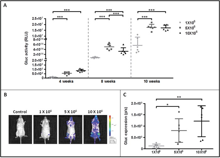
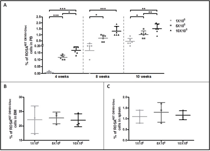
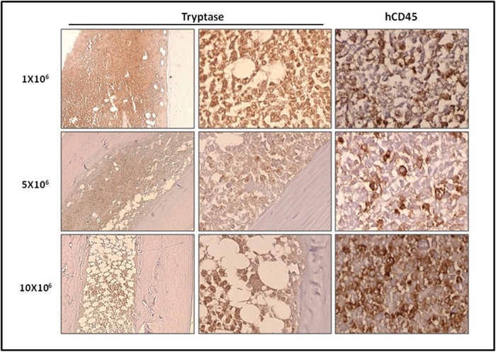
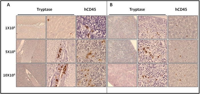
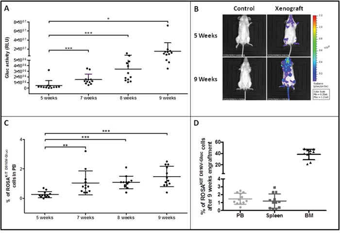
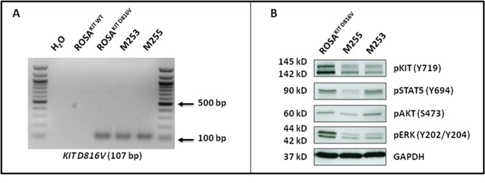
Systemic mastocytosis are rare neoplasms characterized by accumulation of mast cells in at least one internal organ. The majority of systemic mastocytosis patients carry KIT D816V mutation, which activates constitutively the KIT receptor. Patient with advanced forms of systemic mastocytosis, such as aggressive systemic mastocytosis or mast cell leukemia, are poorly treated to date. Unfortunately, the lack of in vivo models reflecting KIT D816V+ advanced disease hampers pathophysiological studies and preclinical development of new therapies for such patients. Here, we describe a new in vivo model of KIT D816V+ advanced systemic mastocytosis developed by transplantation of the human ROSAKIT D816V-Gluc mast cell line in NOD-SCID IL-2R γ-/- mice, using Gaussia princeps luciferase as a reporter. Intravenous injection of ROSAKIT D816V-Gluc cells led, in 4 weeks, to engraftment in all injected primary recipient mice. Engrafted cells were found at high levels in bone marrow, and at lower levels in spleen, liver and peripheral blood. Disease progression was easily monitored by repeated quantification of Gaussia princeps luciferase activity in peripheral blood. This quantification evidenced a linear relationship between the number of cells injected and the neoplastic mast cell burden in mice. Interestingly, the secondary transplantation of ROSAKIT D816V-Gluc cells increased their engraftment capability. To conclude, this new in vivo model mimics at the best the features of human KIT D816V+ advanced systemic mastocytosis. In addition, it is a unique and convenient tool to study the kinetics of the disease and the potential in vivo activity of new drugs targeting neoplastic mast cells.

Keywords: KIT D816V mutant; NSG mice; ROSAKIT D816V cell line; advanced systemic mastocytosis; gluc reporter.


Conflict of interest statement

CONFLICTS OF INTEREST

The authors declare no competing financial interest.

Figures

Figure 1
Figure 1. Generation of ROSAKIT D816V-Gluc cell line and comparison with the parental ROSAKIT D816V cell line
A. ROSAKIT D816V-Gluc GFP+/CFP+ sorted cells using flow cytometry. B. Comparison of ROSAKIT D816V and ROSAKIT D816V-Gluc cell proliferation by the use of the MTT method. Cells were seeded in 96-well plate for 5 days (1 plate/day). At each day (d0 - d4), 20 μL of MTT were added in each well and the cells were incubated for 3 additional hours at 37°C. After adding 100 μL of acidified isopropanol, the number of living cells was measured for each condition by reading the absorbance at 570 nm. Data are presented as the mean ± SD (n = 3). C. Phenotypic comparison of the cytological aspect of ROSAKIT D816V and ROSAKIT D816V-Gluc. MGG-stained cytospin preparations of ROSAKIT D816V (left panel) and ROSAKIT D816V-Gluc (right panel). Magnification is x50 (top), x100 (bottom). D. Comparative expression of CD117 (KIT) by in ROSAKIT D816V (left) and ROSAKIT D816V-Gluc (right) analyzed by flow cytometry.
Figure 2
Figure 2. Effects of transduction of ROSAKIT D816V cells with LV-Gluc-CFP on KIT activation and response to TKIs
A. Analysis of KIT D816V gene expression in ROSAKIT D816V-Gluc cell line using agarose gel electrophoresis of conventional RT-PCR products. Reactions were compared to the levels of expression of mRNA for HPRT. The image is an inverted form of the original picture. B. Detection of spontaneous phosphorylation of KIT receptor by western blotting in ROSAKIT D816V-Gluc. Cell lysates from ROSAKIT WT cells stimulated with SCF, or from unstimulated ROSAKIT D816V and ROSAKIT D816V-Gluc cells, were subjected to electrophoresis and stained with antibody against p-KIT Y719. An anti-human glyceraldehyde-3-phosphate dehydrogenase (GAPDH) was used as a loading control. The lanes were run on the same gel. C. Gluc activity in cell culture supernatants of ROSAKIT D816V (grey line) and ROSAKIT D816V-Gluc (black line). Signals were calculated as RLU; relative luminescent units. Data present the mean ± SD (n = 3). D, E. Effect of Imatinib or Midostaurin on the proliferation of ROSAKIT D816V (grey line) and ROSAKIT D816V-Gluc cells (black line). Each cell lines were seeded for 72h in 96-well plate in the presence of various concentrations (0.001 - 10 μM) of Imatinib (D) or Midostaurin (E). Data are presented as the mean ± SD (n = 3) and are expressed as percent of proliferation in each condition relative to the control (untreated cells) considered as 100% proliferation.
Figure 3
Figure 3. Monitoring of ROSAKIT D816V-Gluc cells engraftment in injected mice by measurement of Gluc activity in blood and by IVIS
A. Gluc activity in plasma of mice after various times of engraftment. Different numbers of ROSAKIT D816V-Gluc cells [1x10⁶ (●), 5x10⁶ (■) and 10x106 (▲)] were injected intravenously in mice and engraftment was monitored over time (4, 8 and 10 weeks). Each point represents an individual mouse. B. IVIS showing localization of Gluc in injected mice after 10 weeks. Indicated numbers of ROSAKIT D816V-Gluc cells were IV injected in mice (n = 6). Units in rainbow color scales are photons per second per cm2 per steradian (p/sec/cm2/sr). Results shown are from one representative mouse for each group. C. Total relative units (RLU) per second were calculated for Gluc intensity shown in (B) by ROI analysis after 10 weeks of engraftment in the three groups of mice. Each point represents an individual mouse.
Figure 4
Figure 4. Evaluation of ROSAKIT D816V-Gluc cells numbers by flow cytometry in peripheral blood, bone marrow and spleen
A. Percentage of ROSAKIT D816V-Gluc cells in PB after increasing times of engraftment (4, 8 and 10 weeks). The percentage of ROSAKIT D816V-Gluc cells was determined in the three groups of mice: 1x10⁶ (●), 5x10⁶ (■) and 10x106 (▲) by flow cytometry using monoclonal antibodies against human CD45 and human CD117. B, C. Percentages of ROSAKIT D816V-Gluc cells after 10 weeks of engraftment in BM and spleen respectively in the three groups of mice: 1x10⁶ (●), 5x10⁶ (■) and 10x106(▲). Each point represents an individual mouse.
Figure 5
Figure 5. Localization of primary engrafted ROSAKIT D816V-Gluc cells in mice BM by IHC detection of tryptase and hCD45
Ten weeks after engraftment, BM sections from the three groups of mice were stained by IHC with an anti-human tryptase antibody (left and middle panels) antibody and an anti-human CD45 antibody (right panel). Staining was visualized by Histomouse Kit, showing human MCs in brownish staining. Magnification is x10 (left panel) and x50 (middle and right panels). Results are from one representative mouse from each group.
Figure 6
Figure 6. Localization of primary engrafted ROSAKIT D816V-Gluc cells in mice spleen and liver by IHC detection of tryptase and hCD45
Ten weeks after engraftment, spleen A. and liver B. sections from the three groups of mice were stained by IHC with an anti-human tryptase (left and middle panels) antibody and an anti-human CD45 (right panel) showing human MCs in brownish staining. Magnification is x10 (left panel) and x50 (middle and right panels). Results are from one representative mouse from each group.
Figure 7
Figure 7. Reconstitution capacity of the human ROSAKIT D816V-Gluc cells in NSG mice after secondary transplantation
Ten weeks after engraftment in primary recipient mice, human cells were isolated from BM samples and injected into secondary recipient mice (n = 12). Engraftment was assessed 5, 7, 8 and 9 weeks after injection. A. Time-dependent increase of Gluc activity in plasma after secondary transplantation with ROSAKIT D816V-Gluc cells. Each point represents an individual mouse. B. In vivo imaging of Gluc activity in secondary injected mice (right lane) after 5 weeks (top lane) and 9 weeks (bottom lane) versus negative control (left lane). Results shown are from one representative mouse. C. Time-dependent increase in the percentages of ROSAKIT D816V-Gluc cells in PB after secondary transplantation. D. Percentage of ROSAKIT D816V-Gluc cells after secondary transplantation in PB (●), spleen (■) and BM (▲). The percentage of hCD45+/hCD117+ was measured using flow cytometry in C and D.
Figure 8
Figure 8. Localization of previously xenotransplanted ROSAKIT D816V-Gluc cells in mice BM using IHC detection of hCD45 after secondary transplantation
BM sections (left panel), spleen sections (middle panel), and liver sections (right panel) stained by IHC with anti-human CD45 antibody showing human cells in brownish staining. Magnification is x10 (top lane) and x50 (bottom lane). Each result shown is from one representative mouse having received previously xenotransplanted ROSAKIT D816V-Gluc cells.
Figure 9
Figure 9. ROSAKIT D816V-Gluc cells express in vivo KIT D816V mutant leading to constitutive phosphorylation of KIT and of downstream signaling pathways after secondary transplantation
A. Expression of the KIT D816V gene in BM samples of injected mice using allele-specific RT-PCR. KIT D816V allele-specific PCR was performed using ROSAKIT D816V cells as a positive control. The image is an inverted form of the original picture. B. Spontaneous phosphorylation of the KIT receptor and of its downstream signaling pathways (STAT5, AKT and ERK) revealed by western blotting in ROSAKIT D816V cells and BM cells of secondary transplanted mice. An anti-human glyceraldehyde-3-phosphate dehydrogenase (GAPDH) was used as a loading control. The lanes were run on the same gel.

Similar articles

Cited by

References

    1. Kirshenbaum AS, Kessler SW, Goff JP, Metcalfe DD. Demonstration of the origin of human mast cells from CD34+ bone marrow progenitor cells. J Immunol. 1991;146:1410–5. - PubMed
    1. Krishnaswamy G, Ajitawi O, Chi DS. The human mast cell: an overview. Methods Mol Biol. 2006;315:13–34. - PubMed
    1. Blume-Jensen P, Hunter T. Oncogenic kinase signalling. Nature. 2001;411:355–65. - PubMed
    1. Galli SJ, Tsai M, Wershil BK, Tam SY, Costa JJ. Regulation of mouse and human mast cell development, survival and function by stem cell factor, the ligand for the c-kit receptor. Int Arch Allergy Immunol. 1995;107:51–3. - PubMed
    1. Kitamura Y, Oboki K, Ito A. Molecular mechanisms of mast cell development. Immunol Allergy Clin North Am. 2006;26:387–405. v. - PubMed

MeSH terms

Substances