

Quantitative analysis of the role of fiber length on phagocytosis and inflammatory response by alveolar macrophages
- PMID: 27784615
- PMCID: PMC5228597
- DOI: 10.1016/j.bbagen.2016.09.031
Quantitative analysis of the role of fiber length on phagocytosis and inflammatory response by alveolar macrophages
Abstract
Background: In the lung, macrophages attempt to engulf inhaled high aspect ratio pathogenic materials, secreting inflammatory molecules in the process. The inability of macrophages to remove these materials leads to chronic inflammation and disease. How the biophysical and biochemical mechanisms of these effects are influenced by fiber length remains undetermined. This study evaluates the role of fiber length on phagocytosis and molecular inflammatory responses to non-cytotoxic fibers, enabling development of quantitative length-based models.
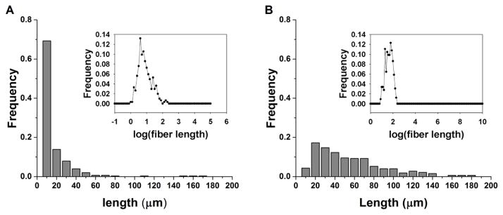
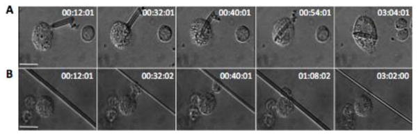
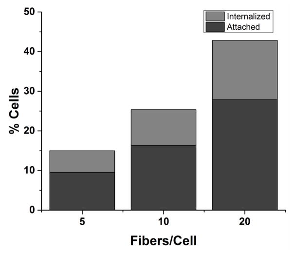
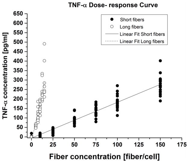
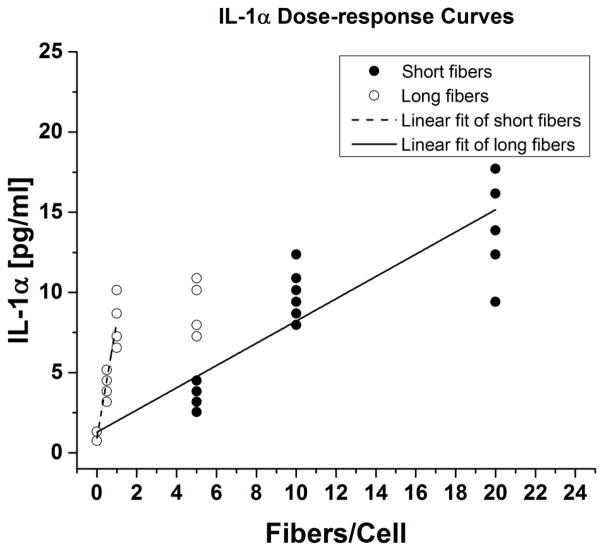
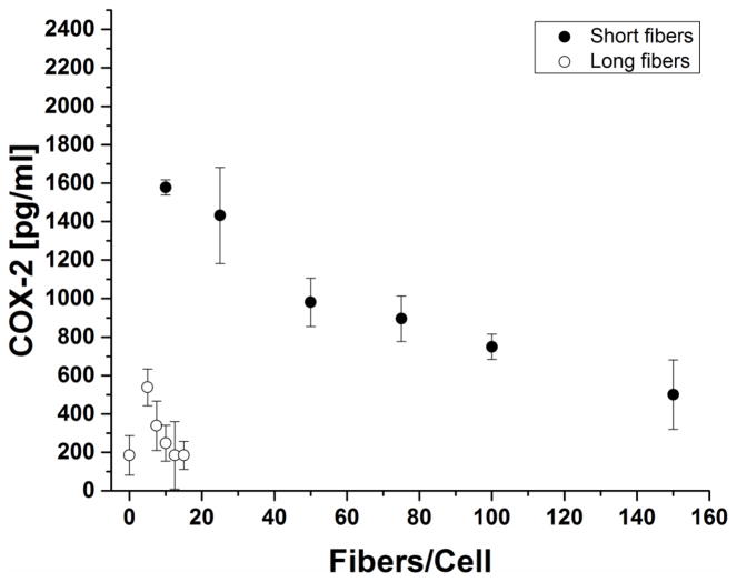
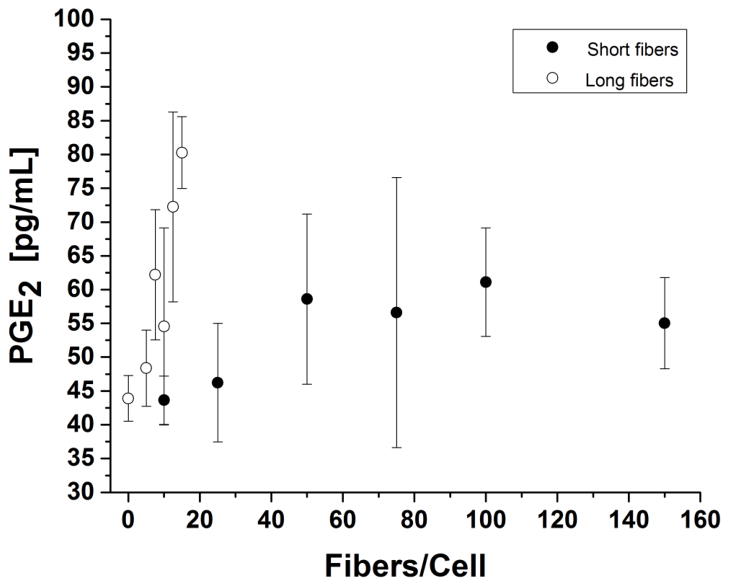
Methods: Murine alveolar macrophages were exposed to short and long populations of JM-100 glass fibers, produced by successive sedimentation and repeated crushing, respectively. Interactions between fibers and macrophages were observed using time-lapse video microscopy, and quantified by flow cytometry. Inflammatory biomolecules (TNF-α, IL-1α, COX-2, PGE2) were measured.
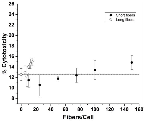
Results: Uptake of short fibers occurred more readily than for long, but long fibers were more potent stimulators of inflammatory molecules. Stimulation resulted in dose-dependent secretion of inflammatory biomolecules but no cytotoxicity or strong ROS production. Linear cytokine dose-response curves evaluated with length-dependent potency models, using measured fiber length distributions, resulted in identification of critical fiber lengths that cause frustrated phagocytosis and increased inflammatory biomolecule production.
Conclusion: Short fibers played a minor role in the inflammatory response compared to long fibers. The critical lengths at which frustrated phagocytosis occurs can be quantified by fitting dose-response curves to fiber distribution data.
General significance: The single physical parameter of length can be used to directly assess the contributions of length against other physicochemical fiber properties to disease endpoints.
Keywords: Frustrated phagocytosis; Glass fibers; Length; Macrophage; TNF-α: Tumor necrosis factor-α.
Copyright © 2016 Elsevier B.V. All rights reserved.
Figures
References
-
- Aderem A, Underhill D. Mechanisms of phagocytosis in macrophages. Annu Rev Immunol. 1999;17:593–623. - PubMed
-
- Shukla A, Gulumian M, Hei TK, Kamp D, Rahman Q, Mossman BT. Multiple roles of oxidants in the pathogenesis of asbestos-induced diseases. Free Radic Biol Med. 2003;34:1117–1129. - PubMed
-
- Ault JG, Cole RW, Jensen CG, Jensen LCW, Bachert LA, Reider CL. Behavior of Crocidolite Asbestos during Mitosis in Living Vertebrate Lung Epithelial Cells. Cancer Res. 1995;55:792–798. - PubMed
Publication types
MeSH terms
Substances
Grants and funding
LinkOut - more resources
Full Text Sources
Other Literature Sources
Research Materials
