. 2017 Jan 1;312(1):F77-F83.
doi: 10.1152/ajprenal.00367.2016. Epub 2016 Oct 26.

Impaired urinary osteopontin excretion in Npt2a-/- mice

Affiliations

Impaired urinary osteopontin excretion in Npt2a-/- mice

Daniel Caballero et al. Am J Physiol Renal Physiol. .

Abstract

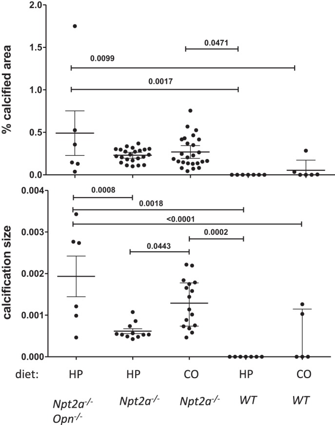
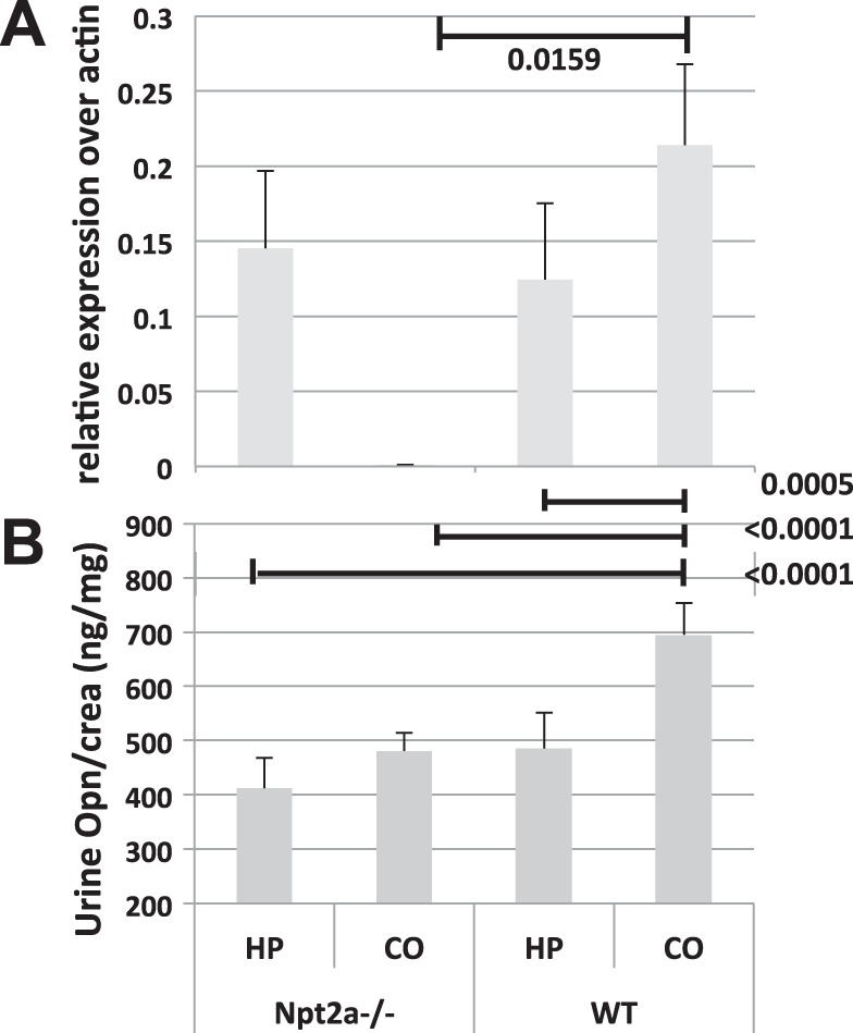
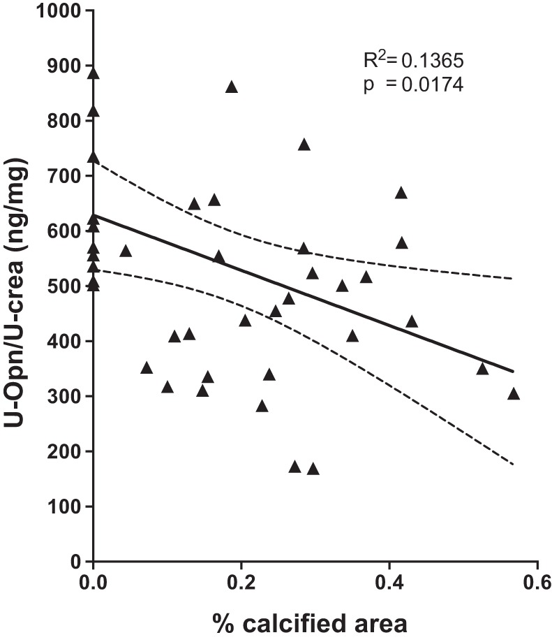
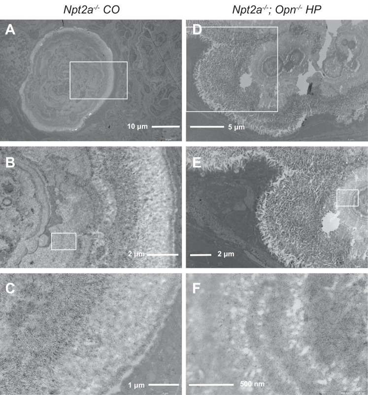
Mutations in the renal sodium-dependent phosphate cotransporters NPT2a and NPT2c have been reported in patients with renal stone disease and nephrocalcinosis. Oral phosphate supplementation is currently thought to reduce risk by reversing the hypercalciuria, but the exact mechanism remains unclear and the relative contribution of modifiers of mineralization such as osteopontin (Opn) to the formation of renal mineral deposits in renal phosphate wasting disorders has not been studied. We observed a marked decrease of renal gene expression and urinary excretion of Opn in Npt2a-/- mice, a mouse model of these disorders, at baseline. Following supplementation with phosphate Opn gene expression was restored to wild-type levels in Npt2a-/- mice; however, urine excretion of the protein remained low. To further investigate the role of Opn, we used a double-knockout strategy, which provides evidence that loss of Opn worsens the nephrocalcinosis and nephrolithiasis observed in these mice on a high-phosphate diet. These studies suggest that impaired Opn gene expression and urinary excretion in Npt2a-/- mice may be an additional risk factor for nephrolithiasis, and normalizing urine Opn levels may improve the therapy of phosphaturic disorders.

Keywords: NPT2a; hypophosphatemia; nephrocalcinosis; osteopontin; rickets.


Figures

Fig. 1.
Fig. 1.
Ablation of Opn worsens renal mineralization size seen in Npt2a−/− mice on HP diet. Histomorphometric analysis of renal mineralization [% calcified area = 100 × mineralization area/tissue area (A) and mineralization size = mineralization area/number of mineralizations, μm2 (B)] in 10 μm sections of kidneys from mice feed different diets for 10 wk (see legend of Table 1 for composition of diets). The data represent individual animals (●) with the means ± SE. P values shown above the lines of comparisons were calculated by one-way ANOVA and Tukey’s test for multiple comparisons.
Fig. 2.
Fig. 2.
Renal osteopontin mRNA expression and urinary excretion are decreased in Npt2a−/− mice. A: Opn gene expression measured by quantitative reverse transcription PCR corrected over β-actin in total kidney RNA. B: urinary excretion of Opn protein (U-Opn) corrected for urine creatinine (U-Crea) from mice feed different diets for 10 wk (see legend of Table 1 for composition of diets). The data represent means ± SE. P values shown above the lines of comparisons were calculated by one-way ANOVA and Tukey’s test for multiple comparisons.
Fig. 3.
Fig. 3.
Urinary Opn excretion is significantly associated with renal mineralization in a combined bivariate linear regression analysis of WT and Npt2a−/− mice fed different diets. WT and Npt2a−/− mice from Table 1 (n = 42) were evaluated by linear regression analysis to determine the association of renal mineralization with the ratio of urine osteopontin/urine creatinine (U-Opn/U-crea). Data points represent values of individual animals. Results of the linear regression analysis are shown as solid line with 95% confidence interval (stippled lines), R2, and P values.
Fig. 4.
Fig. 4.
Cortical and medullary renal mineralization. Light micrographs of 10-μm renal sections, prepared from paraffin-embedded kidneys: von Kossa, methylene green staining, 4X (A); von Kossa, hematoxylin and eosin staining, 40X (B). See legend of Table 1 for composition of CO and HP diets.
Fig. 5.
Fig. 5.
Transmission electron microscopy of mouse kidneys reveals thin submicrometer, plate-like crystals starting at center and organizing into microspheres. Cross section shows concentrically arranged microspherules microspherules that appear to be growing by addition of crystal layers at the periphery. White box indicates approximate location of the image with next higher magnification. Letters indicate the following: a–c Npt2a−/− CO diet; d–f Npt2a−/− and Opn−/− HP diet.

References

    1. Albright RA, Stabach P, Cao W, Kavanagh D, Mullen I, Braddock AA, Covo MS, Tehan M, Yang G, Cheng Z, Bouchard K, Yu ZX, Thorn S, Wang X, Folta-Stogniew EJ, Negrete A, Sinusas AJ, Shiloach J, Zubal G, Madri JA, De La Cruz EM, Braddock DT. ENPP1-Fc prevents mortality and vascular calcifications in rodent model of generalized arterial calcification of infancy. Nat Commun 6: 10006, 2015. doi:10.1038/ncomms10006. - DOI - PMC - PubMed
    1. Alon U, Donaldson DL, Hellerstein S, Warady BA, Harris DJ. Metabolic and histologic investigation of the nature of nephrocalcinosis in children with hypophosphatemic rickets and in the Hyp mouse. J Pediatr 120: 899–905, 1992. doi:10.1016/S0022-3476(05)81957-2. - DOI - PubMed
    1. Arcidiacono T, Mingione A, Macrina L, Pivari F, Soldati L, Vezzoli G. Idiopathic calcium nephrolithiasis: a review of pathogenic mechanisms in the light of genetic studies. Am J Nephrol 40: 499–506, 2014. doi:10.1159/000369833. - DOI - PubMed
    1. Barros NM, Hoac B, Neves RL, Addison WN, Assis DM, Murshed M, Carmona AK, McKee MD. Proteolytic processing of osteopontin by PHEX and accumulation of osteopontin fragments in Hyp mouse bone, the murine model of X-linked hypophosphatemia. J Bone Miner Res 28: 688–699, 2013. doi:10.1002/jbmr.1766. - DOI - PubMed
    1. Beck L, Karaplis AC, Amizuka N, Hewson AS, Ozawa H, Tenenhouse HS. Targeted inactivation of Npt2 in mice leads to severe renal phosphate wasting, hypercalciuria, and skeletal abnormalities. Proc Natl Acad Sci USA 95: 5372–5377, 1998. doi:10.1073/pnas.95.9.5372. - DOI - PMC - PubMed

Publication types

MeSH terms

Substances