Review
. 2016 Oct 31;60(3):255-273.
doi: 10.1042/EBC20160016.

Photosynthesis

Affiliations
Review

Photosynthesis

Matthew P Johnson. Essays Biochem. .

Erratum in

  • Correction: Photosynthesis.
    Johnson MP. Johnson MP. Essays Biochem. 2017 Oct 31;61(4):429. doi: 10.1042/EBC20160016_COR. Print 2017 Oct 31. Essays Biochem. 2017. PMID: 29089380 Free PMC article. No abstract available.

Abstract

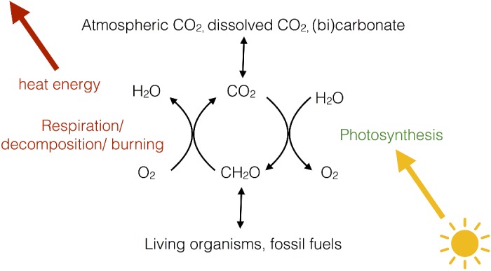
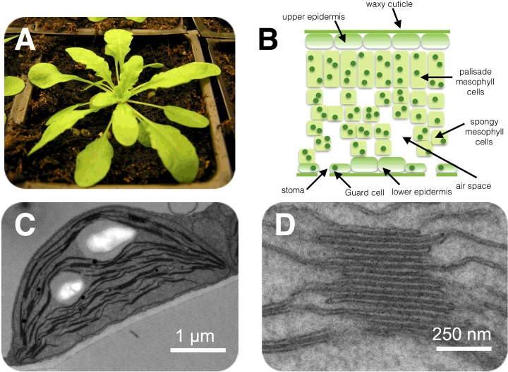
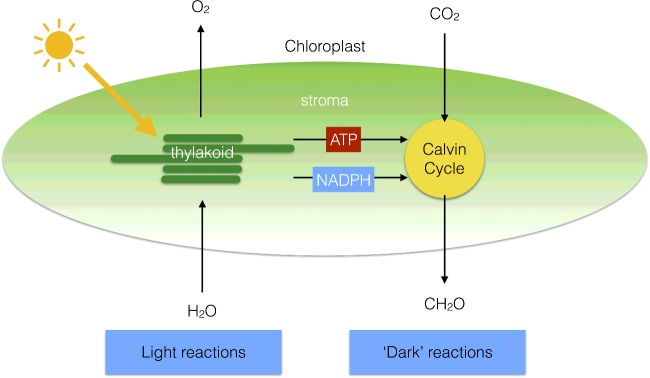
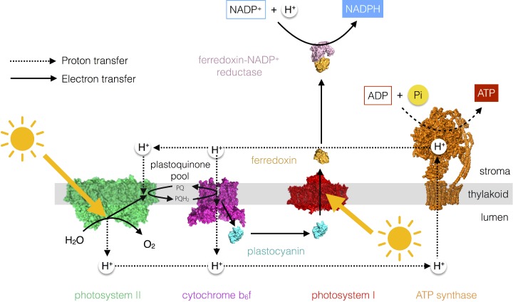
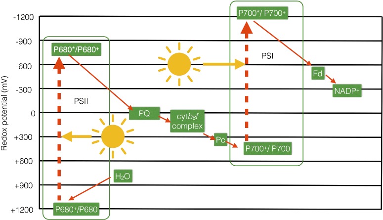
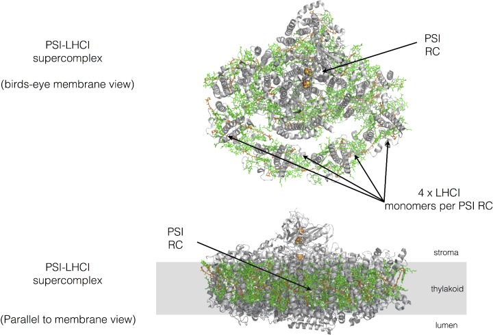
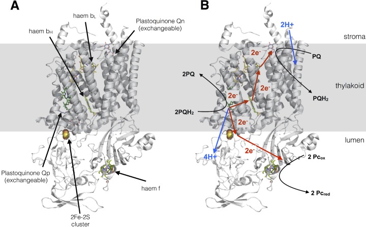
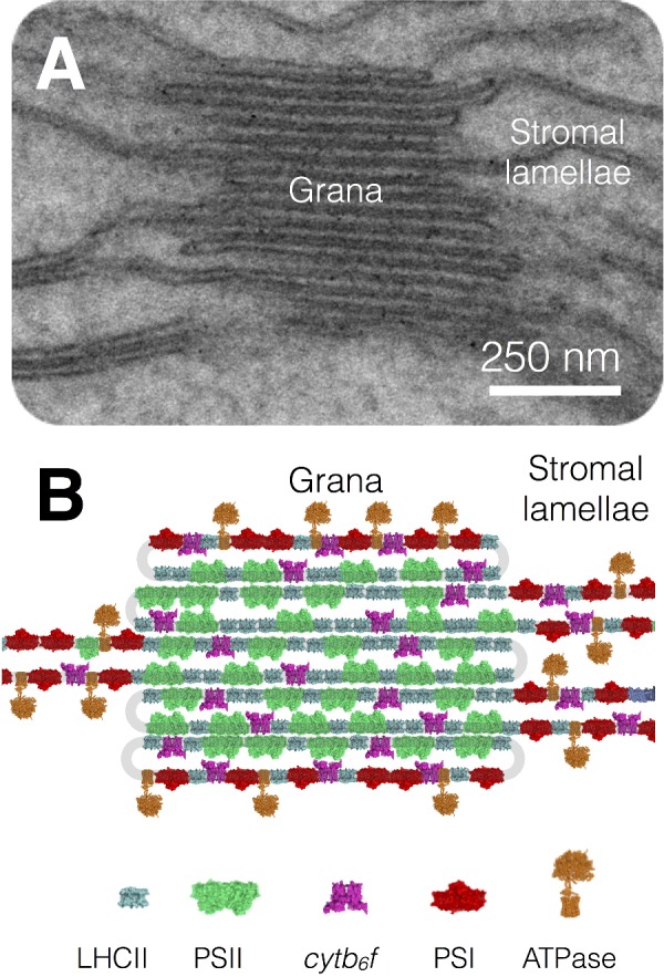
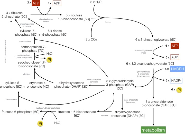
Photosynthesis sustains virtually all life on planet Earth providing the oxygen we breathe and the food we eat; it forms the basis of global food chains and meets the majority of humankind's current energy needs through fossilized photosynthetic fuels. The process of photosynthesis in plants is based on two reactions that are carried out by separate parts of the chloroplast. The light reactions occur in the chloroplast thylakoid membrane and involve the splitting of water into oxygen, protons and electrons. The protons and electrons are then transferred through the thylakoid membrane to create the energy storage molecules adenosine triphosphate (ATP) and nicotinomide-adenine dinucleotide phosphate (NADPH). The ATP and NADPH are then utilized by the enzymes of the Calvin-Benson cycle (the dark reactions), which converts CO2 into carbohydrate in the chloroplast stroma. The basic principles of solar energy capture, energy, electron and proton transfer and the biochemical basis of carbon fixation are explained and their significance is discussed.

Keywords: membrane; photosynthesis; thylakoid.


Figures

Figure 1
Figure 1. The global carbon cycle
The relationship between respiration, photosynthesis and global CO2 and O2 levels.
Figure 2
Figure 2. Location of the photosynthetic machinery
(A) The model plant Arabidopsis thaliana. (B) Basic structure of a leaf shown in cross-section. Chloroplasts are shown as green dots within the cells. (C) An electron micrograph of an Arabidopsis chloroplast within the leaf. (D) Close-up region of the chloroplast showing the stacked structure of the thylakoid membrane.
Figure 3
Figure 3. Division of labour within the chloroplast
The light reactions of photosynthesis take place in the thylakoid membrane, whereas the dark reactions are located in the chloroplast stroma.
Figure 4
Figure 4. The photosynthetic electron and proton transfer chain
The linear electron transfer pathway from water to NADP+ to form NADPH results in the formation of a proton gradient across the thylakoid membrane that is used by the ATP synthase enzyme to make ATP.
Figure 5
Figure 5. Z-scheme of photosynthetic electron transfer
The main components of the linear electron transfer pathway are shown on a scale of redox potential to illustrate how two separate inputs of light energy at PSI and PSII result in the endergonic transfer of electrons from water to NADP+.
Figure 6
Figure 6. Major photosynthetic pigments in plants
The chemical structures of the chlorophyll and carotenoid pigments present in the thylakoid membrane. Note the presence in each of a conjugated system of carbon–carbon double bonds that is responsible for light absorption.
Figure 7
Figure 7. Basic absorption spectra of the major chlorophyll and carotenoid pigments found in plants
Chlorophylls absorb light energy in the red and blue part of the visible spectrum, whereas carotenoids only absorb light in the blue/green.
Figure 8
Figure 8. Jablonski diagram of chlorophyll showing the possible fates of the S1 and S2 excited states and timescales of the transitions involved
Photons with slightly different energies (colours) excite each of the vibrational substates of each excited state (as shown by variation in the size and colour of the arrows).
Figure 9
Figure 9. Basic mechanism of excitation energy transfer between chlorophyll molecules
Two chlorophyll molecules with resonant S1 states undergo a mirror transition resulting in the non-radiative transfer of excitation energy between them.
Figure 10
Figure 10. Basic structure of a photosystem
Light energy is captured by the antenna pigments and transferred to the special pair of RC chlorophylls which undergo a redox reaction leading to reduction of an acceptor molecule. The oxidized special pair is regenerated by an electron donor.
Figure 11
Figure 11. Basic structure of the PSII–LHCII supercomplex from spinach
The organization of PSII and its light-harvesting antenna. Protein is shown in grey, with chlorophylls in green and carotenoids in orange. Drawn from PDB code 3JCU
Figure 12
Figure 12. S-state cycle of water oxidation by the manganese cluster (shown as circles with roman numerals representing the manganese ion oxidation states) within the PSII oxygen-evolving complex
Progressive extraction of electrons from the manganese cluster is driven by the oxidation of P680 within PSII by light. Each of the electrons given up by the cluster is eventually repaid at the S4 to S0 transition when molecular oxygen (O2) is formed. The protons extracted from water during the process are deposited into the lumen and contribute to the protonmotive force.
Figure 13
Figure 13. Basic structure of the PSI–LHCI supercomplex from pea
The organization of PSI and its light-harvesting antenna. Protein is shown in grey, with chlorophylls in green and carotenoids in orange. Drawn from PDB code 4XK8.
Figure 14
Figure 14. Cytochrome b6f complex
(A) Structure drawn from PDB code 1Q90. (B) The protonmotive Q-cycle showing how electrons from plastoquinol are passed to both plastocyanin and plastoquinone, doubling the protons deposited in the lumen for every plastoquinol molecule oxidized by the complex.
Figure 15
Figure 15. Lateral heterogeneity in thylakoid membrane organization
(A) Electron micrograph of the thylakoid membrane showing stacked grana and unstacked stromal lamellae regions. (B) Model showing the distribution of the major complexes of photosynthetic electron and proton transfer between the stacked grana and unstacked stromal lamellae regions.
Figure 16
Figure 16. The Calvin–Benson cycle
Overview of the biochemical pathway for the fixation of CO2 into carbohydrate in plants.
Figure 17
Figure 17. Rubisco
(A) Structure of the Rubisco enzyme (the large subunits are shown in blue and the small subunits in green); four of each type of subunit are visible in the image. Drawn from PDB code 1RXO. (B) Activation of the lysine residue within the active site of Rubisco occurs via elevated stromal pH and Mg2+ concentration as a result of the activity of the light reactions.
Figure 18
Figure 18. Diagram of a C4 plant leaf showing Kranz anatomy
Figure 19
Figure 19. The C4 pathway (NADP+–malic enzyme type) for fixation of CO2

Comment in

  • Editorial Note: Photosynthesis.
    [No authors listed] [No authors listed] Essays Biochem. 2021 Jul 26;65(2):405. doi: 10.1042/EBC-2016-0016C_EDN. Epub 2021 Jul 16. Essays Biochem. 2021. PMID: 34309653 Free PMC article. No abstract available.

References

    1. Blankenship R.E. Early evolution of photosynthesis. Plant Physiol. 2010;154:434–438. doi: 10.1104/pp.110.161687. - DOI - PMC - PubMed
    1. Blankenship R.E. Molecular Mechanisms of Photosynthesis. Chichester: Wiley–Blackwell Publishing; 2014.
    1. Nelson N., Ben Shem A. The complex architecture of oxygenic photosynthesis. Nat. Rev. 2004;5:1–12. doi: 10.1038/nrm1525. - DOI - PubMed
    1. Raines C. The Calvin cycle revisited. Photosynth. Res. 2003;75:1–10. doi: 10.1023/A:1022421515027. - DOI - PubMed
    1. Ruban A.V. Evolution under the sun: optimizing light harvesting in photosynthesis. J. Exp. Bot. 2015;66:7–23. doi: 10.1093/jxb/eru400. - DOI - PubMed

MeSH terms

Substances