Review
. 2017 Jan;49(1):7-19.
doi: 10.1002/lsm.22600. Epub 2016 Oct 27.

Reflectance confocal microscopy of skin in vivo: From bench to bedside

Affiliations
Review

Reflectance confocal microscopy of skin in vivo: From bench to bedside

Milind Rajadhyaksha et al. Lasers Surg Med. 2017 Jan.

Abstract

Following more than two decades of effort, reflectance confocal microscopy (RCM) imaging of skin was granted codes for reimbursement by the US Centers for Medicare and Medicaid Services. Dermatologists in the USA have started billing and receiving reimbursement for the imaging procedure and for the reading and interpretation of images. RCM imaging combined with dermoscopic examination is guiding the triage of lesions into those that appear benign, which are being spared from biopsy, against those that appear suspicious, which are then biopsied. Thus far, a few thousand patients have been spared from biopsy of benign lesions. The journey of RCM imaging from bench to bedside is certainly a success story, but still much more work lies ahead toward wider dissemination, acceptance, and adoption. We present a brief review of RCM imaging and highlight key challenges and opportunities. The success of RCM imaging paves the way for other emerging optical technologies, as well-and our bet for the future is on multimodal approaches. Lasers Surg. Med. 49:7-19, 2017. © 2016 Wiley Periodicals, Inc.

Keywords: melanoma; non-melanoma; reflectance confocal microscopy; reimbursement codes; skin; skin cancer.


Conflict of interest statement

Conflict of Interest Disclosures: All authors have completed and submitted the ICMJE Form for Disclosure of Potential Conflicts of Interest and have disclosed the following: Milind Rajadhyaksha is a former employee of, and owns equity in, Caliber Imaging and Diagnostics (formerly, Lucid Inc.), the company that manufactures and sells the VivaScope reflectance confocal microscope. The VivaScope is the commercial version of an original laboratory prototype that was developed by Rajadhyaksha when he was at Wellman Laboratories of Photomedicine, Massachusetts General Hospital, Harvard Medical School. Allan Halpern serves on the scientific advisory board of Caliber Imaging and Diagnostics. None for Ashfaq Marghoob, Anthony Rossi and Kishwer Nehal.

Figures

Fig. 1
Fig. 1
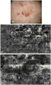
A female patient, 51 years old, noticed a pigmented lesion on her left cheek, which had not changed during the past year, but she was concerned due to previous history of melanoma. The dermoscopic picture (a, yellow circled area) appeared nebulous with no specific features. RCM imaging was performed to rule out malignant melanoma of lentigo maligna type. Mosaics (b) and magnified sub-mosaics (yellow square area, c) in the supra-basal layer showed shows regular honeycomb network of spinous cells with focal cobblestone pattern. No bright atypical pagetoid dendritic cells were identified in the superficial epidermis or around the hair follicle, which ruled out the diagnosis of lentigo maligna. Mosaics (d) and magnified sub-mosaics (yellow square area, e) at the dermal-epidermal junction showed branching tubular structures with bulbous projections or cord-like rete-ridges, lined by bright keratinocytes along with solar elastosis in the surrounding dermis. In this case, RCM imaging spared biopsy of a benign lesion and addressed the patient's concern. Follow-up imaging will be performed in 6 months to monitor the lesion. (All patient cases and figures for this paper were prepared by Drs. Manu Jain, Miguel Cordova, and Kivanc Kose in our Dermatology Service at MSKCC).
Fig. 2
Fig. 2
On a 76-year-old female patient, with a history of melanoma and squamous cell carcinoma, skin examination revealed an irregular 1.3 cm-sized pigmented lesion on the left posterior lower leg. Dermoscopy (a) revealed leaf-like structures, concentric globules, and shiny white blotches and strands. Dermoscopic examination was supplemented with RCM imaging to further guide and confirm diagnosis. Mosaics (b) and magnified sub-mosaics (yellow rectangular area, c) revealed streaming of nuclei with tumor nests and vessels, indicating the presence of superficial and nodular basal cell carcinoma (BCC). Based on RCM imaging, the option of proceeding to complete excision versus biopsy and subsequent topical (Imiquimod) treatment was discussed. The patient chose the latter option, and the saucerized specimen confirmed the presence of superficial and nodular BCC with clear margins. In this case, the impact of RCM imaging was to confirm the diagnosis of BCC and guide decision on definitive treatment.
Fig. 3
Fig. 3
On a 38-year-old female patient with a history of melanoma and atypical mole syndrome, skin examination with total body photography revealed changes in a 1.3 cm-sized lesion on her right abdomen, relative to an earlier baseline examination. The lesion developed a focal darker area and appeared more pink incolor. Dermoscopy (a) revealed focal atypical networkinthe darker area and shiny white lines and dotted vessels in the pink area. Dermoscopic examination was supplemented with RCM imaging, given the patient's history and high risk for developing melanoma, to determine whether a nevus could be differentiated from melanoma, and perhaps avoid performing an unnecessary biopsy. Mosaics (b) and magnified sub-mosaics (yellow square area, c) revealed widespread pagetoid cells, epidermal atypia, and disarray of the spinous layers, perifollicular infiltration of atypical cells and non-edged dermal papillae. All of these features were highly suggestive of melanoma, leading to a complete excision of the lesion. The pathology confirmed the presence of melanoma in situ arising in association with a melanocytic nevus. In this case, the impact of RCM imaging was to confirm the need to perform an excisional biopsy of this lesion.
Fig. 4
Fig. 4
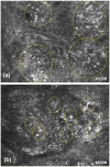
A pitfall of RCM imaging is highlighted in these two cases, each showing a compound melanocytic lesion. In one lesion (a), bright dendritic cells with roundish nuclear morphology were seen in the basal layer at the dermal-epidermal junction (yellow circled areas). Pathology and immunostaining revealed proliferation of melanocytes as either solitary cells or small and large nests situated at dermal-epidermal junction, in the overlying basal and spinous layers and underlying papillary dermis. This lesion turned out to be a melanoma. In the other lesion (b), bright pleomorphic and dendritic cells were seen in pagetoid patterns (yellow circled areas). Here, pathology and immunostaining revealed proliferation of melanocytes along dermal-epidermal junction as well as scattered Langerhans cells in the overlying epidermis. This lesion turned out to be a lentiginous compound melanocytic nevus with some architectural disorder, slight atypia, and partial regression. Details of this study, including the pathology and immunostaining correlation results, are available in reference [70]. Bright cells in pagetoid patterns in RCM images may suggest the presence of pagetoid melanocytes and is a useful feature to guide diagnosis of melanoma, but this observation can be confounded with the presence of similar appearing Langerhans cells. Bright cells in pagetoid patterns often tend to be Langerhans cells in benign nevi whereas they are usually melanocytes in melanomas.

References

    1. Current Procedural Terminology, Professional Edition. Chicago IL: American Medical Association; 2016. The preliminary physician fee schedule for 2017 is available at https://www.cms.gov/Medicare/Medicare-Fee-for-Service-Payment/PhysicianF....
    1. Rogers HW, Weinstock MA, Harris AR, Hinckley MR, Feldman SR, Fleischer AB, Coldiron BM. Incidence estimate of nonmelanoma skin cancer in the United States 2006. Arch Dermatol. 2010;146:283–287. - PubMed
    1. Lomas A, Leonardi-Bee J, Bath-Hextall F. A systematic review of worldwide incidence of nonmelanoma skin cancer. Br J Dermatol. 2012;166:1069–1080. - PubMed
    1. Nikolaou V, Stratigos AJ. Emerging trends in the epidemiology of melanoma. Br J Dermatol. 2014;170:11–19. - PubMed
    1. Rigel DS, Russak J, Friedman R. The evolution of melanoma diagnosis: 25 years beyond the ABCDs. CA Cancer J Clin. 2010;60:301–316. - PubMed