

Determining putative vectors of the Bogia Coconut Syndrome phytoplasma using loop-mediated isothermal amplification of single-insect feeding media
- PMID: 27786249
- PMCID: PMC5082361
- DOI: 10.1038/srep35801
Determining putative vectors of the Bogia Coconut Syndrome phytoplasma using loop-mediated isothermal amplification of single-insect feeding media
Abstract
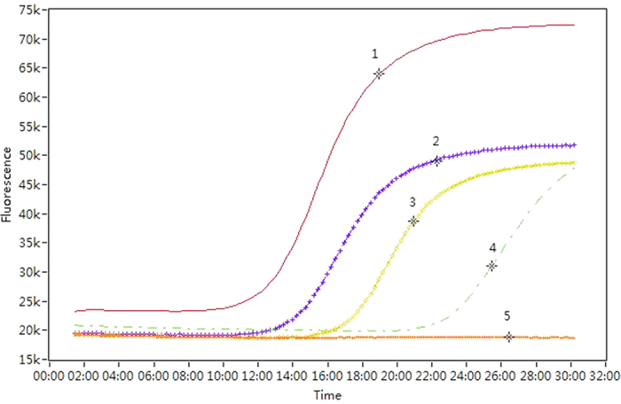
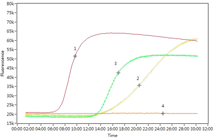
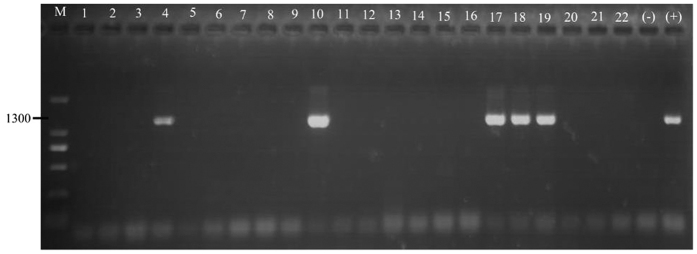
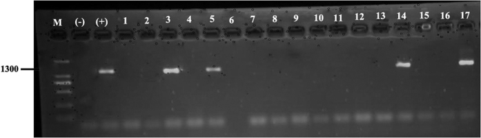
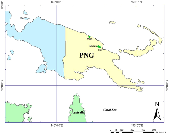
Phytoplasmas are insect vectored mollicutes responsible for disease in many economically important crops. Determining which insect species are vectors of a given phytoplasma is important for managing disease but is methodologically challenging because disease-free plants need to be exposed to large numbers of insects, often over many months. A relatively new method to detect likely transmission involves molecular testing for phytoplasma DNA in sucrose solution that insects have fed upon. In this study we combined this feeding medium method with a loop-mediated isothermal amplification (LAMP) assay to study 627 insect specimens of 11 Hemiptera taxa sampled from sites in Papua New Guinea affected by Bogia coconut syndrome (BCS). The LAMP assay detected phytoplasma DNA from the feeding solution and head tissue of insects from six taxa belonging to four families: Derbidae, Lophopidae, Flatidae and Ricaniidae. Two other taxa yielded positives only from the heads and the remainder tested negative. These results demonstrate the utility of combining single-insect feeding medium tests with LAMP assays to identify putative vectors that can be the subject of transmission tests and to better understand phytoplasma pathosystems.
Figures
References
-
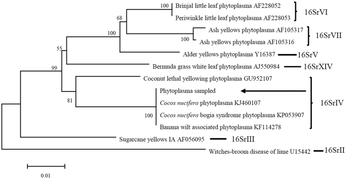
- Kelly P. et al.. First report of a phytoplasma identified in coconut palms (Cocos nucifera) with lethal yellowing-like symptoms in Papua New Guinea. New Dis. Rep. 23, 9 (2011).
-
- Harrison N., Cordova I., Richardson P. & Dibonito R. In Current Advances in Coconut Biotechnology Vol. 35 (eds Oropeza C. et al..) 183–196 (Springer, 1999).
-
- Danyo G. Review of scientific research into the Cape Saint Paul Wilt Disease (CSPWD) of coconut in Ghana. Afr. J. Agr. Res. 6, 4567–4578 (2011).
-
- Nipah J., Jones P. & Dickinson M. Detection of lethal yellowing phytoplasma in embryos from coconut palms infected with Cape St Paul wilt disease in Ghana. Plant Pathol. 56, 777–784 (2007).
-
- Kembu A., Yasinge W., Tenakanai D. & Bangulass N. Delimiting Survey of an Infectious Coconut Syndrome in Bogia District of Madang Province: a Preliminary Report. 43pp (PNG Cocoa Coconut Institute, 2009).
Publication types
MeSH terms
LinkOut - more resources
Full Text Sources
Other Literature Sources
