. 2016 Oct 27:7:13188.
doi: 10.1038/ncomms13188.

The fungal-specific β-glucan-binding lectin FGB1 alters cell-wall composition and suppresses glucan-triggered immunity in plants

Affiliations

The fungal-specific β-glucan-binding lectin FGB1 alters cell-wall composition and suppresses glucan-triggered immunity in plants

Stephan Wawra et al. Nat Commun. .

Abstract

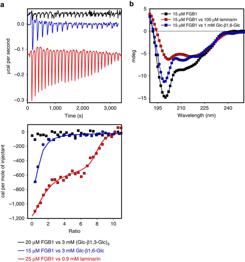
β-glucans are well-known modulators of the immune system in mammals but little is known about β-glucan triggered immunity in planta. Here we show by isothermal titration calorimetry, circular dichroism spectroscopy and nuclear magnetic resonance spectroscopy that the FGB1 gene from the root endophyte Piriformospora indica encodes for a secreted fungal-specific β-glucan-binding lectin with dual function. This lectin has the potential to both alter fungal cell wall composition and properties, and to efficiently suppress β-glucan-triggered immunity in different plant hosts, such as Arabidopsis, barley and Nicotiana benthamiana. Our results hint at the existence of fungal effectors that deregulate innate sensing of β-glucan in plants.


Figures

Figure 1
Figure 1. The small secreted P. indica protein FGB1 binds to fungal cell walls and harbours a fungal-specific lectin domain.
(a) Protein sequence logo of FGB1 (PIIN_03211) obtained after alignment of the mature protein sequence with homologous sequences from different fungi. (b) P. indica FGB1 transcript levels in barley (Hordeum vulgare) and Arabidopsis Col-0 roots grown on ½ MS medium inoculated with spores and harvested 3, 7 and 14 dpi and on control ½ MS medium (7 dpi) relative to transcription elongation factor (TEF, PIIN_03008). Error bars are s.d.'s calculated from three biological replicate samples (eight technical replicates each). (c) FGB1:GFP localizes to the fungal septa (arrowheads) and cell wall in colonized A. thaliana Col-0 (7 dpi) and Ws-0 roots (6 dpi), as well as barley roots (7 dpi). The asterisks show the entry point of the hyphae into the root cell. The chitin dye Alexa Fluor 594 labelled wheat germ agglutinin is shown in red (see Supplementary Figs 1 and 3 for further details). Scale bars, 10 μm. Scale bar on the detail image (top right corner) is 3 μm. (d) SDS–PAGE showing the outcome of a pull down experiment probing the binding ability of FGB1 to protein free cell wall extracts of P. indica, F. oxysporum and barley roots. P, insoluble pellet fraction; S, supernatant. Incubation of FGB1 (20 μM) with the insoluble polysaccharides (10 mg each) was carried out for 1 h at room temperature in 25 mM sodium-acetate buffer (pH 5.0) containing 500 mM NaCl.
Figure 2
Figure 2. FGB1 specifically binds to β-glucan and to the disaccharide gentiobiose.
(a) ITC profiles showing titrations of laminarihexaose [(Glc-β-1,3-Glc)3] (black line), gentiobiose (Glc-β-1,6-Glc) (blue line) and laminarin (red line) to FGB1. The laminarin titration was fitted using a model that allows binding to two equivalent and independent sites. Assuming a molecular mass of laminarin of 5 kDa the following parameters were obtained: N1=6.8±1.09, K1=2.14 × 10⁶±1.97 × 10⁷ M−1, ΔH1=−508±128 cal mol−1, ΔS1=27.2 cal mol−1 per degree and N2=1.02±0.9, K2=1.19 × 10⁷±6.11 × 10⁷ M−1, ΔH2=−1971±251 cal mol−1, ΔS2=25.7 cal mol−1 per degree. The titration of gentiobiose to FGB1 was fitted to a single-side-binding model (N=0.854±0.243, K=8.79 × 10⁴±6.88 × 10⁵ M−1, ΔH=−563.9±241 cal mol−1, ΔS=19.4 cal mol−1 per degree). No binding of FGB1 to laminarihexaose was observed. Concentrations of the stock solutions are indicated. All titrations were baseline corrected and substracted with the corresponding control titration of ligand into water. Errors correspond to the s.d. of the nonlinear least-squares fit of the data points of the titration curve. (b) The circular dichroism-spectrum of 15 μM FGB1 (black square) shows a significant shift in the secondary structure in the presence of 1 mM gentiobiose (blue square) or 100 μM laminarin (red square). Spectra were corrected with the spectra obtained for the individual ligands (see Supplementary Fig. 4 for further details).
Figure 3
Figure 3. FGB1 confers resistance to Congo red-mediated cell wall stress and increases plant colonization by fungi.
(a) Drop dilution series of U. maydis SG200 strains expressing either GFP or FGB1:GFP. Growth of three independent strains were tested on complete medium (CM) and CM supplemented with 150 μg ml−1 CR. U. maydis FGB1:GFP strains show an increased sensitivity towards chitinase activity (Trichoderma viride) compared with GFP expressing control strains. Protoplastation was carried out in 20 mM sodium citrate containing 1 M sorbitol, pH 5.8 for 15 min at 37 °C using 20 mg ml−1 chitinase. Data were obtained from four independent biological replicates carried out in three technical replicates using three independent strains by counting protoplasts and non-protoplasted cells. Error bars show s.d. Significance was calculated using the paired t-test algorithm of SigmaPlot. (b) To test if expression of PromPiFGB1FGB1:GFP in P. indica also increases resistance to CR, plugs of identical size from the active growth zone of 7-day old culture plates were transferred onto CM with and without CR. Growth of P. indicaPromPiFGB1FGB1:GFP strains was compared with that of the homokaryotic control strain and the dikaryotic WT strain. P. indicaPromPiFGB1FGB1:GFP strains are more resistant to CR compared with the controls. At least four biological repetitions were done. No significant differences in barley colonization by P. indicaPromPiFGB1FGB1:GFP strains compared with the homokaryotic control strain was observed at 7 dpi (see Supplementary Figs 6–8 for further details). Bars show the average of five independent biological experiments each done with two technical replicates. Error bars show the s.d. (c) Left: addition of 9 μM native FGB1 leads to a ∼4 fold increased P. indica colonization of barley roots at 3 dpi. Error bars show the standard error of the mean from eight independent biological replicates performed with three independently purified FGB1 batches. Right: Relative expression levels of barley PR10, a PR gene significantly induced during root colonization by P. indica. Asterisks indicate significant difference calculated using the paired t-test algorithm of SigmaPlot.
Figure 4
Figure 4. FGB1 supresses β-glucan-induced oxidative burst in barley and A. thaliana Ws-0.
(a) Barley leaf disks react with a strong ROS burst after elicitation with 600 μM complex laminarin (red square). Incubation with 10 μM FGB1 supress ROS burst (black square). Neither 10 μM FGB1 (blue square) nor the mock water control (grey square) induce ROS production. (b) Leaf disks of A. thaliana ecotype Ws-0 react with ROS production after elicitation with 600 μM laminarin. Comparable to barley, Ws-0 ROS production was supressed by 10 μM FGB1. At least four independent repetitions with three different FGB1 batches were carried out and showed similar results. Error bars represent the standard error of the mean of 8–12 technical replicates (see Supplementary Fig. 10 for further details).
Figure 5
Figure 5. Model displaying the potential dual function of FGB1.
Binding of FGB1 to the fungal cell wall (CW) after secretion is mediated through β-1,6-glycosidic linkages. FGB1 modulates CW polysaccharides composition. FGB1 does not protect the CW against β-1,3-endo- and β-1,3-exo-glucanase activities. Host glucanases activity release β-glucan fragments that can be sensed by a specific plant β-glucan receptor complex. Recognition leads to activation of basal defence mechanisms, such as the production of reactive oxygen species (ROS). Based on the observation that FGB1 is able to suppress β-glucan induced ROS production at substoichiometric concentrations, we hypothize that FGB1/β-glucan complexes have higher affinities to the plant β-glucan receptors than free β-glucan fragments. Binding of FGB1/β-glucan complexes to the β-glucan receptor would possibly prevent receptor/co-receptor association and defense downstream signalling.

References

    1. Spoel S. H. & Dong X. N. How do plants achieve immunity? Defence without specialized immune cells. Nat. Rev. Immunol. 12, 89–100 (2012). - PubMed
    1. Jones J. D. & Dangl J. L. The plant immune system. Nature 444, 323–329 (2006). - PubMed
    1. Limpens E., van Zeijl A. & Geurts R. Lipochitooligosaccharides modulate plant host immunity to enable endosymbioses. Annu. Rev. Phytopathol. 53, 311–334 (2015). - PubMed
    1. Nurnberger T., Brunner F., Kemmerling B. & Piater L. Innate immunity in plants and animals: striking similarities and obvious differences. Immunol. Rev. 198, 249–266 (2004). - PubMed
    1. Sanchez-Vallet A., Mesters J. R. & Thomma B. P. The battle for chitin recognition in plant-microbe interactions. FEMS Microbiol. Rev. 39, 171–183 (2015). - PubMed

Publication types

MeSH terms