. 2017 Jul 3;12(7):527-539.
doi: 10.1080/15592294.2016.1248007. Epub 2016 Oct 27.

De novo methylation in male germ cells of the common marmoset monkey occurs during postnatal development and is maintained in vitro

Affiliations

De novo methylation in male germ cells of the common marmoset monkey occurs during postnatal development and is maintained in vitro

Daniel Langenstroth-Röwer et al. Epigenetics. .

Abstract

The timing of de novo DNA methylation in male germ cells during human testicular development is yet unsolved. Apart from that, the stability of established imprinting patterns in vitro is controversially discussed. This study aimed at determining the timing of DNA de novo methylation and at assessing the stability of the methylation status in vitro. We employed the marmoset monkey (Callithrix jacchus) as it is considered the best non-human primate model for human testicular development. We selected neonatal, pre-pubertal, pubertal, and adult animals (n = 3, each) and assessed germ cell global DNA methylation levels by 5-methyl cytosine staining, and Alu elements and gene-specific methylation (H19, LIT1, SNRPN, MEST, OCT4, MAGE-A4, and DDX-4) by pyrosequencing. De novo methylation is progressively established during postnatal primate development and continues until adulthood, a process that is different in most other species. Importantly, once established, methylation patterns remained stable, as demonstrated using in vitro cultures. Thus, the marmoset monkey is a unique model for the study of postnatal DNA methylation mechanisms in germ cells and for the identification of epimutations and their causes.

Keywords: de novo methylation; germ cell development; primate-specific DNA methylation patterns; spermatogonia.


Figures
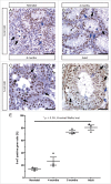
Figure 1.
Figure 1.
Global DNA methylation is gradually established in germ cells during postnatal development. (A-D) Representative micrographs show 5mC stainings of testicular cross sections from neonatal, 4-months-old, 8-months-old, and adult animals. Arrowheads indicate gonocytes, arrows spermatogonia, empty arrows spermatocytes and empty arrow heads round spermatids. Positive cells are indicated by black and negative cells by blue symbols. No staining was detected in corresponding IgG controls (Fig. S1). Scale bars represent 50 µm. (E) Proportion of 5mC positive germ cells in histological stainings (n = 3 per age group). Data are presented as mean with standard deviation. Individual values for each biological replicate are indicated by dots. Statistically significant differences between all 4 groups were found applying the non-parametric Kruskal Wallis test (P < 0.05).
Figure 2.
Figure 2.
Global DNA methylation pattern during postnatal development in specific germ cell types. The proportion of 5mC positive cells was determined separately for each cell type in neonatal, 4-months-old, 8-months-old, and adult marmosets (n = 3 per age group). Significant differences between values for spermatogonia at the different developmental stages and for the different cell types in the adult testis were found applying the non-parametric Kruskal Wallis test (P < 0.05). Gon: gonocytes, Spg: spermatogonia, Spc: spermatocytes, Rs: round spermatids, Es: elongated spermatids.
Figure 3.
Figure 3.
DNA promoter methylation levels of repetitive elements and imprinted, pluripotency, and germ cell marker genes during testicular maturation. (A-D) DNA methylation at selected CpG sites of the repetitive element Alu, the maternally imprinted genes LIT1, SNRPN, MEST, and the paternally imprinted gene H19 in DNA from testicular tissues of neonatal, 4-months-old, 8-months-old, and adult marmosets (n = 3 per age group). (E,F) Calculated proportion of DNA promoter methylation for H19 in germ cells from the respective age groups based on MEST and LIT1 methylation levels. Note that the H19 methylation increases gradually during postnatal development. (G-I) DNA methylation at selected CpG sites of the pluripotency marker OCT4 and the germ cell marker genes MAGE-A4 and DDX-4. Dotted lines indicate means of reference measurements which were performed with sperm and blood DNA (n = 7, each) as well as with DNA from pluripotent stem cells (PSC; n = 4).
Figure 4.
Figure 4.
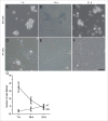
Marmoset testicular cells in vitro. Representative micrographs of (A-C) supernatant and (D-F) attached cells from adult marmoset monkeys (n = 6) on days 7, 14, and 21. (G) Cell numbers of floating and attached cells. Note that SN cells and AT cells exhibit a different morphology and different dynamics in cell numbers. SN: germ cell-enriched supernatant fraction, AT: attached cell fraction. Scale bar represent 50 µm.
Figure 5.
Figure 5.
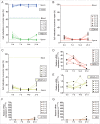
DNA methylation is stable in germ cells in vitro. (A-C) DNA methylation of imprinted genes H19 and MEST (A) and germ cell marker genes DDX-4 (B) and MAGE-A4 (C) in cultured supernatant (SN) cells. Dotted lines indicate means of reference measurements established with sperm and blood DNA (n = 7 each). (D-G) mRNA expression levels for the germ cell marker genes DDX-4 (D) and MAGE-A4 (E) and the somatic marker genes ACTA2 (F) and VIM (G) in SN cells. In all graphs the results for the biological replicates are presented as separate data points. Note that the DNA methylation profile of all samples remains germ cell specific throughout culture, except for 2 samples that exhibit increased somatic marker expression at day 21. C.j. 1-6: Cultures initiated from Callithrix jacchus (marmoset) material.
Figure 6.
Figure 6.
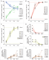
DNA methylation of adherently growing mixed fractions of germ cells and somatic cells is dynamic. (A-C) DNA promoter methylation of imprinted genes H19 and MEST (A) and germ cell marker genes DDX-4 (B) and MAGE-A4 (C) in attached culture fractions. Dotted lines indicate means of reference measurements that were performed with sperm and blood DNA (n = 7 each). (D-G) mRNA expression levels for the germ cell marker genes DDX-4 (D) and MAGE-A4 (E) and the somatic marker genes ACTA2 (F) and VIM (G) in the attached fractions. In all graphs, the results for the biological replicates are presented as separate data points. Cultures initiated from Callithrix jacchus (marmoset) material.
Figure 7.
Figure 7.
Scheme illustrating the dynamics of germ cell-specific and somatic DNA methylation profiles during in vivo maturation and in vitro culture of germ cells. (A) In neonatal marmoset testes, most of the germ cells are unmethylated (white nucleus) and only a few germ cells have already obtained full DNA methylation at paternally imprinted genes (black nucleus). In contrast, the somatic cells exhibit intermediate DNA methylation levels (gray nucleus). During maturation from 4-months-old via 8-months-old to adult marmosets, the proportion of somatic cells with intermediate DNA methylation levels decreases and the proportion of germ cells with established paternal imprints increases. (B) Cultured marmoset testicular cells maintain germ cell-specific DNA-methylation patterns. Since the supernatant (SN) fraction predominantly consists of germ cells, its DNA methylation profile remains germ cell-specific. In contrast, somatic cells overgrow germ cells in the attached (AT) fraction, resulting in a dynamic DNA methylation profile which approaches levels of somatic cells.

References

    1. Monk D. Germline-derived DNA methylation and early embryo epigenetic reprogramming: The selected survival of imprints. Int J Biochem Cell Biol 2015; 67:128-138; PMID:25966912; https://doi.org/10.1016/j.biocel.2015.04.014 - DOI - PubMed
    1. Seisenberger S, Peat JR, Hore TA, Santos F, Dean W, Reik W. Reprogramming DNA methylation in the mammalian life cycle: Building and breaking epigenetic barriers. Philos Trans R Soc Lond B Biol Sci 2013; 368:20110330; PMID:23166394; https://doi.org/10.1098/rstb.2011.0330 - DOI - PMC - PubMed
    1. Reik W, Walter J. Genomic imprinting: Parental influence on the genome. Nat Rev Genet 2001; 2:21-32; PMID:11253064; https://doi.org/10.1038/35047554 - DOI - PubMed
    1. Guibert S, Forne T, Weber M. Global profiling of DNA methylation erasure in mouse primordial germ cells. Genome Res 2012; 22:633-41; PMID:22357612; https://doi.org/10.1101/gr.130997.111 - DOI - PMC - PubMed
    1. Hackett JA, Sengupta R, Zylicz JJ, Murakami K, Lee C, Down TA, Surani MA. Germline DNA demethylation dynamics and imprint erasure through 5-hydroxymethylcytosine. Science 2013; 339:448-52; PMID:23223451; https://doi.org/10.1126/science.1229277 - DOI - PMC - PubMed

Publication types