Multicenter Study
. 2016 Sep 26;13(1):279.
doi: 10.1186/s12974-016-0717-1.

MOG-IgG in NMO and related disorders: a multicenter study of 50 patients. Part 1: Frequency, syndrome specificity, influence of disease activity, long-term course, association with AQP4-IgG, and origin

Affiliations
Multicenter Study

MOG-IgG in NMO and related disorders: a multicenter study of 50 patients. Part 1: Frequency, syndrome specificity, influence of disease activity, long-term course, association with AQP4-IgG, and origin

Sven Jarius et al. J Neuroinflammation. .

Abstract

Background: Antibodies to myelin oligodendrocyte glycoprotein (MOG-IgG) have been suggested to play a role in a subset of patients with neuromyelitis optica and related disorders.

Objective: To assess (i) the frequency of MOG-IgG in a large and predominantly Caucasian cohort of patients with optic neuritis (ON) and/or myelitis; (ii) the frequency of MOG-IgG among AQP4-IgG-positive patients and vice versa; (iii) the origin and frequency of MOG-IgG in the cerebrospinal fluid (CSF); (iv) the presence of MOG-IgG at disease onset; and (v) the influence of disease activity and treatment status on MOG-IgG titers.

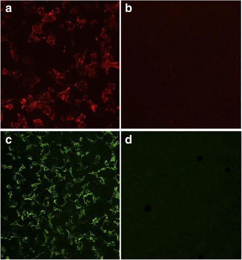
Methods: 614 serum samples from patients with ON and/or myelitis and from controls, including 92 follow-up samples from 55 subjects, and 18 CSF samples were tested for MOG-IgG using a live cell-based assay (CBA) employing full-length human MOG-transfected HEK293A cells.

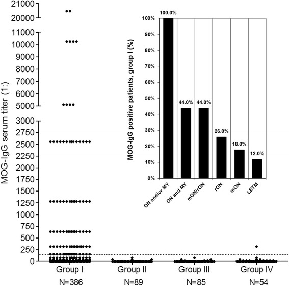
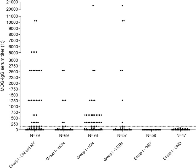
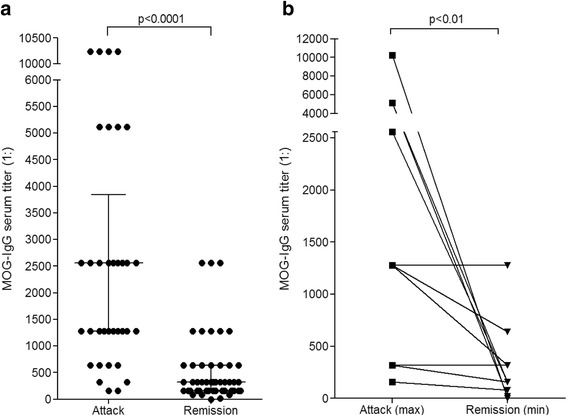
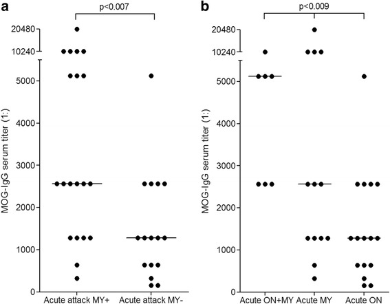
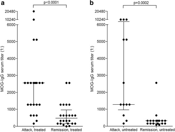
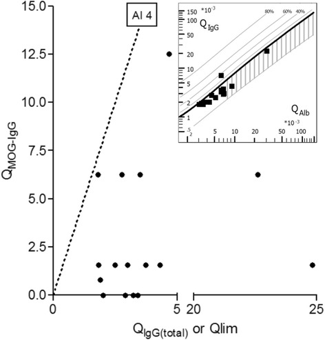
Results: MOG-IgG was detected in 95 sera from 50 patients with ON and/or myelitis, including 22/54 (40.7 %) patients with a history of both ON and myelitis, 22/103 (21.4 %) with a history of ON but no myelitis and 6/45 (13.3 %) with a history of longitudinally extensive transverse myelitis but no ON, and in 1 control patient with encephalitis and a connective tissue disorder, all of whom were negative for AQP4-IgG. MOG-IgG was absent in 221 further controls, including 83 patients with AQP4-IgG-seropositive neuromyelitis optica spectrum disorders and 85 with multiple sclerosis (MS). MOG-IgG was found in 12/18 (67 %) CSF samples from MOG-IgG-seropositive patients; the MOG-IgG-specific antibody index was negative in all cases, indicating a predominantly peripheral origin of CSF MOG-IgG. Serum and CSF MOG-IgG belonged to the complement-activating IgG1 subclass. MOG-IgG was present already at disease onset. The antibodies remained detectable in 40/45 (89 %) follow-up samples obtained over a median period of 16.5 months (range 0-123). Serum titers were higher during attacks than during remission (p < 0.0001), highest during attacks of simultaneous myelitis and ON, lowest during acute isolated ON, and declined following treatment.

Conclusions: To date, this is the largest cohort studied for IgG to human full-length MOG by means of an up-to-date CBA. MOG-IgG is present in a substantial subset of patients with ON and/or myelitis, but not in classical MS. Co-existence of MOG-IgG and AQP4-IgG is highly uncommon. CSF MOG-IgG is of extrathecal origin. Serum MOG-IgG is present already at disease onset and remains detectable in the long-term course. Serum titers depend on disease activity and treatment status.

Keywords: Antibody index; Aquaporin-4 antibodies (AQP4-IgG); Autoantibodies; Cell-based assays; Cerebrospinal fluid; Devic’s syndrome; Longitudinally extensive transverse myelitis (LETM); Multiple sclerosis; Myelin oligodendrocyte glycoprotein antibodies (MOG-IgG); Neuromyelitis optica antibodies (NMO-IgG); Neuromyelitis optica spectrum disorders (NMOSD); Neuromyelitis optica (NMO); Optic neuritis; Transverse Myelitis.


Figures

Fig. 1
Fig. 1
MOG-IgG as detected by two independent cell-based assays (CBA): typical findings. a, b Binding of serum IgG from a group I patient (a) but not from a control patient (b) to live HEK293A cells transfected with human full-length MOG. c, d Binding of serum IgG from a group I patient to formalin-fixed HEK293 cells transfected with full-length MOG (c) but not to their mock-transfected counterpart (d) in a commercial CBA. Bound MOG-IgG was visualized in the live-cell assay using a Cy3-conjugated goat anti-human IgG antibody and in the fixed-cell assay by use of a fluorescein isothiocyanate (FITC)-labeled goat anti-human IgG antibody
Fig. 2
Fig. 2
Frequency and titers of MOG-IgG in 614 serum samples from 522 subjects as detected using a live-cell CBA. MOG-IgG was detected in 95/386 (24.6 %) samples in group I but was almost completely absent among 228 control samples (groups II–IV), including 89 samples from AQP4-IgG-positive patients, 85 samples from patients with MS according to the McDonald criteria (group III), and 54 samples from healthy controls and OND patients (group IV). While all low-titer samples (1:160–1:320) in group I were positive also in the fixed-cell CBA, the only positive control sample (from group IV) was negative in the fixed-cell CBA, suggesting a false-positive test result. The horizontal dashed line indicates the assay-specific cut-off (> = 1:160)
Fig. 3
Fig. 3
MOG-IgG serum titers in 386 samples from 300 patients included in group I. Diagnoses are given as provided by the referring centers. ON and MY = optic neuritis and myelitis; mON = monophasic optic neuritis; rON = recurrent ON; LETM = longitudinally extensive transverse myelitis; MS = multiple sclerosis; OND = other neurological disorders
Fig. 4
Fig. 4
MOG-IgG titers and disease activity. Titers were significantly higher during acute attacks than during remission in the total cohort (a) as well as in individual patients with available follow-up sera (b). Horizontal lines and whiskers in panel a indicate median titers and interquartile ranges, respectively. The median interval between samples in the right panel was 16.5 months (range 2–103). Note that panel b shows maximum titers detected during acute attacks and minimum titers detected in follow-up sera. The difference was also significant if not the remission sample with the lowest titer but that with the longest time interval since attack onset was used (median 1:1280 vs. 1:320; p < 0.009; not shown)
Fig. 5
Fig. 5
MOG-IgG titers and clinical presentation. Titers were higher during attacks involving myelitis than in attacks not involving myelitis (a), and higher during attacks involving simultaneous ON and myelitis than in attacks of isolated myelitis or isolated ON (b). The horizontal lines indicate median titers. ON = optic neuritis; MY = myelitis
Fig. 6
Fig. 6
MOG-IgG titers and treatment status. While median MOG-IgG titers were lower during remission than during acute attacks in the treated subgroup (a), a similarly significant difference was also observed in the untreated subgroup (b). By contrast, no significant difference in median titers was observed between treated and untreated patients, neither during acute attacks nor during remission (not shown)
Fig. 7
Fig. 7
MOG-specific antibody index (AI). Calculation of the MOG-specific AI in 17 paired CSF/serum samples from 15 MOG-IgG-positive patients did not reveal evidence for intrathecal synthesis of MOG-IgG. The dotted line indicates the upper limit of the reference range (AI = 4). Inset: Reiber diagram [40] demonstrating absence of total IgG intrathecal synthesis in 16 samples from 14 patients and presence of blood-CSF barrier dysfunction in 6/17 samples. QIgG = CSF/serum total IgG ratio; QMOG-IgG = CSF/serum MOG-IgG ratio; QAlb = CSF/serum albumin ratio; Qlim = upper reference range of QIgG (see methods section for details)
Fig. 8
Fig. 8
MOG-IgG1 as detected in the fixed-cell CBA. a, b Binding of serum IgG1 antibodies (from a patient with recurrent optic neuritis) to HEK293 cells transfected with human full-length MOG (a), but not to mock-transfected HEK293 cells (b). c Negative control serum (from a patient with RRMS) binding neither to the MOG-transfected cells (upper panel) nor to the mock-transfected control cells (lower panel). Bound patient IgG1 was detected by successive incubation with an unlabeled sheep anti-human IgG1 secondary antibody and an AlexaFluore®568-labeled donkey anti-sheep IgG antibody (red fluorescence). Cell nuclei were stained with 4’,6-diamidino-2-phenylindole (blue fluorescence)

References

    1. Lennon VA, Kryzer TJ, Pittock SJ, Verkman AS, Hinson SR. IgG marker of optic-spinal multiple sclerosis binds to the aquaporin-4 water channel. J Exp Med. 2005;202:473–7. doi: 10.1084/jem.20050304. - DOI - PMC - PubMed
    1. Jarius S, Paul F, Franciotta D, Waters P, Zipp F, Hohlfeld R, Vincent A, Wildemann B. Mechanisms of Disease: Aquaporin-4 antibodies in neuromyelitis optica. Nat Clin Pract Neurol. 2008;4:202–14. - PubMed
    1. Jarius S, Wildemann B, Paul F. Neuromyelitis optica: clinical features, immunopathogenesis and treatment. Clin Exp Immunol. 2014;176:149–64. doi: 10.1111/cei.12271. - DOI - PMC - PubMed
    1. Levy M, Wildemann B, Jarius S, Orellano B, Sasidharan S, Weber MS, Stuve O. Immunopathogenesis of neuromyelitis optica. Adv Immunol. 2014;121:213–42. doi: 10.1016/B978-0-12-800100-4.00006-4. - DOI - PubMed
    1. Jarius S, Wildemann B. AQP4 antibodies in neuromyelitis optica: diagnostic and pathogenetic relevance. Nat Rev Neurol. 2010;6:383–92. doi: 10.1038/nrneurol.2010.72. - DOI - PubMed

Publication types