. 2016 Oct 27;16(1):18.
doi: 10.1186/s12900-016-0068-2.

Combined small angle X-ray solution scattering with atomic force microscopy for characterizing radiation damage on biological macromolecules

Affiliations

Combined small angle X-ray solution scattering with atomic force microscopy for characterizing radiation damage on biological macromolecules

Luca Costa et al. BMC Struct Biol. .

Abstract

Background: Synchrotron radiation facilities are pillars of modern structural biology. Small-Angle X-ray scattering performed at synchrotron sources is often used to characterize the shape of biological macromolecules. A major challenge with high-energy X-ray beam on such macromolecules is the perturbation of sample due to radiation damage.

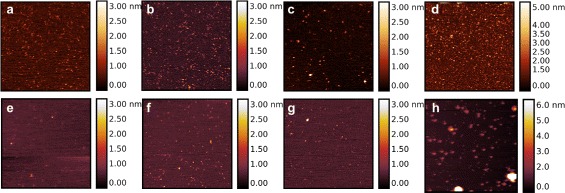
Results: By employing atomic force microscopy, another common technique to determine the shape of biological macromolecules when deposited on flat substrates, we present a protocol to evaluate and characterize consequences of radiation damage. It requires the acquisition of images of irradiated samples at the single molecule level in a timely manner while using minimal amounts of protein. The protocol has been tested on two different molecular systems: a large globular tetremeric enzyme (β-Amylase) and a rod-shape plant virus (tobacco mosaic virus). Radiation damage on the globular enzyme leads to an apparent increase in molecular sizes whereas the effect on the long virus is a breakage into smaller pieces resulting in a decrease of the average long-axis radius.

Conclusions: These results show that radiation damage can appear in different forms and strongly support the need to check the effect of radiation damage at synchrotron sources using the presented protocol.

Keywords: Atomic force microscopy (AFM); Radiation damage; Small angle x-ray scattering (SAXS); Tobacco mosaic virus; β-Amylase.


Figures

Fig. 1
Fig. 1
β-Amylase SAXS data. a SAXS curve of β-Amylase (black dots) obtained after a standard exposure with fit to a mixture of monomers and tetramers (orange line). Inlay: Guinier plot and associated linear fit at low- q 2 (q R g < 1.3). b Model of tetrameric β-Amylase (pdb entry 1FA2) [28]. The bounding box size of tetrameric β-Amylase is 12.4 × 12.4 × 7.5 nm 3. c SAXS data for different exposures time: standard, 5 min and 30 min exposures are plotted in blue, red and green, respectively; very small variations with the exposure time can be observed
Fig. 2
Fig. 2
AFM imaging of β-Amylase using tapping mode in liquid environment. Top row (a, b, c, d) corresponds to images obtained when β-Amylase was deposited on Nickel pre-treated mica whereas the bottom row (e, f, g, h) corresponds to bare mica. Non-irradiated β-Amylase is shown in (a, e) whereas increasing exposure to X-ray beam is shown in (b, f) for 10 s exposure, (c, g) 5 min exposure, (d, h) 30 min exposure. The scan size is 1 μm with each line made of 512 pixels. A clear increase in height (and diameter) can be easily seen upon increase exposure (radiation damage) with no apparent differences between control and standard exposure (10 s) time. A total of 13 AFM images have been used for statistical analysis representing a total of 3693 and 948 particles measured on nickel pre-treated mica and bare mica, respectively
Fig. 3
Fig. 3
Average long-axis radius of β-Amylase estimated from their distribution in AFM images. Particles were identified using the threshold or the Otsu’s method when β-Amylase was deposited on bare mica (a) or Nickel pre-treated mica (b). Control represents β-Amylase that was not exposed to X-ray. Standard exposure is about 10 s whereas over-exposed corresponds to a 30 min exposure to X-ray. Long-axis radii were determined with standard parameters of the Grain distribution section of Gwyddion. Upon increase exposure time in X-ray beam, a slight increase in the long-axis radius of β-Amylase is observed which could be interpreted as aggregation of β-Amylase monomers or consolidation of β-Amylase tetramer after radiation damage (see text). The number of identified particles on bare mica was 139, 122, 457 and 230 for over-exposed, exposed 5 min, standard exposure and control, respectively; whereas on nickel pre-treated mica the number of particles was 1877, 136, 1453 and 277 for over-exposed, exposed 5 min, standard exposure and control, respectively
Fig. 4
Fig. 4
Evolution of the radius of gyration for the β-Amylase upon X-ray exposure obtained from SAXS data. While there is no significant increase for low exposure time, R g increases once proteins are over-exposed: this is consistent with the AFM results presented in Fig. 3
Fig. 5
Fig. 5
TMV SAXS data. a SAXS curve of TMV (black dots) obtained during a standard exposure with fit to a three shell cylinder model (orange line). Inlay: Cross-sectional Guinier plot and fit. b Radial pair distance distribution function of TMV using the virus diameter found by AFM as constrained for D max. c Schematic of the three-shell electron density distribution used as a model in a). d Atomistic model of the TMV cross-section with the RNA in orange. Based on pdb entry 1VTM. e SAXS data for different exposures time: standard, 5 min and 30 min exposures are plotted in blue, red and green, respectively
Fig. 6
Fig. 6
AFM imaging of TMV using tapping mode in liquid environment. Top row (a, b, c) is TMV deposited on HOPG whereas bottom row (d, e, f) corresponds to TMV deposited on Nickel pre-treated mica. Non-irradiated TMV is shown in (a, d). Increasing exposure to X-ray beam is shown in (b, e) for 10 s exposure, and (c, f) 30 min exposure. The scan size is 5 μm with each line made of 512 pixels. A clear fragmentation of the 300 nm-long TMV can be observed upon increase in exposure time (radiation damage). To the contrary of β-Amylase (Fig. 2), even at standard exposure time, a beginning of fragmentation is observed for TMV. A total of 14 AFM images has been used for statistical analysis representing 6539 particles on HOPG at 10 μm scan size, 2007 particles on HOPG at 5 μm scan size, and 808 particles on nickel pre-treated mica
Fig. 7
Fig. 7
Average long-axis radius of TMV estimated from their distribution in AFM images. Particles were identified using the Otsu’s method when TMV was deposited on HOPG (a, b) or mica (c). AFM data have been acquired on unexposed samples (control), after a standard exposure as well as after 30 min exposure (over-exposed). Upon increasing exposure time in X-ray beam, a consistent decrease in the long-axis radius of TMV is observed which corresponds to a fragmentation of TMV particles upon radiation damage. The number of identified particles on HOPG 10 μm was 3912, 2273 and 354 for over-exposed, standard exposure and control, respectively; on HOPG 5 μm the number of identified particles was 1906, 68 and 33 for over-exposed, standard exposure and control, respectively; on mica the number of identified particles was 347, 157 and 304 for over-exposed, standard exposure and control, respectively

References

    1. Ward AB, Sali A, Wilson IA. Integrative structural biology. Science. 2013;339(6122):913–5. doi: 10.1126/science.1228565. - DOI - PMC - PubMed
    1. Graewert MA, Svergun DI. Impact and progress in small and wide angle x-ray scattering (saxs and waxs) Curr Opin Struct Biol. 2013;23(5):748–54. doi: 10.1016/j.sbi.2013.06.007. - DOI - PubMed
    1. Kikhney AG, Svergun DI. A practical guide to small angle x-ray scattering (saxs) of flexible and intrinsically disordered proteins. FEBS Lett. 2015;589(19):2570–7. doi: 10.1016/j.febslet.2015.08.027. - DOI - PubMed
    1. Putnam CD, Hammel M, Hura GL, Tainer JA. X-ray solution scattering (saxs) combined with crystallography and computation: defining accurate macromolecular structures, conformations and assemblies in solution. Q Rev Biophys. 2007;40(03):191–285. doi: 10.1017/S0033583507004635. - DOI - PubMed
    1. Jacques DA, Trewhella J. Small angle scattering for structural biology expanding the frontier while avoiding the pitfalls. Protein Sci. 2010;19(4):642–57. doi: 10.1002/pro.351. - DOI - PMC - PubMed

Publication types

LinkOut - more resources