Case Reports
. 2017 Jan:94:65-74.
doi: 10.1016/j.bone.2016.10.023. Epub 2016 Oct 24.

Compound heterozygous variants in NBAS as a cause of atypical osteogenesis imperfecta

Affiliations
Case Reports

Compound heterozygous variants in NBAS as a cause of atypical osteogenesis imperfecta

M Balasubramanian et al. Bone. 2017 Jan.

Abstract

Background: Osteogenesis imperfecta (OI), the commonest inherited bone fragility disorder, affects 1 in 15,000 live births resulting in frequent fractures and reduced mobility, with significant impact on quality of life. Early diagnosis is important, as therapeutic advances can lead to improved clinical outcome and patient benefit.

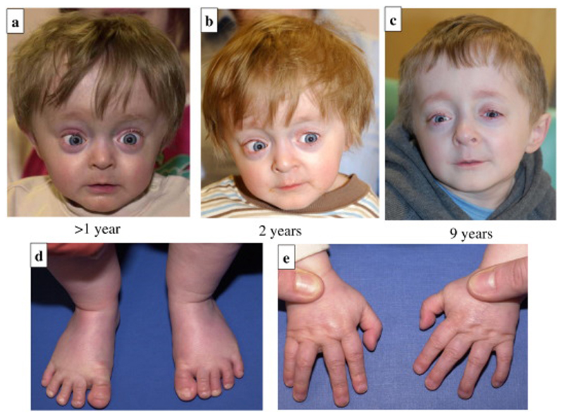
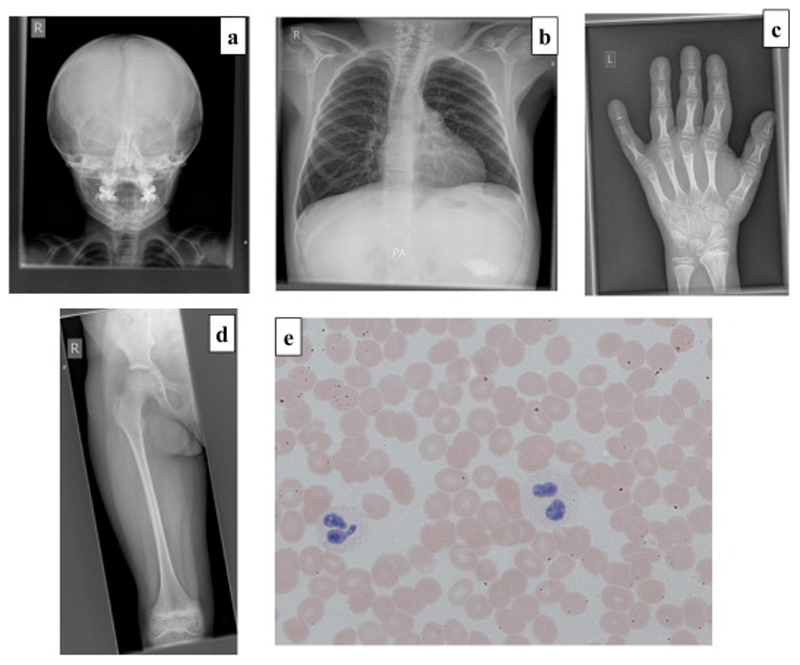
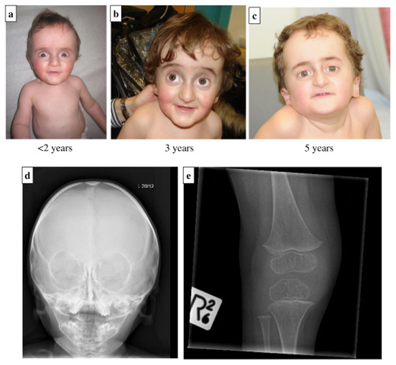
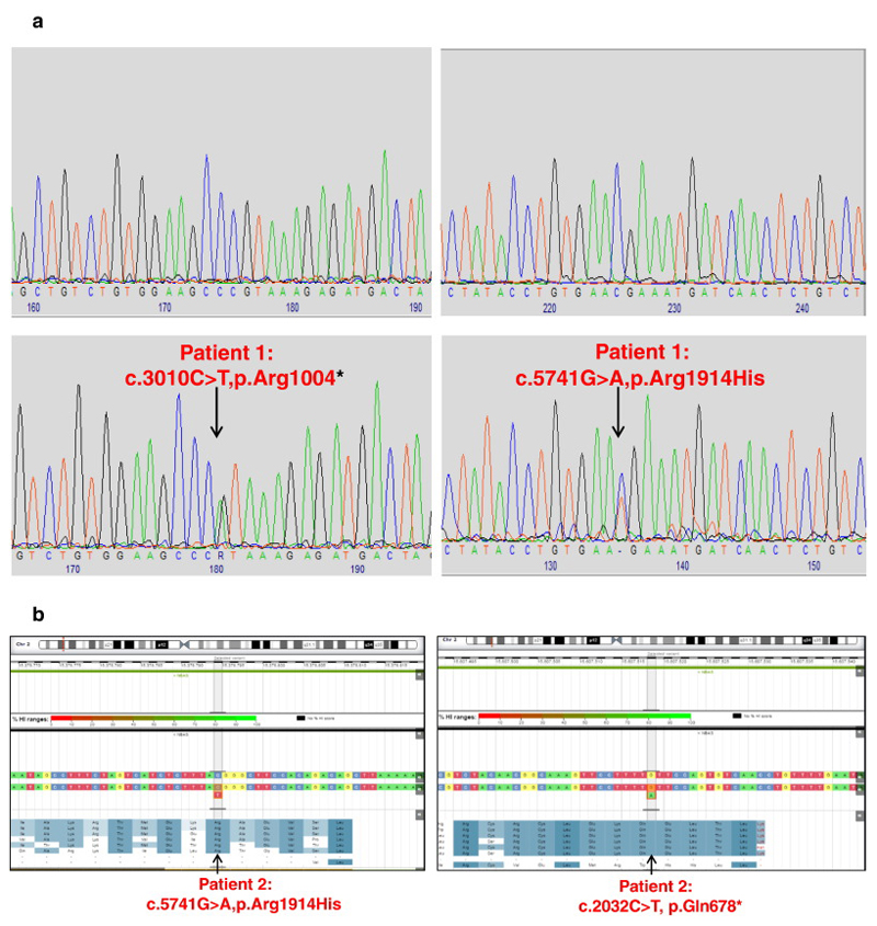
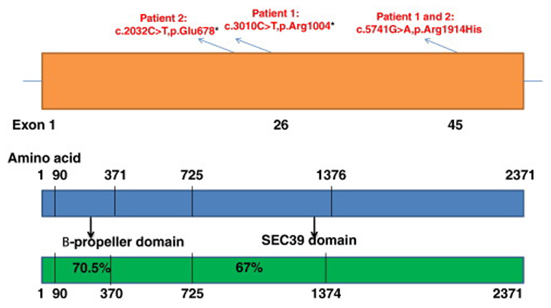
Report: Whole exome sequencing in patients with OI identified, in two patients with a multi-system phenotype, compound heterozygous variants in NBAS (neuroblastoma amplified sequence). Patient 1: NBAS c.5741G>A p.(Arg1914His); c.3010C>T p.(Arg1004*) in a 10-year old boy with significant short stature, bone fragility requiring treatment with bisphosphonates, developmental delay and immunodeficiency. Patient 2: NBAS c.5741G>A p.(Arg1914His); c.2032C>T p.(Gln678*) in a 5-year old boy with similar presenting features, bone fragility, mild developmental delay, abnormal liver function tests and immunodeficiency.

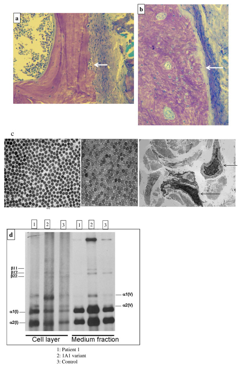
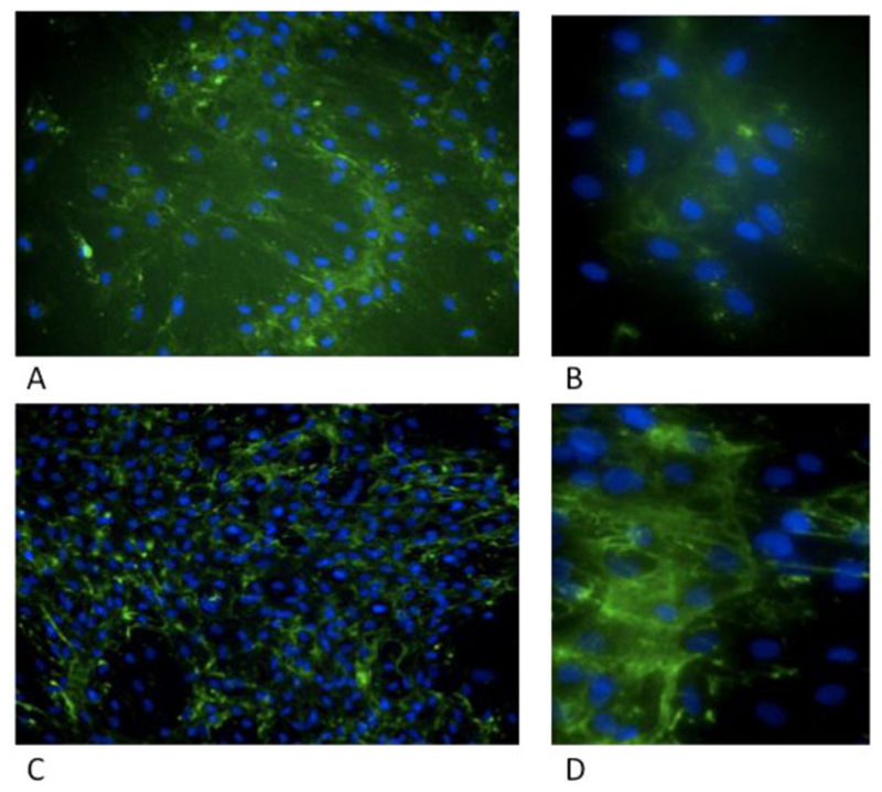
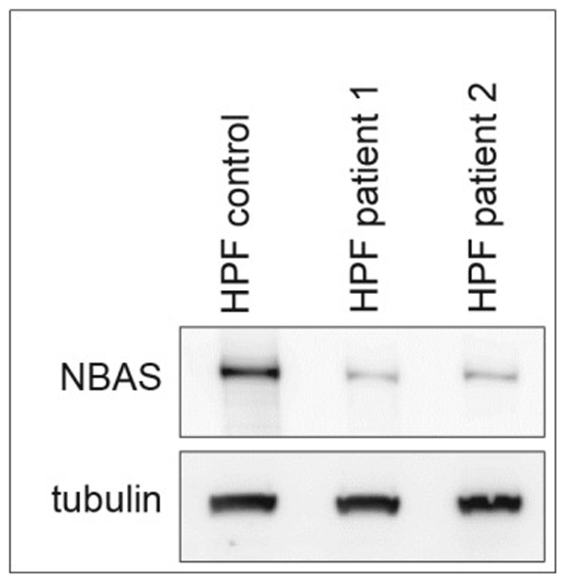
Discussion: Homozygous missense NBAS variants cause SOPH syndrome (short stature; optic atrophy; Pelger-Huet anomaly), the same missense variant was found in our patients on one allele and a nonsense variant in the other allele. Recent literature suggests a multi-system phenotype. In this study, patient fibroblasts have shown reduced collagen expression, compared to control cells and RNAseq studies, in bone cells show that NBAS is expressed in osteoblasts and osteocytes of rodents and primates. These findings provide proof-of-concept that NBAS mutations have mechanistic effects in bone, and that NBAS variants are a novel cause of bone fragility, which is distinguishable from 'Classical' OI.

Conclusions: Here we report on variants in NBAS, as a cause of bone fragility in humans, and expand the phenotypic spectrum associated with NBAS. We explore the mechanism underlying NBAS and the striking skeletal phenotype in our patients.

Keywords: Bone; Collagen expression; Fragility; NBAS; Nonsense mediated decay (NMD); Osteogenesis imperfecta; Secretory pathway.


Conflict of interest statement

Competing Interests

No competing interest to declare.

Figures

Figure 1
Figure 1
a-e: 1a-c: Facial features as an infant and aged >1, 2 and 9 years showing grey sclerae, broad forehead, bilateral low-set ears, proptosis and progeric appearance; 1d-e: Hands and feet at 2-years of age.
Figure 2
Figure 2
a-d: Radiographs demonstrating slender ribs, tubular long bones with thin cortices and osteopenia consistent with a diagnosis of OI. 2a: AP skull radiograph (aged 6 years) There are multiple Wormian bones; the clavicles are slender and the erupted teeth are relatively dense. The anterior fontanelle remains open. 2b-d: Selected images from a full dysplasia skeletal survey (aged 9 years 8 months) 2b: Left hand. There is significant periarticular osteopenia (see Fig 2c). The metacarpals (and less marked) the proximal phalanges are overmodelled and there is an ivory epiphysis of the terminal phalanx of the fifth finger. The terminal tufts are prominent. 2c: AP Chest. The ribs and clavicles are slender; however, there are no fractures and vertebral body height is preserved. Note the presence of a gastrostomy. 2d. AP Right Femur: Overmodelled with slender diaphysis and relatively flared distal metaphysis. Periarticular osteopenia is again noted (see Fig 2a) 2e: Peripheral blood film in Patient 1 demonstrating a hypolobulated neutrophil (left) and a Pelger-Huet cell (right).
Figure 3
Figure 3
a-b: 3a: Toluidine blue-stained section of an undecalcified trans-iliac bone biopsy, original magnification x400 demonstrating cortex, with periosteum on the right showing high turnover osteopenia with marked sub-periosteal bone resorption (arrow) and normal lamellar bone matrix structure in Patient-1, aged 9 years; 3b: appearance in ‘Classical OI’ with abnormal matrix pattern and increased periosteal bone formation surface (arrow). Toluidine blue; Original magnification of 400x. c-d: 3c: Electron Microscopy of skin biopsy from Patient 1(left image) compared to normal control (middle image) showing normal collagen in mid-reticular dermis with mildly reduced mean collagen fibril diameter (CFD); Original magnification of 20,000x; Lower magnification (right image) with arrows indicating deep reticular dermal fibroblast with expanded protein filled rough endoplasmic reticulum; Original magnification x2,600. 3d: normal collagen species analysis with no deviation from control (1 and 3) in comparison to patient with a COL1A1 variant (2).
Figure 4
Figure 4
a-e: 4a-c: Facial appearance of Patient 2 aged <2, 3 and 5 years demonstrating similar facial dysmorphism to Patient 1 with progeric appearance, grey sclerae, broad forehead, bilateral low-set ears, proptosis; 4d-e: Skull and right lower limb X-rays demonstrating thin skull vault, slender bones and osteopenia.
Figure 5
Figure 5
a and b: Electropherograms (forward sequence) demonstrating NBAS variants in Patient 1 compared to normal control and Patient 2 (sequence variant plot).
Figure 6
Figure 6
NBAS patient 1 cultured fibroblasts (A,B) and control sample (C,D) were grown for 3 days in 96 well plates, fixed and stained with anti-Col1A1 antibody (green) and Hoechst (blue) and imaged using a high content microscope. (A, B) show increased diffuse cytoplasmic staining. Collagen bundles from control (C, D).
Figure 7
Figure 7
Western blot on Patient 1 and 2 cultured fibroblasts showing reduced NBAS protein levels compared to controls; HPF: Human primary fibroblasts.
Figure 8
Figure 8
Schematic representation of NBAS structure with known protein domains in human (blue) and zebrafish (green) protein (shaded boxes represent the regions of sequence conservation within proteins; the level of conservation is indicated in percentage).

References

    1. Scott DK, Board JR, Lu X, Pearson AD, Kenyon RM, Lunec J. The neuroblastoma amplified gene, NAG: genomic structure and characterisation of the 7.3 kb transcript predominantly expressed in neuroblastoma. Gene. 2003;307:1–11. - PubMed
    1. Wimmer K, Zhu XX, Lamb BJ, Kuick R, Ambros PF, Kovar H, Thoraval D, Motyka S, Alberts JR, Hanash SM. Co-amplification of a novel gene, NAG, with the N-myc gene in neuroblastoma. Oncogene. 1999;18(1):233–8. - PubMed
    1. Longman D, Plasterk RH, Johnstone IL, Cáceres JF. Mechanistic insights and identification of two novel factors in the C. elegans NMD pathway. Genes Dev. 2007;21(9):1075–85. - PMC - PubMed
    1. Anastasaki C, Longman D, Capper A, Patton EE, Cáceres JF. Dhx34 and Nbas function in the NMD pathway and are required for embryonic development in zebrafish. Nucleic Acids Res. 2011 May;39(9):3686–94. - PMC - PubMed
    1. Longman D, Hug N, Keith M, Anastasaki C, Patton EE, Grimes G, Cáceres JF. DHX34 and NBAS form part of an autoregulatory NMD circuit that regulates endogenous RNA targets in human cells, zebrafish and Caenorhabditis elegans. Nucleic Acids Res. 2013;41(17):8319–31. - PMC - PubMed

Publication types