. 2016 Dec 9;291(50):25853-25863.
doi: 10.1074/jbc.M116.752782. Epub 2016 Oct 27.

The Salmonella Effector SpvD Is a Cysteine Hydrolase with a Serovar-specific Polymorphism Influencing Catalytic Activity, Suppression of Immune Responses, and Bacterial Virulence

Affiliations

The Salmonella Effector SpvD Is a Cysteine Hydrolase with a Serovar-specific Polymorphism Influencing Catalytic Activity, Suppression of Immune Responses, and Bacterial Virulence

Grzegorz J Grabe et al. J Biol Chem. .

Abstract

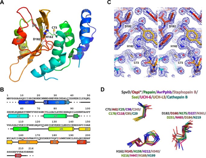
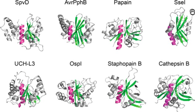
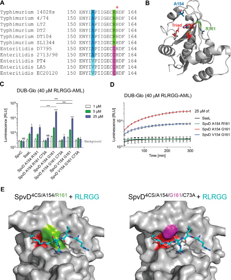
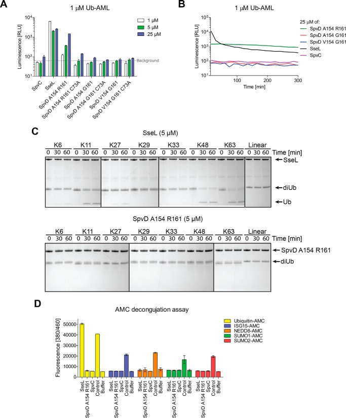
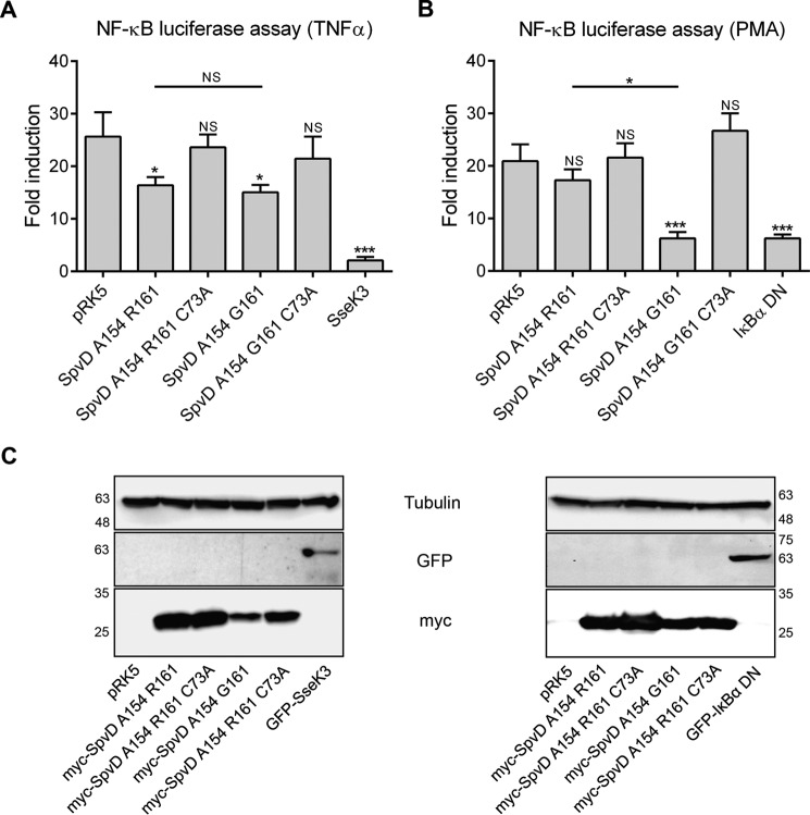
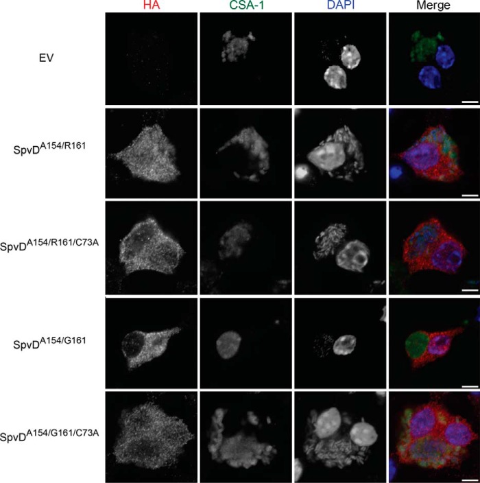
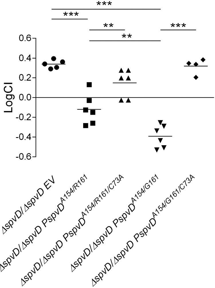
Many bacterial pathogens secrete virulence (effector) proteins that interfere with immune signaling in their host. SpvD is a Salmonella enterica effector protein that we previously demonstrated to negatively regulate the NF-κB signaling pathway and promote virulence of S. enterica serovar Typhimurium in mice. To shed light on the mechanistic basis for these observations, we determined the crystal structure of SpvD and show that it adopts a papain-like fold with a characteristic cysteine-histidine-aspartate catalytic triad comprising Cys-73, His-162, and Asp-182. SpvD possessed an in vitro deconjugative activity on aminoluciferin-linked peptide and protein substrates in vitro A C73A mutation abolished SpvD activity, demonstrating that an intact catalytic triad is required for its function. Taken together, these results strongly suggest that SpvD is a cysteine protease. The amino acid sequence of SpvD is highly conserved across different S. enterica serovars, but residue 161, located close to the catalytic triad, is variable, with serovar Typhimurium SpvD having an arginine and serovar Enteritidis a glycine at this position. This variation affected hydrolytic activity of the enzyme on artificial substrates and can be explained by substrate accessibility to the active site. Interestingly, the SpvDG161 variant more potently inhibited NF-κB-mediated immune responses in cells in vitro and increased virulence of serovar Typhimurium in mice. In summary, our results explain the biochemical basis for the effect of virulence protein SpvD and demonstrate that a single amino acid polymorphism can affect the overall virulence of a bacterial pathogen in its host.

Keywords: Salmonella enterica; bacterial pathogenesis; crystal structure; cysteine protease; effector; structure-function.


Figures

FIGURE 1.
FIGURE 1.
SpvD adopted a papain-like fold with Cys-73, His-162, and Asp-182 catalytic triad. A, crystal structure of S. typhimurium SpvD4CS colored starting from N terminus (blue) and ending at C terminus (red). Secondary structure elements and catalytic triad (sticks) are labeled in black. Cys-73/His-162/Asp-182 catalytic triad is shown in sticks. The image was generated using PyMOL software. B, secondary structure elements mapped according to their respective amino acid sequence and colored as in A. In the sequence, the catalytic triad residues are colored in red. Surface cysteines mutated to serines are underlined. C, walleye stereo view of the 2FoFc electron density map of the active site region of the SpvD structure. D, superimposition of catalytic triad components of SpvD, OspIC62A (PDB code 3W31), papain (PDB code 1PPN), AvrPphB (PDB code 1UKF), staphopain B (PDB code 1Y4H), SseI (PDB code 4G29), UCH-6 (PDB code 1VJV), UCH-L3 (PDB code 1XD3), and cathepsin B (PDB code 3AI8).
FIGURE 2.
FIGURE 2.
SpvD structure shared papain-like fold features. Schematic representation of SpvDA154/R161, OspIC62A (PDB code 3W31), papain (PDB code 1PPN), AvrPphB (PDB code 1UKF), staphopain B (PDB code 1Y4H), SseI (PDB code 4G29), UCH-L3 (PDB code 1XD3), and cathepsin B (PDB code 3AI8) is shown. The central α-helix containing the catalytic cysteine is colored in purple, and the main anti-parallel β-sheet is colored in green. Structures were oriented according to their superimposed catalytic triads.
FIGURE 3.
FIGURE 3.
SpvD had a deconjugative activity that varied between serovars. A, zoom on SpvD amino acid alignment highlighting the two main (154 and 161) polymorphisms occurring in Enteritidis and Typhimurium serovars. The red asterisk indicates the catalytic histidine His-162. B, both Ala-154 (blue stick) and Arg-161 (green stick) positions are situated in the vicinity of the CHD catalytic triad (red sticks). C, DUB-GloTM assay with three (1, 5, and 25 μm) concentrations of tested proteins. Results are expressed as mean ± S.E. of three independent experiments. Asterisks above the blue bars represent significant differences when compared with the buffer control (background) as measured by the Student's t test. Comparison between SpvD variants at 25 μm concentration was made using one-way ANOVA statistical analysis with Bonferroni's multiple comparison test. *, p < 0.05; ***, p < 0.005; NS, not significant. RLU, relative light units. D, DUB-GloTM signal over time for the highest (25 μm) concentration tested for SseL, SpvDA154/R161, SpvDA154/G161, and SpvDV154/G161. Signal intensities were measured every 15 min for which ± S.E. of three independent experiments are shown. E, modeling of the RLRGG peptide onto the surface of SpvD4CS/A154/R161 (left) or SpvD4CS/A154/G161/C73A (right) using the C-terminal ubiquitin tail (RLRGG; cyan sticks) from the UCH-L3 ubiquitin hydrolase complexed with ubiquitin (PDB code 1XD3). The catalytic triad region of SpvD is colored in red, Arg-161 is in green, and Gly-161 is in purple.
FIGURE 4.
FIGURE 4.
SpvDR161 was active on a ubiquitin-AML substrate. A, a modification of the DUB-GloTM assay with RLRGG substrate replaced by a full-length ubiquitin conjugated to aminoluciferin (Ub-AML). A representative assay result is shown. B, Ub-AML deconjugation assay over time measured for the highest concentration (25 μm) of the tested proteins. Signals were taken every 15 min. C, in vitro deubiquitination assay done with 5 μm of either SseL (top) or SpvDA154/R161 (bottom). Diubiquitin linkages were incubated with tested proteins for 0, 30, and 60 min followed by SDS-PAGE separation and Coomassie staining. Detectable cleavage product in the form of a single ubiquitin band is indicated with an arrow. D, AMC deconjugation assay using ubiquitin-AMC (yellow), ISG15-AMC (blue), NEDD8-AMC (orange), SUMO1-AMC (green), and SUMO2-AMC (red). Results are shown as mean fluorescence values with S.D. of three technical repeats obtained 1 h after start of the experiment. Isopeptidase T was used as a positive control for ubiquitin-AMC and ISG15-AMC. SENP2 was used as a control protease for SUMO1-AMC and SUMO2-AMC substrates, whereas NEDP1 was used in case of NEDD8-AMC.
FIGURE 5.
FIGURE 5.
SpvD variants had differential NF-κB inhibitory activities in PMA- but not in TNFα-stimulated cells. NF-κB luciferase assay from transfected HEK293 cell line after 16 h stimulation with TNFα (A) or PMA (B). Results are shown as -fold activation in relation to unstimulated cells. Values are expressed as the mean ± S.E. of at least four independent experiments. Statistical significances were calculated using one-way ANOVA followed by Bonferroni's multiple comparison test against pRK5- or SpvDA154/R161-transfected cells (*, p < 0.05; ***, p < 0.005; NS, not significant). C, protein expression levels measured by SDS-PAGE and immunoblotting with anti-tubulin (top), anti-GFP (mid), and anti-myc (bottom) antibodies. Numbers on the sides correspond to molecular weight protein ladder in kDa.
FIGURE 6.
FIGURE 6.
Catalytically dead variants of SpvD were translocated from bacteria in iBMDMs. Cells were infected for 18 h with S. typhimurium spvD strain carrying either an empty pWSK29 plasmid (EV) or a pWSK29 plasmid encoding C-terminally 2×HA-tagged variants of SpvD under SPI-2 promoter. In the last 2 h of infection, cells were treated with 10 μm MG-132 proteasomal inhibitor. DNA, Salmonella, and HA-tagged SpvD were stained using DAPI dye, CSA-1, and HA11 antibodies, respectively. The white scale bar represents 5 μm.
FIGURE 7.
FIGURE 7.
SpvDA154/G161 conferred increased virulence over SpvDA154/R161. C57BL/6 mice were inoculated by intraperitoneal injection with equal numbers (5 × 10² cfu of each of the two strains) of the indicated bacteria. Bacteria were recovered from infected spleens 3 days post-inoculation. CI values were calculated as described under “Experimental Procedures.” The scatter plot displays values obtained for individual mice and the mean are indicated (line). Statistical significances were calculated using one-way ANOVA followed by Bonferroni's multiple comparison test. **, p < 0.01; ***, p < 0.005.

References

    1. Figueira R., and Holden D. W. (2012) Functions of the Salmonella pathogenicity island 2 (SPI-2) type III secretion system effectors. Microbiology 158, 1147–1161 - PubMed
    1. Beninger P. R., Chikami G., Tanabe K., Roudier C., Fierer J., and Guiney D. G. (1988) Physical and genetic mapping of the Salmonella dublin virulence plasmid pSDL2: relationship to plasmids from other Salmonella strains. J. Clin. Invest. 81, 1341–1347 - PMC - PubMed
    1. Gulig P. A., and Curtiss R. 3rd (1988) Cloning and transposon insertion mutagenesis of virulence genes of the 100-kilobase plasmid of Salmonella typhimurium. Infect. Immun. 56, 3262–3271 - PMC - PubMed
    1. Jones G. W., Rabert D. K., Svinarich D. M., and Whitfield H. J. (1982) Association of adhesive, invasive, and virulent phenotypes of Salmonella typhimurium with autonomous 60-megadalton plasmids. Infect. Immun. 38, 476–486 - PMC - PubMed
    1. Libby S. J., Adams L. G., Ficht T. A., Allen C., Whitford H. A., Buchmeier N. A., Bossie S., and Guiney D. G. (1997) The spv genes on the Salmonella dublin virulence plasmid are required for severe enteritis and systemic infection in the natural host. Infect. Immun. 65, 1786–1792 - PMC - PubMed

MeSH terms