. 2017 Feb 1;215(3):420-430.
doi: 10.1093/infdis/jiw511.

Selective Destruction of Interleukin 23-Induced Expansion of a Major Antigen-Specific γδ T-Cell Subset in Patients With Tuberculosis

Affiliations

Selective Destruction of Interleukin 23-Induced Expansion of a Major Antigen-Specific γδ T-Cell Subset in Patients With Tuberculosis

Hongbo Shen et al. J Infect Dis. .

Erratum in

  • Corrigendum.
    [No authors listed] [No authors listed] J Infect Dis. 2019 Oct 22;220(11):1862. doi: 10.1093/infdis/jiz384. J Infect Dis. 2019. PMID: 31613317 Free PMC article. No abstract available.

Abstract

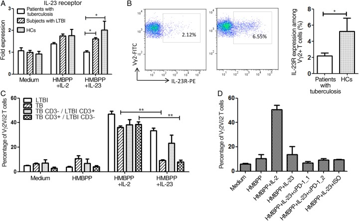
A loss of antigen-specific T-cell responses due to defective cytokine signaling during infections has not been reported. We hypothesize that tuberculosis can destroy signaling effects of selective cytokine(s) and induce exhaustion of antigen-specific T cells. To test this hypothesis, mechanistic studies were performed to examine whether and how tuberculosis blocked interleukin 23 (IL-23) and interleukin 2 (IL-2) signaling effects on a major human γδ T-cell subpopulation, phosphoantigen HMBPP-specific Vγ2Vδ2 T cells. IL-23 and IL-2 significantly expanded HMBPP-stimulated Vγ2Vδ2 T cells from subjects with latent tuberculosis infection, and IL-2 synergized the effect of IL-23. IL-23-induced expansion of Vγ2Vδ2 T cells involved STAT3. Surprisingly, patients with tuberculosis exhibited a selective destruction of IL-23-induced expansion of these cells. The tuberculosis-driven destruction of IL-23 signaling coincided with decreases of expression and phosphorylation of STAT3. Interestingly, impairing of STAT3 was linked to marked increases in the microRNAs (miRNAs) hsa-miR-337-3p and hsa-miR-125b-5p in Vγ2Vδ2 T cells from patients with tuberculosis. Downregulation of hsa-miR-337-3p and hsa-miR-125b-5p by miRNA sponges improved IL-23-mediated expansion of Vγ2Vδ2 T cells and restored the ability of these cells to produce anti-tuberculosis cytokines. These results support our hypothesis that tuberculosis can selectively impair a cytokine effect while sparing another and can induce exhaustion of T cells in response to the respective cytokine.

Keywords: JAK2/STAT3; T-cell exhaustion; Vγ2Vδ2 T cells; cytokine signaling; miRNA; tuberculosis.


Figures

Figure 1.
Figure 1.
Interleukin 23 (IL-23) and interleukin 2 (IL-2) signals significantly expanded HMBPP-stimulated Vγ2Vδ2 T cells from subjects with latent tuberculosis infection (LTBI), and IL-2 facilitates the IL-23–mediated expansion. A, Representative flow cytometry histograms and bar graph of pooled data show that IL-23, as well as IL-2, reproducibly expanded Vγ2Vδ2 T cells from subjects with LTBI, on coculture with HMBPP. Peripheral blood mononuclear cells (PBMCs) from subjects with LTBI were cultured for 7 days with medium, HMBPP, HMBPP plus IL-2, and HMBPP plus IL-23 and then subjected to surface staining and flow cytometry. B, Endogenous IL-2 produced by IL-23/HMBPP-activated Vγ2Vδ2 T cells sustained IL-23–induced expansion of the Vγ2Vδ2 T-cell subset. Note that blockade experiments using anti-IL-2 neutralizing antibody but not those using controls clearly reduced the magnitude of IL-23–induced expansion of HMBPP-stimulated Vγ2Vδ2 T cells. C, The transient first signaling model of T-cell receptor stimulation revealed that exogenous IL-2 synergized or enhanced IL-23–induced expansion of HMBPP-specific Vγ2Vδ2 T cells. In the transient first signaling model, PBMCs were incubated with HMBPP for 3 hours, HMBPP was washed out, and IL-2 alone, IL-23 alone, or IL-23 with different concentrations of IL-2 (0.5 ng/mL and 0.05 ng/mL) were added in the culture. IL-23 could not effectively expand Vγ2Vδ2 T cells after transient HMBPP stimulation, but a low concentration of IL-2 (0.5 ng/mL) helped IL-23 expand transiently stimulated Vγ2Vδ2 T cells. Data are means ± standard errors of the mean derived from 10 subjects with LTBI in 3 independent experiments. *P < .05 and **P < .01.
Figure 2.
Figure 2.
Patients with tuberculosis exhibited selective impairment of the effect of interleukin 23 (IL-23) signaling but not interleukin 2 (IL-2) signaling and showed exhaustion of the HMBPP-specific Vγ2Vδ2 T-cell subpopulation in response to IL-23. A, IL-23 could not expand HMBPP-stimulated Vγ2Vδ2 T cells from patients with tuberculosis, compared with those from subjects with latent tuberculosis infection (LTBI) and BCG-vaccinated healthy controls (HCs). Note that the effect of IL-2 signaling on γδ T cells from patients with tuberculosis was not impaired. Data are means ± standard errors of the mean (SEM), pooled from 40 subjects with LTBI, 40 HCs, and 40 patients with tuberculosis in 5 independent experiments. **P < .01. B, IL-23/HMBPP costimulation of Vγ2Vδ2 T cells from patients with tuberculosis but not HCs failed to induce an effector function for production of the anti-tuberculosis cytokines interferon γ (IFN-γ), tumor necrosis factor α (TNF-α), interleukin 17A (IL-17A), and interleukin 22 (IL-22). Enzyme-linked immunosorbent assays were used to measure cytokines in culture supernatants of Vγ2Vδ2 T cells after stimulation for 3 days in peripheral blood mononuclear cells (PBMCs) treated with medium, HMBPP, HMBPP + IL-2, or HMBPP + IL-23. Data are means ± SEM, pooled from 43 subjects with LTBI, 40 HCs, and 45 patients with tuberculosis in 7 independent experiments. *P < .05 and **P < .01. C, Short-term (ie, 1-month) treatment of patients with tuberculosis with antituberculosis antibiotics did not effectively reconstitute the expansion capability of Vγ2Vδ2 T cells in response to IL-23/HMBPP in vitro (top panels), but such transient therapy partially restored the ability of HMBPP-specific Vγ2Vδ2 T cells to mount an effector function for cytokine production in response to IL-23 (lower panels). Data are means ± SEM, pooled from 20 patients with tuberculosis in 3 independent experiments. *P < .05, **P < .01, and ***P < .001. Abbreviation: NS, not significant.
Figure 3.
Figure 3.
Selective impairment of the effects of interleukin 23 (IL-23) signaling during tuberculosis could not be attributed to nonexpression of IL-23R, dysfunction of antigen-presenting cells (APCs), or regulation of the programmed cell death 1 (PD-1) pathway. A, IL-23R expression was detectable in the absence or presence of HMBPP/IL-23 costimulation of Vγ2Vδ2 T cells in peripheral blood mononuclear cells (PBMCs) from patients with tuberculosis, although the HMBPP/IL-23–driven expression of IL-23R in patients with tuberculosis was lower than that in subjects with latent tuberculosis infection (LTBI) or healthy controls (HCs). Messenger RNA (mRNA) extracted from treated PBMCs was used for complementary DNA synthesis and real-time quantitative polymerase chain reaction (qPCR) analysis of IL-23R gene expression, with β-actin as a control. Expression levels are shown as fold expression, verified by β-actin. Data are means ± standard errors of the mean (SEM), pooled from 10 subjects with LTBI, 10 HCs, and 10 patients with tuberculosis in 3 independent experiments. *P < .05. B, Representative flow cytometry–determined histograms and bar graph show that the HMBPP/IL-23–driven expression of IL-23R in patients with tuberculosis was lower than that in HCs (P = .0286). *P < .05. C, APCs from HCs could not reconstitute the ability of Vγ2Vδ2 T cells from patients with tuberculosis to undergo expansion in response to HMBPP/IL-23 costimulation. In contrast, APCs from patients with tuberculosis (TB) could still induce HMBPP/IL-23–induced expansion of Vγ2Vδ2 T cells from subjects with LTBI (LTBI), which was similar to findings in the setting of autologous APCs. Data are means ± SEM, pooled from 10 patients with tuberculosis and 10 subjects with LTBI in 3 independent experiments. **P < .01. D, Blockade experiments using anti-PD-1 antibody showed that PD-1 antibody could not rescue the nonexpansion of Vγ2Vδ2 T cells from patients with tuberculosis in response to IL-23/HMBPP costimulation. Two sources of anti-human PD-1 antibodies (αPD-1_1 [clone EH12.2H7; Biolegend] and αPD-1_2 [R&D]) or isotype control were added to PBMC culture in the presence of IL-23 plus HMBPP. These 2 anti-PD-1 antibodies were shown to blockade PD-1–mediated exhaustion of αβ T cells (data not shown) [23, 24, 43]. Data are means ± SEM, derived from 18 patients with tuberculosis in 3 independent experiments. **P < .01.
Figure 4.
Figure 4.
Interleukin 23 (IL-23)–induced expansion of Vγ2Vδ2 T cells involves STAT3. Vγ2Vδ2 T cells in patients with tuberculosis showed decreased expression and phosphorylation of STAT3, with overexpression of antagonizing SOCS3. A, At top, hypothetical IL-23 signaling via the JAK2 and Tyk2 pathway resulted in phosphorylation of STAT3 in Vγ2Vδ2 T cells, a modification based on IL-23 engagement of CD4+ T cells [33, 44]. Note that the phosphorylated STAT3 dimer subsequently activates transcription of proliferating/cytokine-responsive genes in the nucleus [31, 32]. Below, the STAT3-targeted inhibitor cucurbitacin I (CUI) blocked IL-23–induced expansion of HMBPP-stimulated Vγ2Vδ2 T cells from subjects with latent tuberculosis infection (LTBI). CUI at a concentration of 2 nM significantly reduced IL-23–mediated expansion of HMBPP-stimulated Vγ2Vδ2 T cells but spared the effect of IL-2 signaling on these cells. B, Flow cytometry–based analysis showed that phosphorylation levels of STAT3 at pS727 and pY705 sites in Vγ2Vδ2 T cells were significantly decreased in fresh, unstimulated peripheral blood mononuclear cells (PBMCs) from patients with tuberculosis as compared to healthy controls (HCs). At left are representative flow cytometry–based histograms displaying percentage numbers of Vγ2+pY705+ cells (upper) and Vγ2+pS727+ cells (middle) among CD3+ T cells, as well as pY705+pS727+ cells among Vγ2+ cells (lower panels) from PBMCs of patients with tuberculosis and HCs. Representative flow panel of the isotype control is shown at top left. At right are bar graphs comparing frequencies of corresponding cell subpopulations between patients with tuberculosis and HCs. In accordance with manufacturer instructions, fresh PBMCs were briefly treated for 15 minutes with PMA and interleukin 6 (IL-6) and then stained for phosphorylated STAT3, using anti-Stat3 (pS727)–PE and anti-Stat3 (pY705)–Alexa Fluor 647 after surface staining for CD3 and Vγ2. C, The bar graph shows that, 72 hours after IL-23/HMBPP costimulation, the frequency of the Vγ2+pY705+pS727+ cell subpopulation among Vγ2+ T cells in patients with tuberculosis was also significantly lower than that in HCs. In contrast, IL-2/HMBPP costimulation did not cause significant changes in phosphorylated STAT3 between patients with TB and HCs. PBMCs were stimulated for 3 days with medium, HMBPP, HMBPP plus IL-2, and HMBPP plus IL-23 and then were assessed for percentages of pY705+pS727+ cells among CD3+Vγ2+ cells, using flow cytometry. D, Expression levels of STAT3 and SOCS3 genes in Vγ2Vδ2 T cells among patients with tuberculosis and HCs. Vγ2Vδ2 T cells were purified from PBMCs by flow cytometry sorting and subjected to RNA extraction and real-time quantitative polymerase chain reaction analysis. Expression of the STAT3 gene in Vγ2Vδ2 T cells from patients with tuberculosis decreased to 16% of the level seen in Vγ2Vδ2 T cells from HCs. Expression of SOCS3 in Vγ2Vδ2 T cells from TB patients is more than 4 times of that in HCs. Data in panels BD are means ± standard errors of the mean, pooled from 10 HCs and 10 patients with tuberculosis in 3 independent experiments. *P < .05, **P < .01, and ***P < .001. Abbreviation: DMSO, dimethyl sulfoxide.
Figure 5.
Figure 5.
Impairment of the STAT3 pathway coincided with marked increases in hsa-miR-337-3p and hsa-miR-125b-5p expression in Vγ2Vδ2 T cells from patients with tuberculosis. A, Among the microRNAs (miRNAs) capable of targeting STAT3 expression, hsa-miR-337-3p and hsa-miRNA-125b-5p were upregulated by approximately 6-fold in Vγ2Vδ2 T cells from patients with tuberculosis, compared with those from healthy controls (HCs). Expression of 2 other STAT3-targeted miRNAs, hsa-miR-124-3p and has-miR-223-3p, was also increased, although individual variations were seen. Total miRNA was extracted from freshly purified Vδ2+ T cells. The relative expression level of miRNAs was measured by real-time quantitative polymerase chain reaction (qPCR) analysis and normalized by U6 or 5s. BCG-vaccinated HCs were used as a calibrator. Data are means ± SEM standard errors of the mean (SEM), pooled from 16 patients with tuberculosis and 16 HCs in 3 independent experiments. **P < .01. B, Mycobacterial infection of human cells significantly increased the expression of hsa-miR-337-3p and hsa-miRNA-125b-5p. Human A549 cells were infected with Mycobacterium bovis BCG and assessed for expression of miRNAs, using real-time qPCR analysis. Data are fold changes expressed as means ± SEM. ***P < .001.
Figure 6.
Figure 6.
Downregulation of hsa-miR-337-3p and hsa-miR-125b-5p by microRNA (miRNA) sponges improved the interleukin 23 (IL-23)–mediated expansion of HMBPP-stimulated Vγ2Vδ2 T cells in patients with tuberculosis and restored the ability of these cells to produce cytokines. The pCMV-LV-GV234-GFP vector system was used to develop LV-has-125b-5p, LV-has-337-3p, and control miRNA sponge constructs to manipulate decreases in hsa-miR-337-3p and/or hsa-miRNA-125b-5p expression in Vγ2Vδ2 T cells. A, Microscopic fluorescence (left) and optical (right) pictures show that most peripheral blood mononuclear cells (PBMCs) containing Vγ2Vδ2 T cells give rise to green fluorescent protein–associated florescence after transduction with the miRNA sponges. B, Representative flow cytometry–based histograms (left panels) and bar graph data (right panel) show that transduction with a combination of LV-has-125b-5p and LV-has-337-3p but not controls significantly improved IL-23–induced expansion of HMBPP-stimulated Vγ2Vδ2 T cells in PBMCs from 12 patients with tuberculosis in 4 independent experiments (mean values [ ± SD] were 10.89% ± 1.17% for LV-has-125b-5p and LV-has-337-3p and 7.86% ± 2.08% for control; P = .013136). C, Transduction with LV-has-125b-5p and LV-has-337-3p combined also restored increases in the expression levels of T-cell receptor Vγ2 and the anti–tuberculosis cytokines interferon γ (IFN-γ) and interleukin 17A (IL-17A). D, Representative flow cytometry–based histograms and bar graph data show that transduction with a combination of LV-has-125b-5p and LV-has-337-3p but not controls significantly improved production of IL-17A and IFN-γ by IL-23–expanded, HMBPP-stimulated Vγ2Vδ2 T cells from patients with tuberculosis. Data are means ± standard errors of the mean, pooled from 12 patients with tuberculosis in 4 independent experiments. *P < .05 and **P < .01.

Similar articles

Cited by

References

    1. Penaloza-MacMaster P, Kamphorst AO, Wieland A et al. . Interplay between regulatory T cells and PD-1 in modulating T cell exhaustion and viral control during chronic LCMV infection. J Exp Med 2014; 211:1905–18. - PMC - PubMed
    1. Severson JJ, Serracino HS, Mateescu V et al. . PD-1+ Tim-3+ CD8+ T lymphocytes display varied degrees of functional exhaustion in patients with regionally metastatic differentiated thyroid cancer. Cancer Immunol Res 2015; 3:620–30. - PMC - PubMed
    1. Qiu Y, Chen J, Liao H et al. . Tim-3-Expressing CD4+ and CD8+ T cells in human tuberculosis (TB) exhibit polarized effector memory phenotypes and stronger anti-TB effector functions. PLoS Pathog 2012; 8:e1002984. - PMC - PubMed
    1. Posch SKC, Barthel SR, Mueller H et al. . Melanoma cell-intrinsic PD-1 receptor functions promote tumor growth. Cell 2015; 162:1242–56. - PMC - PubMed
    1. Li Q, Skinner PJ, Duan S-JHL et al. . Visualizing antigen-specific and infected cells in situ predicts outcomes in early viral infection. Science 2009; 323:1726–9. - PMC - PubMed

Publication types