. 2016 Dec 1;48(12):912-921.
doi: 10.1152/physiolgenomics.00091.2016. Epub 2016 Oct 27.

Impact of MYH6 variants in hypoplastic left heart syndrome

Affiliations

Impact of MYH6 variants in hypoplastic left heart syndrome

Aoy Tomita-Mitchell et al. Physiol Genomics. .

Abstract

Hypoplastic left heart syndrome (HLHS) is a clinically and anatomically severe form of congenital heart disease (CHD). Although prior studies suggest that HLHS has a complex genetic inheritance, its etiology remains largely unknown. The goal of this study was to characterize a risk gene in HLHS and its effect on HLHS etiology and outcome. We performed next-generation sequencing on a multigenerational family with a high prevalence of CHD/HLHS, identifying a rare variant in the α-myosin heavy chain (MYH6) gene. A case-control study of 190 unrelated HLHS subjects was then performed and compared with the 1000 Genomes Project. Damaging MYH6 variants, including novel, missense, in-frame deletion, premature stop, de novo, and compound heterozygous variants, were significantly enriched in HLHS cases (P < 1 × 10⁻⁵). Clinical outcomes analysis showed reduced transplant-free survival in HLHS subjects with damaging MYH6 variants (P < 1 × 10⁻²). Transcriptome and protein expression analyses with cardiac tissue revealed differential expression of cardiac contractility genes, notably upregulation of the β-myosin heavy chain (MYH7) gene in subjects with MYH6 variants (P < 1 × 10⁻³). We subsequently used patient-specific induced pluripotent stem cells (iPSCs) to model HLHS in vitro. Early stages of in vitro cardiomyogenesis in iPSCs derived from two unrelated HLHS families mimicked the increased expression of MYH7 observed in vivo (P < 1 × 10⁻²), while revealing defective cardiomyogenic differentiation. Rare, damaging variants in MYH6 are enriched in HLHS, affect molecular expression of contractility genes, and are predictive of poor outcome. These findings indicate that the etiology of MYH6-associated HLHS can be informed using iPSCs and suggest utility in future clinical applications.

Keywords: cardiomyocyte-autonomous; genetics; hypoplastic left heart syndrome; transplant-free survival outcome; upregulation of contractility genes.


Figures

Fig. 1.
Fig. 1.
Five-stage study design. Stage 1 analyzed the pedigree of family F MYH6-R443P. Stage 2 was a case-control association study assessing rare α-myosin heavy chain gene (MYH6) variants in 190 unrelated hypoplastic left heart syndrome (HLHS) subjects. Stage 3 assessed the clinical outcome of MYH6 variant carriers compared with non-MYH6 variant carriers. Stage 4 utilized transcriptome sequencing and Western blotting to compare gene expression in HLHS subjects with and without MYH6 variants. Stage 5 employed induced pluripotent stem cells (iPSCs) to model HLHS disease in 2 unrelated families with MYH6 variants.
Fig. 2.
Fig. 2.
Pedigree of family MYH6: R443P, variant filtering scheme and MYH6 variants in a case-control study. A: black squares and circles denote patients with left ventricular outflow tract obstructions (LVOTO; III:1, III:9, IV:1, IV:3). Striping denotes ventricular septal defect, unrelated heart defect to HLHS. Gray-shaded symbols denote subjects with uncertain or no diagnosis of congenital heart disease (CHD). White squares/circles denote patients without CHD. Genotypes denoted R443P indicate heterozygous carriers of an MYH6 variant. WT, wild type; symbols without a genotype are of unknown genotype. Squares, male; circles, female; triangles, fetal demise. B: identification and filtering variants in affected siblings and a distant relative followed by subtracting variants in the unaffected mother identified 20 candidate genes. Among them, MYH6 was the only candidate highly expressed in the heart. C: loci of 19 distinct rare MYH6 variants discovered in 190 HLHS subjects. Left to right: loci of variants in the MYH6 gene encoding the head, neck, and coiled-coil (tail) regions of α-MHC protein. Dots denote variants in HLHS subjects.
Fig. 3.
Fig. 3.
Transplant-free survival analysis. Kaplan-Meier survival curve constructed from 72 HLHS subjects. Among these, 10 subjects had a rare MYH6 variant (MUT), compared with 62 WT subjects. The Breslow (generalized Wilcoxon) statistic evaluated significance, resulting in rejection of the null hypothesis that these curves are equivalent (P = 6 × 10⁻³).
Fig. 4.
Fig. 4.
Pairwise analysis of gene expression of MYH7 in HLHS patients with and without MYH6 variants. A: gray-shaded and black bars denote HLHS patients with and without MYH6 variants, respectively. The annotation within each gray bar indicates the amino acid change resultant from each MYH6 mutation. Paired bars on the left and right side of the figure, respectively, indicate ventricular and atrial septal samples. Subject information is available in Supplemental Table S2. Sample number, sex status (male or female), and age (m, month; d, day; y, year) are denoted in the x-axis label. MYH7 expression is significantly increased (346%) in HLHS patients with an MYH6 mutation (P < 1 × 10⁻³, Supplemental Table S3). TPM, transcripts per million. B: Western blot showing increased β-MHC protein in the right ventricle and atrial septum of HLHS patients with an MYH6 variant. B, top: representative Western blot selected from 5 technical replicates of 4 pairs of patient samples; pairings are denoted on the x-axis. The entire immunoblot is shown in Supplemental Fig. S2. B, bottom: densitometric analysis of β-MHC levels normalized to GAPDH, reflecting the average of 5 replicate determinations performed on the 4 tissue pairs shown in B, top. Statistical analysis of the aggregate data representing the 4 pairs was statistically significant P < 1 × 10⁻³, generalized linear model/ANOVA, 62%, ± 0.15 SE.
Fig. 5.
Fig. 5.
Increased MYH7 expression in iPSC-derived cardiomyocytes from probands in 2 HLHS families. A: scheme for differentiating cardiomyocytes with small molecule Gsk3 inhibitor (CHIR99021) and wnt inhibitor (IWP). B: immunostaining of α-MHC at day 10 showing defective cardiomyogenesis in iPSCs from the HLHS proband and affected parent (carrier father) of family F MYH6-R443P. C: flow cytometry of cells cultured in parallel with those in B, showing decreased percentages of cardiac troponin T (cTnT)-positive cells at day 10. Data were compiled from 3 iPSC lines derived from each individual. The P values were calculated by Student's t-test (2-tailed, equal variance); vertical lines = ± SE. D: RNA-Seq showing MYH7 expression in iPSC-derived cardiomyocytes (CMs) from family F MYH6-R443P at differentiation day 8. Proband bars represent the average of 2 cell lines; vertical lines = range. Unaffected parent bars represent values from single cell line. E, left: quantitative PCR showing increased MYH7 expression in the HLHS proband of family F MYH6-R443P. E, right: a similar result from iPSC-derived cardiomyocytes of a separate HLHS family (F MYH6-D588A). Bars represent the average of triplicate cultures evaluated in 2 independent iPSC lines (n = 6). The P values were calculated by Student's t-test (2-tailed, equal variance); vertical lines = ± SE. Additional iPSC analysis from family F MYH6-D588A is shown in Supplemental Fig. S4.
Fig. 6.
Fig. 6.
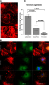
Dysmorphic sarcomeres in CMs derived from iPSCs reprogrammed from the HLHS proband and its affected parent. A: α-actinin immunostaining of mass cultured CMs at differentiation day 65. B: comparative sarcomere organization in individual CMs isolated from the cultures shown in A and subcultured at low density. A minimum of 100 isolated CMs representing each individual were judged to contain dysmorphic sarcomere organization if most of the myocyte area displayed blurred staining in which sarcomeric ladders contained punctate or truncated deposits of α-actinin ladders, rather than the relatively crisp and elongated ladders with relatively wide Z-bands that characterize MYH6+/+ cells. C: data were compiled from quadruplicate dishes representing each iPSC line (1 line from each parent; 2 lines from the proband) evaluated during a single determination. The P values were calculated by Student's t-test (2-tailed, equal variance); vertical lines = ± SE.

References

    1. Arrington CB, Bleyl SB, Matsunami N, Bonnell GD, Otterud BE, Nielsen DC, Stevens J, Levy S, Leppert MF, Bowles NE. Exome analysis of a family with pleiotropic congenital heart disease. Circ Cardiovasc Genet 5: 175–182, 2012. - PMC - PubMed
    1. Berdougo E, Coleman H, Lee DH, Stainier DY, Yelon D. Mutation of weak atrium/atrial myosin heavy chain disrupts atrial function and influences ventricular morphogenesis in zebrafish. Development 130: 6121–6129, 2003. - PubMed
    1. Boneva RS, Botto LD, Moore CA, Yang Q, Correa A, Erickson JD. Mortality associated with congenital heart defects in the United States: trends and racial disparities, 1979–1997. Circulation 103: 2376–2381, 2001. - PubMed
    1. Carniel E, Taylor MR, Sinagra G, Di Lenarda A, Ku L, Fain PR, Boucek MM, Cavanaugh J, Miocic S, Slavov D, Graw SL, Feiger J, Zhu XZ, Dao D, Ferguson DA, Bristow MR, Mestroni L. Alpha-myosin heavy chain: a sarcomeric gene associated with dilated and hypertrophic phenotypes of cardiomyopathy. Circulation 112: 54–59, 2005. - PubMed
    1. Chen J, Kubalak SW, Minamisawa S, Price RL, Becker KD, Hickey R, Ross J Jr, Chien KR. Selective requirement of myosin light chain 2v in embryonic heart function. J Biol Chem 273: 1252–1256, 1998. - PubMed

Publication types

LinkOut - more resources