. 2016 Dec 9;36(6):e00422.
doi: 10.1042/BSR20160457. Print 2016 Dec.

Influence of haem environment on the catalytic properties of the tetrathionate reductase TsdA from Campylobacter jejuni

Affiliations

Influence of haem environment on the catalytic properties of the tetrathionate reductase TsdA from Campylobacter jejuni

Julia M Kurth et al. Biosci Rep. .

Abstract

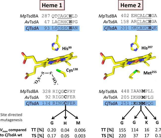
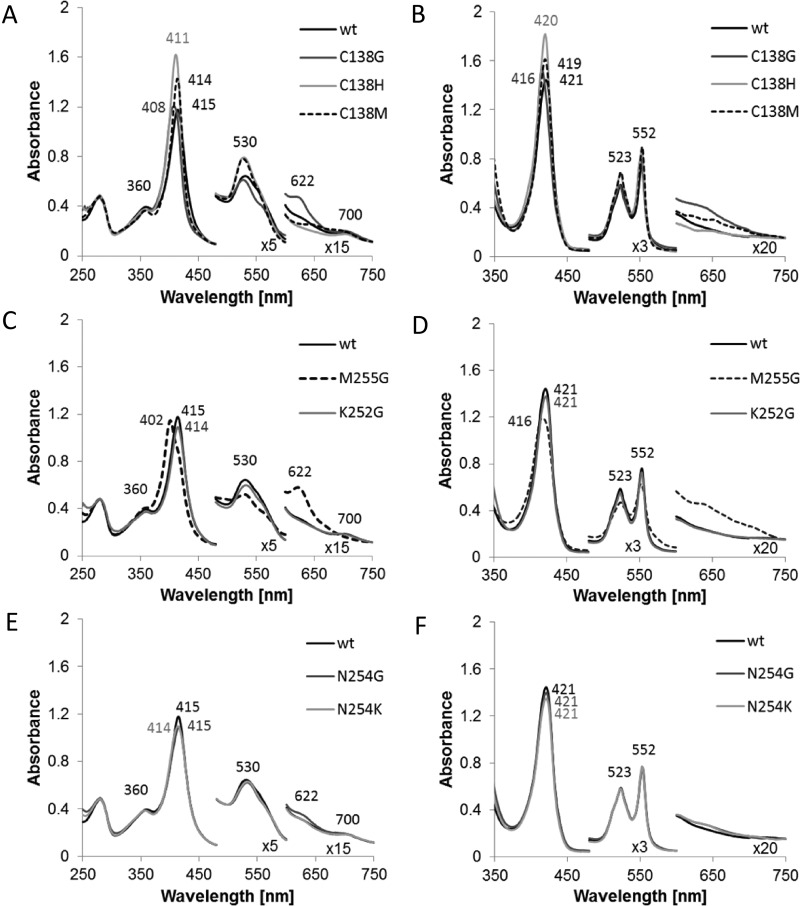
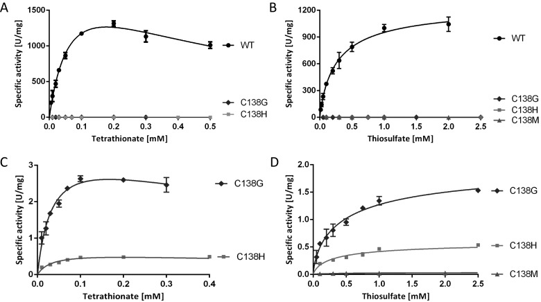
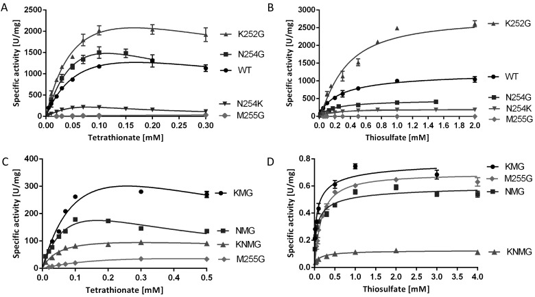
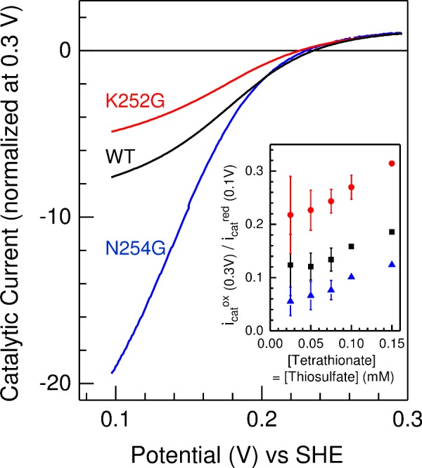
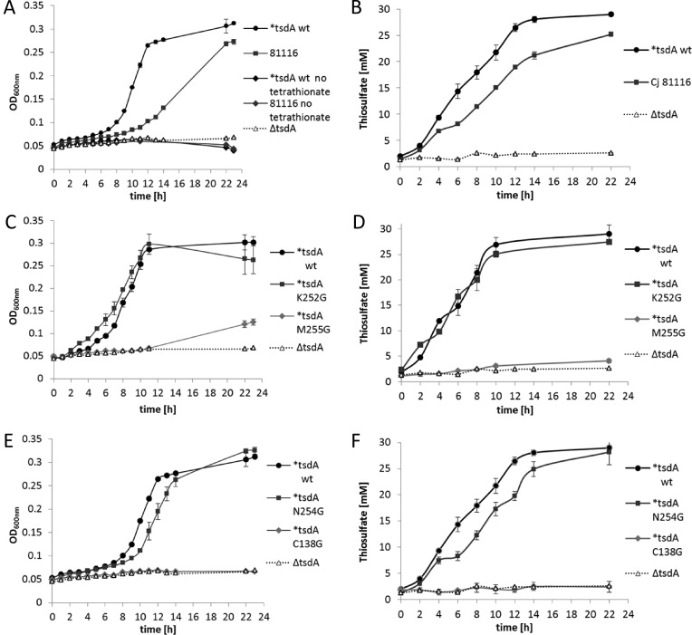
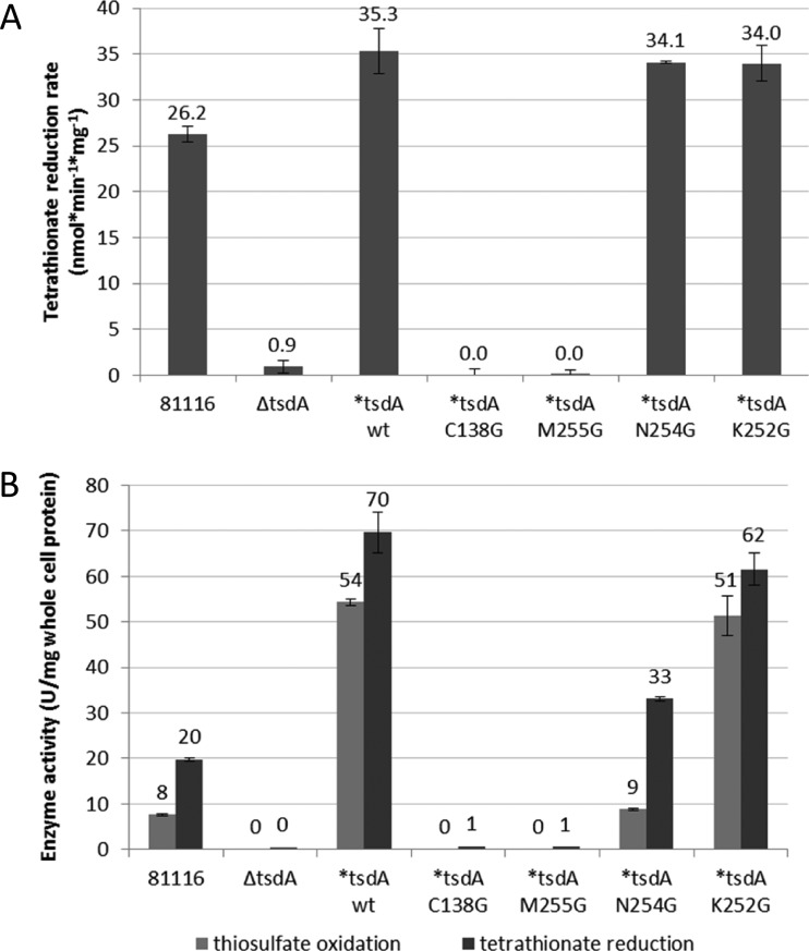
Bifunctional dihaem cytochrome c thiosulfate dehydrogenases/tetrathionate reductases (TsdA) exhibit different catalytic properties depending on the source organism. In the human food-borne intestinal pathogen Campylobacter jejuni, TsdA functions as a tetrathionate reductase enabling respiration with tetrathionate as an alternative electron acceptor. In the present study, evidence is provided that Cys138 and Met255 serve as the sixth ligands of Haem 1 and Haem 2 respectively, in the oxidized CjTsdA wt protein. Replacement of Cys138 resulted in a virtually inactive enzyme, confirming Haem 1 as the active site haem. Significantly, TsdA variants carrying amino acid exchanges in the vicinity of the electron-transferring Haem 2 (Met255, Asn254 and Lys252) exhibited markedly altered catalytic properties of the enzyme, showing these residues play a key role in the physiological function of TsdA. The growth phenotypes and tetrathionate reductase activities of a series of ΔtsdA/*tsdA complementation strains constructed in the original host C. jejuni 81116, showed that in vivo, the TsdA variants exhibited the same catalytic properties as the pure, recombinantly produced enzymes. However, variants that catalysed tetrathionate reduction more effectively than the wild-type enzyme did not allow better growth.

Keywords: axial haem ligation; cytochrome c; reaction directionality; tetrathionate reductase; thiosulfate.


Figures

Figure 1
Figure 1. Schematic overview of Haem 1 and Haem 2 environments in Tsd(B)A proteins
Relevant sequence alignments are shown for TsdA from C. jejuni (CjTsdA), A. vinosum (AvTsdA) and the TsdBA fusion protein from M. purpuratum (MpTsdBA). Amino acid numbers are given for the recombinant proteins without signal peptides. In case of CjTsdA, the sequence of the N-terminal Strep-tag is included. In the central left panel, a tetrathionate molecule is shown in vicinity of the active site Haem 1 iron-ligating cysteine, based on the AvTsdA crystal structure [16]. Amino acid numbers in the central panels refer to CjTsdA. In the lower part of the figure, changes in the environments of Haem 1 and Haem 2 are indicated that were introduced by site-directed mutagenesis. The effects of these changes on maximal reaction velocity are listed as percent of Vmax for the wild-type enzyme in the tetrathionate-reducing (TT) and the thiosulfate-oxidizing (TS) direction.
Figure 2
Figure 2. UV–Vis spectra of CjTsdA wt and variants
UV–Vis spectra of CjTsdA wt are compared with spectra of variants with affected Haem 1 ligation (A and B) and with spectra of variants with affected Haem 2 ligation (CF). As some of the proteins are partly reduced in the ‘as isolated’ state, up to 30 μM ferricyanide was added to record the oxidized spectrum (A, C and E). For full reduction of the proteins, 5–16 mM Na-dithionite was added (B, D and F). Thirty millimolars BisTris buffer (pH 6.5) were used and spectra were normalized to 280 nm and 750 nm. For CjTsdA M255G and CjTsdA C138G, a high-spin feature appears at 622 nm in the oxidized state. The oxidized spectrum of TsdA wt and all variants except of M255G exhibits a 700 nm peak indicating methionine as haem iron ligand. Protein concentration: 8 μM.
Figure 3
Figure 3. Enzyme activity assays with TsdA wt and Haem 1 ligation affected variants
TsdA wt was compared with TsdA C138G, C138H and C138M concerning tetrathionate reduction (A) and thiosulfate oxidation (B). Tetrathionate reduction (C) and thiosulfate oxidation (D) of the TsdA C138 variants are shown in detail in the panels below. Tetrathionate reduction (A and C) was measured under anoxic conditions at 42°C in 100 mM ammonium acetate buffer (pH 5) with 0.3 mM methyl viologen previously reduced with titanium (III) citrate. Control measurements showed that methyl viologen was provided at a saturating concentration in all cases. Tetrathionate reduction activity of TsdA C138M was so low that exact kinetic parameters could not be derived with confidence. Different tetrathionate concentrations (0.01–0.7 mM) were used. Thiosulfate oxidation (B and D) was measured at 42°C in 50 mM BisTris buffer (pH 6.5) with 80 μM horse heart cytochrome c as electron acceptor and 0.05–8 mM thiosulfate. Control measurements showed that horse heart cytochrome c was provided at a saturating concentration in all cases.
Figure 4
Figure 4. Enzyme activity assays with TsdA wt and Haem 2 ligation affected variants
Tetrathionate reduction (A and C) was measured under anoxic conditions at 42°C in 100 mM ammonium acetate buffer (pH 5) with 0.3 mM methyl viologen previously reduced with titanium (III) citrate. Different tetrathionate concentrations (0.01–0.7 mM) were used. Thiosulfate oxidation (B and D) was measured at 42°C in 50 mM BisTris buffer (pH 6.5) with 80 μM horse heart cytochrome c as electron acceptor and 0.05–8 mM thiosulfate.
Figure 5
Figure 5. Baseline subtracted protein film cyclic voltammetry of CjTsdA wild-type (black), N254G (blue) and K252G variant (red) in 0.05 mM tetrathionate and 0.075 mM thiosulfate
Inset: The dependence of icatox/icatred on substrate concentration for equimolar substrate concentrations; wild-type (black), N254G (blue) and K252G variant (red), see text for details. Values are the average of two independent measurements, errors show maximum and minimum values, except for N254G at 0.1 and 0.15 mM where single values are presented. Scan rate 10 mV·s−1, electrode rotation 500 rpm in 100 mM ammonium acetate, 50 mM NaCl, pH 5 at 42°C.
Figure 6
Figure 6. SDS/PAGE of crude cell extracts of C. jejuni 81116 wt, ΔtsdA mutant and complementation strains
C. jejuni cells of 81116 wt (81116), ΔtsdA mutant (ΔtsdA) and the different complementation strains (*tsdA WT, *tsdA C138G, *tsdA M255G, *tsdA N254G, *tsdA K252G) were disrupted by bead beating and the crude cell extracts were used for SDS/PAGE. Forty micrograms of protein per lane were loaded on the gels for Coomassie staining (A) and Western blots (C and D), and 27 μg of protein per lane was used for haem staining (B). For the Western blots, antibody against His-tag was used and the second antibody was a HRP conjugate. The detection was based on the reaction of HRP with chloronaphthol (C) or the interaction between HRP and ECL reagent (D). The 37 kDa protein CjTsdA is marked with an arrow. The approximately 70 kDa haem stained band in panel (B) is the MccA multi-haem sulfite reductase (see text).
Figure 7
Figure 7. Anaerobic growth of C. jejuni 81116 wt, ΔtsdA mutant and complementation strains
C. jejuni cells of 81116 wt (81116), ΔtsdA mutant (ΔtsdA) and the different ΔtsdA/*tsdA complementation strains (*tsdA WT, *tsdA C138G, *tsdA M255G, *tsdA N254G, *tsdA K252G) were grown under oxygen-limited conditions at 42°C in almost completely filled 500 ml shake flasks containing BHI-S medium plus 20 mM sodium formate and 15 mM tetrathionate. In (A, C and E), the absorbance at 600 nm of the wild-type 81116 strain (closed black circles) is compared with that of an 81116 tsdA mutant (open triangles) and the different ΔtsdA/*tsdA complementation strains. In the absence of added tetrathionate [dark grey and black diamonds in (A)], no growth of either strain occurred. In (B, D and F), the conversion of tetrathionate to thiosulfate in each of the culture supernatants at each time point corresponding to the growth curves in (A, C and E) is shown.
Figure 8
Figure 8. Determination of tetrathionate reduction rate with whole cells and enzyme activity assays with crude cell extract
For determination of tetrathionate reduction rate with whole cells (A) C. jejuni cells of 81116 wt (81116), ΔtsdA mutant (ΔtsdA) and the different complementation strains (*tsdA WT, *tsdA C138G, *tsdA M255G, *tsdA N254G, *tsdA K252G) were incubated at 42°C in 14 ml phosphate buffer (pH 7.4) containing 20 mM sodium formate and 2 mM tetrathionate. The thiosulfate concentrations in the supernatants of samples taken at 2 or 5 min intervals were determined directly after the experiment. For enzyme activity assays with crude cell extract (B) C. jejuni cells of 81116 wt (81116), ΔtsdA mutant (ΔtsdA) and the different complementation strains (*tsdA WT, *tsdA C138G, *tsdA M255G, *tsdA N254G, *tsdA K252G) were disrupted by bead beating. Tetrathionate reduction was measured anaerobically at 42°C in 100 mM ammonium acetate buffer (pH 5) with 0.3 mM methyl viologen previously reduced with titanium (III) citrate and 0.1 mM tetrathionate. Thiosulfate oxidation was performed at 42°C in 50 mM BisTris buffer (pH 6.5) with 80 μM horse heart cytochrome c as electron acceptor and 2 mM thiosulfate.

Similar articles

Cited by

References

    1. Kurth J., Dahl C., Butt J.N. Catalytic protein film electrochemistry provides a direct measure of the tetrathionate/thiosulfate reduction potential. J. Am. Chem. Soc. 2015;137:13232–13235. doi: 10.1021/jacs.5b08291. - DOI - PubMed
    1. Liu Y.W., Denkmann K., Kosciow K., Dahl C., Kelly D.J. Tetrathionate stimulated growth of Campylobacter jejuni identifies TsdA as a new type of bi-functional tetrathionate reductase that is widely distributed in bacteria. Mol. Microbiol. 2013;88:173–188. doi: 10.1111/mmi.12176. - DOI - PubMed
    1. Thauer R.K., Jungermann K., Decker K. Energy conservation in chemotrophic anaerobic bacteria. Bacteriol. Rev. 1977;41:100–180. - PMC - PubMed
    1. Winter S.E., Thiennimitr P., Winter M.G., Butler B.P., Huseby D.L., Crawford R.W., Russell J.M., Bevins C.L., Adams L.G., Tsolis R.M., et al. Gut inflammation provides a respiratory electron acceptor for Salmonella. Nature. 2010;467:426–429. doi: 10.1038/nature09415. - DOI - PMC - PubMed
    1. Oltman L.F., Claasen V.P., Kastelein P., Reijinders W.N.M., Stouthammer A.H. The influence of tungstate on the formation and activities of four reductases of Proteus mirabilis: identification of two new molybdoenzymes, chlorate reductase and tetrathionate reductase. FEBS Lett. 1979;106:43–46. doi: 10.1016/0014-5793(79)80691-2. - DOI - PubMed

MeSH terms