. 2016 Oct 13:6:126.
doi: 10.3389/fcimb.2016.00126. eCollection 2016.

SpyB, a Small Heme-Binding Protein, Affects the Composition of the Cell Wall in Streptococcus pyogenes

Affiliations

SpyB, a Small Heme-Binding Protein, Affects the Composition of the Cell Wall in Streptococcus pyogenes

Rebecca J Edgar et al. Front Cell Infect Microbiol. .

Abstract

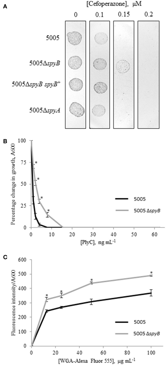
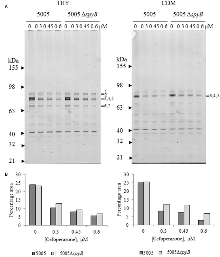
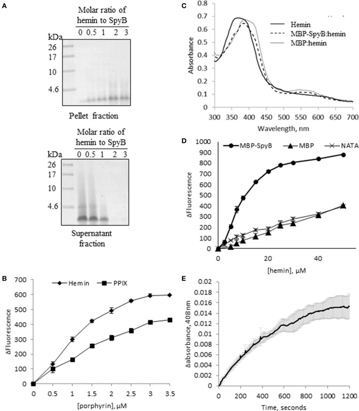
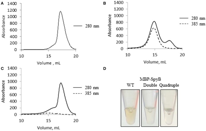
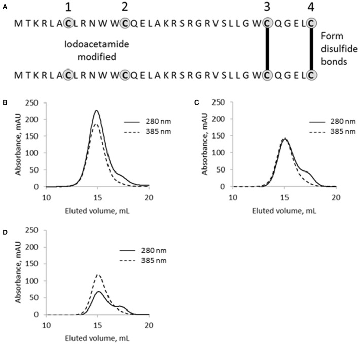
Streptococcus pyogenes (Group A Streptococcus or GAS) is a hemolytic human pathogen associated with a wide variety of infections ranging from minor skin and throat infections to life-threatening invasive diseases. The cell wall of GAS consists of peptidoglycan sacculus decorated with a carbohydrate comprising a polyrhamnose backbone with immunodominant N-acetylglucosamine side-chains. All GAS genomes contain the spyBA operon, which encodes a 35-amino-acid membrane protein SpyB, and a membrane-bound C3-like ADP-ribosyltransferase SpyA. In this study, we addressed the function of SpyB in GAS. Phenotypic analysis of a spyB deletion mutant revealed increased bacterial aggregation, and reduced sensitivity to β-lactams of the cephalosporin class and peptidoglycan hydrolase PlyC. Glycosyl composition analysis of cell wall isolated from the spyB mutant suggested an altered carbohydrate structure compared with the wild-type strain. Furthermore, we found that SpyB associates with heme and protoporphyrin IX. Heme binding induces SpyB dimerization, which involves disulfide bond formation between the subunits. Thus, our data suggest the possibility that SpyB activity is regulated by heme.

Keywords: ADP-ribosyltransferase; Group A carbohydrate; SpyA; SpyB; Streptococcus pyogenes; cell wall; heme.


Figures

Figure 1
Figure 1
Growth of the MGAS5005 (WT), 5005ΔspyB, 5005ΔspyB spyB+, and 5005ΔspyA in THY medium. The data are the mean of three independent experiments ± standard deviation.
Figure 2
Figure 2
SpyB deficiency causes MGAS5005 cells to aggregate. MGAS5005, 5005ΔspyB, 5005ΔspyB spyB+, and 5005ΔspyA strains at middle exponential phase were examined by microscopy. (A) MGAS5005 strains were Gram stained and imaged by light microscopy. (B) MGAS5005 strains were stained with fluorescently labeled vancomycin to visualize the cell walls and septa, and imaged by fluorescence microscopy. (C) Fixed cultures of MGAS5005 and 5005ΔspyB were visualized by transmission electron microscopy or (D) scanning electron microscopy.
Figure 3
Figure 3
Phenotypic analysis of 5005ΔspyB. (A) SpyB deficiency promotes resistance to cefoperazone. MGAS5005 strains were grown in CDM to middle exponential phase, diluted in PBS (10−2 dilution) and then plated on THY agar supplied with 0, 0.1, 0.15, and 0.2 μM cefoperazone in triplicate. This result is representative of five independent experiments (P = 0, t-test). (B) SpyB deficiency decreases the sensitivity of MGAS5005 to the PG hydrolase PlyC. MGAS5005 and 5005ΔspyB at middle exponential phase were measured for growth at 600 nm following 60 min incubation in the presence of various concentrations of PlyC (0–62.5 ng mL−1). The average percentage change in growth ± standard deviation for three replicates is shown, the asterisk indicates values that are statistically different (p < 0.01) from WT 5005. (C) SpyB deficiency alters the affinity of MGAS5005 for GlcNA binding wheat germ agglutinin (WGA). MGAS5005 and 5005ΔspyB at middle exponential phase were incubated for 60 min with various concentrations (0–100 μg mL−1) of WGA conjugated to Alexa Fluor 555. After washing the bacteria were measured for fluorescence (ex 544 nm, em 590 nm) and normalized for growth at 600 nm. Data are the average of three replicates ± standard deviation, the asterisk indicates values that are statistically different (p < 0.01) from WT 5005.
Figure 4
Figure 4
Cefoperazone inhibition of PBPs in MGAS5005 and 5005ΔspyB. (A) Gel-based analysis of PBP expression in MGAS5005 strains grown in THY medium or CDM. Whole cells were treated with various concentrations of cefoperazone and subsequently labeled with Boc-FL. Membrane fractions were separated on 4–12% SDS-PAGE gels and visualized as described in Materials and Methods. (B) Densitometry analysis of bands 3, 4, and 5 for the gels shown in panel (A). Data are representative of biological triplicates.
Figure 5
Figure 5
SpyB is a porphyrin-binding protein. (A) Hemin binding decreases SpyB solubility. SpyB was mixed with hemin in different molar ratios. Soluble and insoluble fractions were separated by centrifugation and resolved on a 16% Tris-Tricine gel. (B) The change in tryptophan fluorescence quenching of SpyB by hemin and protoporphyrin IX (PPIX). A series of hemin or PPIX concentrations (0, 0.5, 1, 1.5, 2, 2.5, 3, and 3.5 μM) was added to 5 μM SpyB, in the presence of 1 mM DTT. The curves are the average of three replicates ± standard deviation. The dissociation constant (KD) was calculated using SigmaPlot software. (C) Absorbance spectra between 300 and 700 nm were collected for 50 μM hemin; 50 μM hemin mixed with 2.5 μM MBP-SpyB; and 50 μM hemin mixed with 2.5 μM MBP, in the presence of 1 mM DTT. (D) The change in tryptophan fluorescence quenching for MBP-SpyB and MBP. A series of hemin concentrations (0, 2.5, 5, 7.5, 10, 15, 20, 25, 30, 40, 50 μM) was added to 2.5 μM MBP-SpyB monomer or MBP, in the presence of 1 mM DTT. A control experiment was performed with 27.5 μM N-acetyltryptophanamide (NATA) under the same conditions. The curves are the average of three replicates ± standard deviation. The KD was calculated using SigmaPlot software. (E) A time course for hemin transfer from MBP-SpyB to apomyoglobin was measured at 408 nm over 20 min. The dissociation rate constant (Koff) was calculated from the change in absorbance at 408 nm using Graphpad Prisim software. Data are the average of three replicates ± standard deviation. The Koff along with the previously measured KD were used to calculate the binding rate constant (Kon) for MBP-SpyB.
Figure 6
Figure 6
Hemin binding induces SpyB oligomerization. (A) The size-exclusion chromatogram of MBP-SpyB monomer. (B) The size-exclusion chromatogram of MBP-SpyB monomer reconstituted with hemin in 1:4 ratio. (C) The size-exclusion chromatogram of MBP-SpyB monomer reconstituted with protoporphyrin IX in 1:4 ratio. The absorbance was monitored at 280 and 385 nm (hemin and protoporphyrin IX detection). (D) The appearance of WT MBP-SpyB, and double (C7A/C13A) and quadruple (C7A/C13A/C30A/C35A) MBP-SpyB mutants, following expression in E. coli Rosetta DE3 cells and purification by Ni-NTA affinity chromatography and size-exclusion chromatography. Data are representative of biological triplicates.
Figure 7
Figure 7
The functional role of cysteine residues in SpyB. (A) Schematic of SpyB modification by iodoacetamide. The size-exclusion chromatogram of (B) MBP-SpyB WT, (C) double (C7A/C13A), and (D) quadruple (C7A/C13A/C30A/C35A) mutants reconstituted with hemin. The absorbance was monitored at 280 and 385 nm (hemin). Data are representative of biological triplicates.

Similar articles

Cited by

References

    1. Agarwal S., Agarwal S., Pancholi P., Pancholi V. (2011). Role of serine/threonine phosphatase (SP-STP) in Streptococcus pyogenes physiology and virulence. J. Biol. Chem. 286, 41368–41380. 10.1074/jbc.M111.286690 - DOI - PMC - PubMed
    1. Albarracin Orio A. G., Pinas G. E., Cortes P. R., Cian M. B., Echenique J. (2011). Compensatory evolution of pbp mutations restores the fitness cost imposed by beta-lactam resistance in Streptococcus pneumoniae. PLoS Pathog. 7:e1002000. 10.1371/journal.ppat.1002000 - DOI - PMC - PubMed
    1. Ascoli F., Fanelli M. R., Antonini E. (1980). Preparation and properties of apohemoglobin and reconstituted hemoglobins. Methods Enzymol. 76, 72–87. - PubMed
    1. Asher C., de Villiers K. A., Egan T. J. (2009). Speciation of ferriprotoporphyrin IX in aqueous and mixed aqueous solution is controlled by solvent identity, pH, and salt concentration. Inorg. Chem. 48, 7994–8003. 10.1021/ic900647y - DOI - PubMed
    1. Atilano M. L., Pereira P. M., Yates J., Reed P., Veiga H., Pinho M. G., et al. . (2010). Teichoic acids are temporal and spatial regulators of peptidoglycan cross-linking in Staphylococcus aureus. Proc. Natl. Acad. Sci. U.S.A. 107, 18991–18996. 10.1073/pnas.1004304107 - DOI - PMC - PubMed

MeSH terms

LinkOut - more resources