Review
. 2016 Oct 13:4:109.
doi: 10.3389/fcell.2016.00109. eCollection 2016.

Sending Out an SOS: Mitochondria as a Signaling Hub

Affiliations
Review

Sending Out an SOS: Mitochondria as a Signaling Hub

Iryna Bohovych et al. Front Cell Dev Biol. .

Abstract

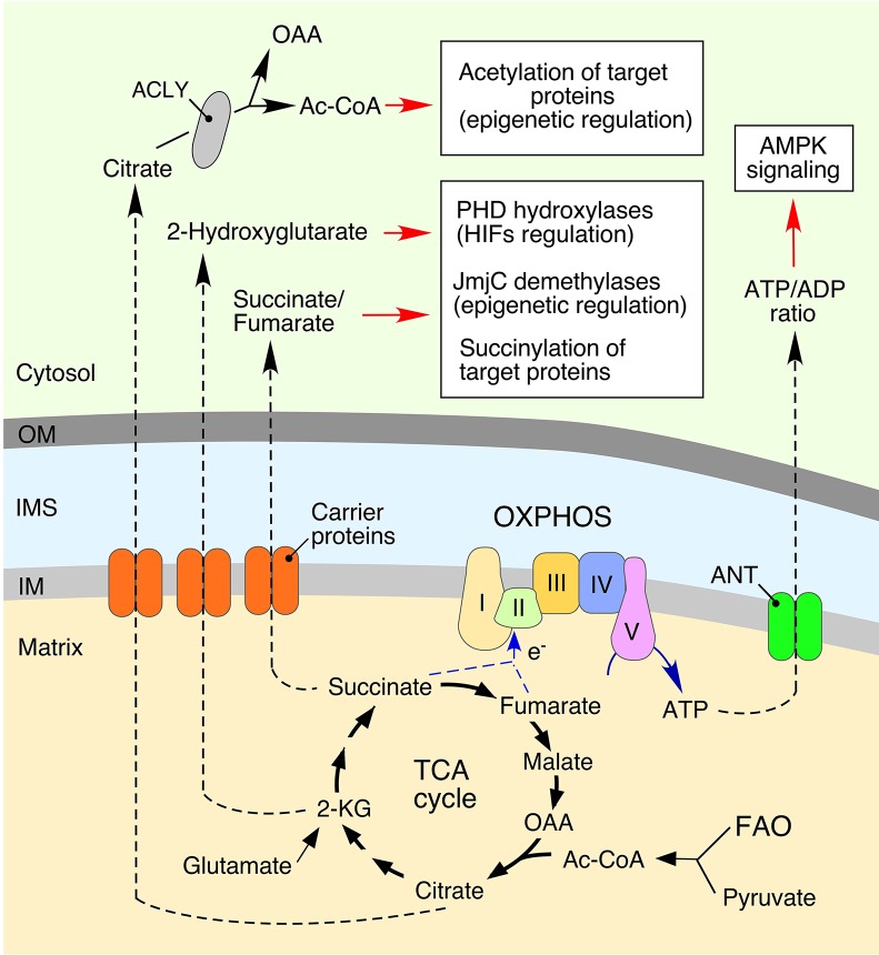
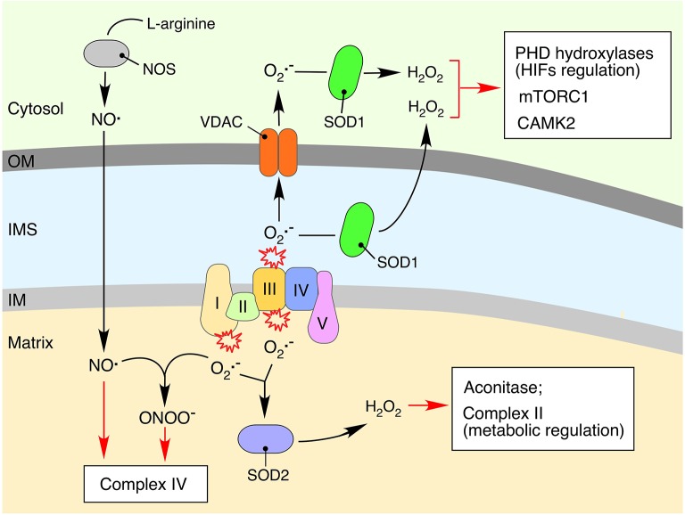
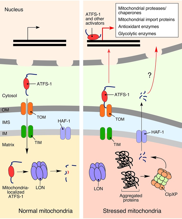
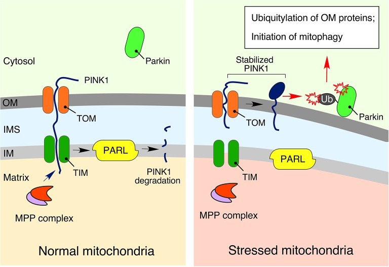
Normal cellular physiology is critically dependent on numerous mitochondrial activities including energy conversion, cofactor and precursor metabolite synthesis, and regulation of ion and redox homeostasis. Advances in mitochondrial research during the last two decades provide solid evidence that these organelles are deeply integrated with the rest of the cell and multiple mechanisms are in place to monitor and communicate functional states of mitochondria. In many cases, however, the exact molecular nature of various mitochondria-to-cell communication pathways is only beginning to emerge. Here, we review various signals emitted by distressed or dysfunctional mitochondria and the stress-responsive pathways activated in response to these signals in order to restore mitochondrial function and promote cellular survival.

Keywords: metabolites; mitochondria; mitochondrial stress; reactive oxygen species; retrograde signaling.


Figures

Figure 1
Figure 1
Mitochondria-derived metabolites regulate various cellular processes. Bioenergetic function of mitochondria is associated with generation of ATP (adenosine triphosphate) via the oxidative phosphorylation (OXPHOS) system. This process involves conversion of various substrates imported from the cytosol via the tricarboxylic acid (TCA) cycle or fatty acid oxidation (FAO). Metabolic intermediates can be exported into the cytosol to impact various signaling cascades. Mitochondria-derived citrate is converted to oxaloacetate (OAA) and acetyl coenzyme A (Ac-CoA) by ATP citrate lyase (ACLY). Subsequently, a fraction of the cytosolic Ac-CoA can be used in protein acetylation, thereby impacting multiple cellular processes (Choudhary et al., ; Spange et al., ; Wellen et al., ; Eisenberg et al., 2014). Another TCA cycle biosynthetic intermediate, 2-ketoglutarate (α-KG), can be converted to 2-Hydroxyglutarate. This metabolite inhibits: (1) Jumonji C domain-containing demethylases (JmjC), affecting epigenetic reprogramming (Pearce et al., ; Zong et al., 2016); and (2) hypoxia-inducible prolyl hydroxylases (PHDs), thus stabilizing the hypoxia-inducible factors (HIFs) and inducing a hypoxic response (Xu et al., ; Pearce et al., ; Zong et al., 2016). The TCA metabolite, fumarate, can also act as a signal-modulating factor via its ability to bind lysine residues of various proteins, resulting in a post-translational modification called succinylation. The adenine nucleotide translocator (ANT) mediates an exchange of ATP and cytosol-derived ADP (adenosine diphosphate) across the inner mitochondrial membrane (IM). Mitochondrial dysfunction derived ATP depletion perturbs ATP/ADP ratios by subsequent activation of adenosine monophosphate-activated protein kinase (AMPK), a master regulator of cellular energy homeostasis (Hall et al., ; Pearce et al., 2013).
Figure 2
Figure 2
Impact of mitochondria-derived free radicals on cellular signaling processes. Reduction of molecular oxygen to the superoxide anion (O2·-) is an unavoidable consequence of electron leakage associated with the bioenergetic function of respiratory complexes (Figueira et al., 2013). The O2·- produced is quickly converted to hydrogen peroxide (H2O2) by the mitochondrial matrix-associated manganese (SOD2) or inner membrane space (IMS)-localized copper-zinc (SOD1) superoxide dismutases (Murphy, ; Figueira et al., 2013). Accumulation of H2O2 can affect functioning of mitochondrial proteins, in particular those containing iron-sulfur clusters. Aconitase, a key component of the TCA cycle, is one of the mitochondrial proteins most susceptible to oxidative damage due to the surface exposed iron-sulfur cluster-containing active sites (Armstrong et al., 2004). Mitochondria-derived O2·- and H2O2 can also impact cytosolic signaling cascades via oxidation of various redox-sensitive proteins including calcium/calmodulin-dependent protein kinase 2 (CaMK2) (Erickson et al., 2008), the mammalian target of rapamycin complex 1 protein kinase (mTORC1), and PHD hydroxylases (Sena and Chandel, 2012). The cytosol-derived nitric oxide radical (NO.), a product of arginine conversion by nitric oxide synthases (NOSs), can permeate mitochondrial membranes and inhibit electron transport chain functioning through competitive inhibition of respiratory complex IV or via condensation with O2·- and formation of a potent oxidant, peroxynitrate (ONOO) (Nisoli et al., ; Antunes et al., ; Figueira et al., 2013).
Figure 3
Figure 3
The mitochondrial unfolded protein response (UPRmt). Activation of UPRmt in response to mitochondrial damage is best characterized in the roundworm Caenorhabditis elegans, where it is regulated by the activating transcription factor associated with stress 1 (ATFS-1). In the absence of stress stimuli, ATFS1 is transported to the mitochondria and degraded by LON protease (Nargund et al., 2012). Conditions of mitochondrial stress stall mitochondrial import of ATFS-1, allowing a fraction of the protein to translocate into the nucleus and promote expression of more than 400 mitochondrial homeostasis-related genes (Lin and Haynes, 2016). Another, not-so-well defined, activation of UPRmt in C. elegans is associated with peptide exporter HAF-1, which translocates signaling peptides generated by the matrix peptidase ClpXP (Haynes et al., 2010).
Figure 4
Figure 4
The PINK1-Parkin signaling relay. Under physiological conditions, phosphatase and tensin homolog (PTEN)-induced ubiquitin kinase 1 (PINK1), a key regulator of mitophagy and mitochondrial turnover, is imported into the mitochondria via the translocases of outer (TOM) and inner (TIM) membranes. It is subsequently degraded by presenilin associated rhomboid-like protease (PARL) in the IM (Jin et al., 2010). However, in response to mitochondrial damage, stabilized PINK1 accumulates on the outer membrane (OM), promoting recruitment, and activation of the E3 ubiquitin ligase parkin, followed by ubiquitylation of OM-associated proteins, and initiation of mitophagy (Kondapalli et al., ; Kane et al., ; Kazlauskaite et al., ; Koyano et al., ; Ordureau et al., 2014). More details are available in the text.

References

    1. Adam J., Hatipoglu E., O'Flaherty L., Ternette N., Sahgal N., Lockstone H., et al. . (2011). Renal cyst formation ih Fh1-deficient mice is independent of the Hif/Phd pathway: roles for fumarate in KEAP1 succination and Nrf2 signaling. Cancer Cell 20, 524–537. 10.1016/j.ccr.2011.09.006 - DOI - PMC - PubMed
    1. Adam J., Yang M., Soga T., Pollard P. J. (2014). Rare insights into cancer biology. Oncogene 33, 2547–2556. 10.1038/onc.2013.222 - DOI - PubMed
    1. Aldridge J. E., Horibe T., Hoogenraad N. J. (2007). Discovery of genes activated by the mitochondrial unfolded protein response (mtUPR) and cognate promoter elements. PLoS ONE 2:e874. 10.1371/journal.pone.0000874 - DOI - PMC - PubMed
    1. Amuthan G., Biswas G., Zhang S. Y., Klein-Szanto A., Vijayasarathy C., Avadhano N. G. (2001). Mitochondria-to-nucleus stress signaling induces phenotypic changes, tumor progression and cell invasion. EMBO J. 20, 1910–1920. 10.1093/emboj/20.8.1910 - DOI - PMC - PubMed
    1. Antunes F., Boveris A., Cadenas E. (2004). On the mechanism and biology of cytochrome oxidase inhibition by nitric oxide. Proc. Natl. Acad. Sci. U.S.A. 101, 16774–16779. 10.1073/pnas.0405368101 - DOI - PMC - PubMed