. 2016 Oct 25;113(43):12150-12155.
doi: 10.1073/pnas.1613055113. Epub 2016 Oct 10.

Context-specific inhibition of translation by ribosomal antibiotics targeting the peptidyl transferase center

Affiliations

Context-specific inhibition of translation by ribosomal antibiotics targeting the peptidyl transferase center

James Marks et al. Proc Natl Acad Sci U S A. .

Abstract

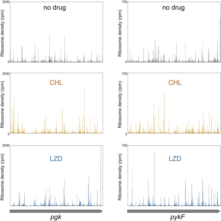
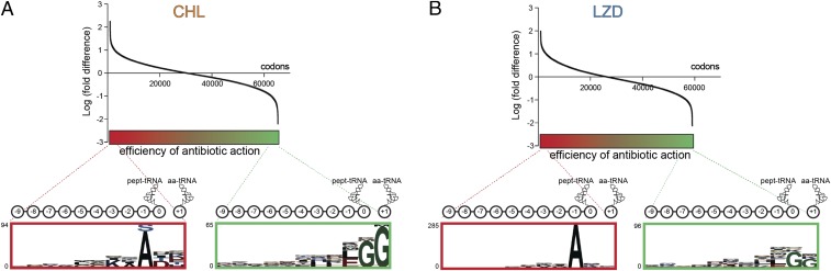
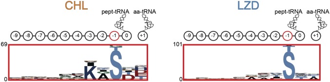
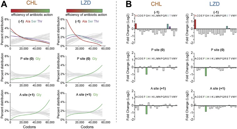
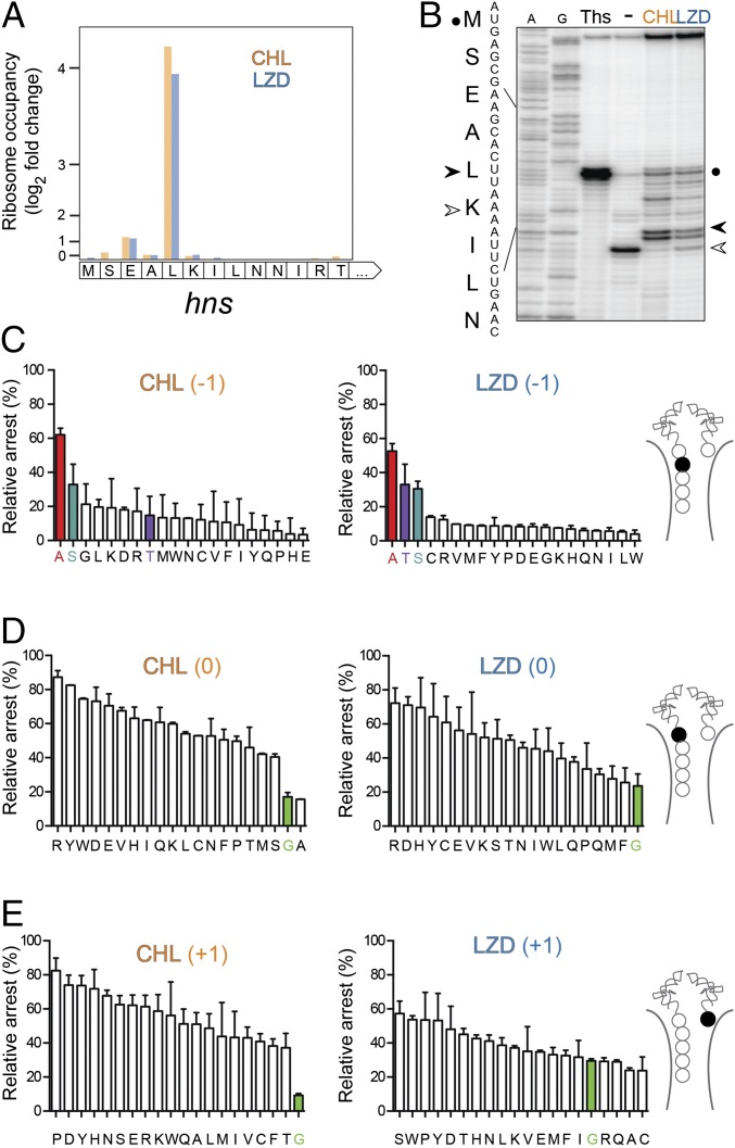
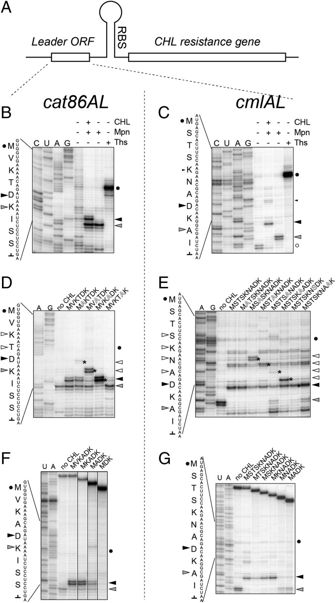
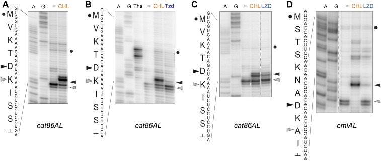
The first broad-spectrum antibiotic chloramphenicol and one of the newest clinically important antibacterials, linezolid, inhibit protein synthesis by targeting the peptidyl transferase center of the bacterial ribosome. Because antibiotic binding should prevent the placement of aminoacyl-tRNA in the catalytic site, it is commonly assumed that these drugs are universal inhibitors of peptidyl transfer and should readily block the formation of every peptide bond. However, our in vitro experiments showed that chloramphenicol and linezolid stall ribosomes at specific mRNA locations. Treatment of bacterial cells with high concentrations of these antibiotics leads to preferential arrest of translation at defined sites, resulting in redistribution of the ribosomes on mRNA. Antibiotic-mediated inhibition of protein synthesis is most efficient when the nascent peptide in the ribosome carries an alanine residue and, to a lesser extent, serine or threonine in its penultimate position. In contrast, the inhibitory action of the drugs is counteracted by glycine when it is either at the nascent-chain C terminus or at the incoming aminoacyl-tRNA. The context-specific action of chloramphenicol illuminates the operation of the mechanism of inducible resistance that relies on programmed drug-induced translation arrest. In addition, our findings expose the functional interplay between the nascent chain and the peptidyl transferase center.

Keywords: antibiotics; nascent peptide; oxazolidinones; protein synthesis; ribosome.


Conflict of interest statement

The research in the laboratory was supported by, among other sources, grants from the pharmaceutical companies Melinta Therapeutics and Cempra Pharmaceuticals.

Figures

Fig. 1.
Fig. 1.
The binding site of CHL and LZD in the peptidyl transferase center of the 50S ribosomal subunit. (Top) The chemical structures of CHL and LZD. (Bottom) When bound to the large ribosomal subunit in the PTC A site, the molecules of CHL (orange) and LZD (blue) would sterically clash with the aminoacyl moiety of the aminoacyl-tRNA (yellow). P-site–bound peptidyl-tRNA esterified with the nascent peptide chain is purple. The images were prepared based on the structures with the Protein Data Bank ID codes 4V7T, 3DLL, and 3J5L (10, 20, 46).
Fig. S1.
Fig. S1.
Ribosome stalling induced by CHL or LZD during in vitro translation. (A) Translation arrest within the E. coli genes osmC, cspA, and mipA was analyzed by toeprinting. The bands representing CHL-induced arrest are marked by orange dots; LZD-induced arrest sites are indicated by blue dots. The control antibiotic clindamycin (Cld) arrests translation at the initiation codon (purple dots). The nucleotide sequences of the relevant genes segments and the encoded amino acids are shown. A- and G-specific sequencing lanes are indicated. CHL and LZD were present in the reactions at the final concentrations of 200 µM and 1 mM, respectively. (B) A list of the CHL- or LZD-induced arrest sites identified by toeprinting analysis in a number of bacterial genes. The sites are aligned according to the codon located in the A site of the arrested ribosome. The amino acids in the P (0) and A (+1) sites, and the penultimate position (−1) of the nascent chain are boxed. The three amino acids preceding the penultimate position (nascent-chain positions −2 to −4) are also shown. Arrowheads indicate those sites of drug-induced arrest where Ala, Ser, or Thr are present in the penultimate position of the nascent peptide (see Fig. 2 in the main text and Fig. S4).
Fig. S2.
Fig. S2.
Time dependence of translation inhibition by CHL or LZD. Antibiotic hypersusceptible E. coli cells growing in defined medium lacking methionine were exposed to a 100-fold excess over the minimal inhibitory concentration of the drugs for varying time periods before addition of l-[35S]methionine. After 1 min of addition of methionine, the incorporation of radioactivity into the trichloracetic acid-precipitable material was determined by scintillation counting.
Fig. S3.
Fig. S3.
CHL and LZD cause redistribution of ribosomes during translation of E. coli genes. Distribution of ribosomes along the two sample genes (pgk on the panels on the left side and pykF genes on the panels on the right side) in the absence (“no drug”) or presence of CHL or LZD. Exponentially growing E. coli cells were exposed for 2.5 min to 100-fold MIC of CHL (100 µg/mL) or LZD (800 µg/mL), and distribution of ribosomes along actively translated genes was analyzed by ribosome profiling. See Materials and Methods for experimental details.
Fig. 2.
Fig. 2.
Context specificity of the action of CHL and LZD in vivo. (A and B) Ranking of genomic sites according to the relative fold difference in the ribosome occupancy in cells treated with CHL (A) or LZD (B) vs. the untreated control. The higher relative fold difference values (left side of the spectrum, colored in red) correspond to the sites of more pronounced inhibition of translation by the antibiotics. The low fold difference values (right side of the spectrum, shown in green) represent the sites where antibiotics were least efficient. The magnified panels at the Bottom show the pLogo analysis of amino acid bias within the top 1,000 (red frame) and bottom 1,000 (green frame) sites of action of CHL or LZD. The 10 C-terminal residues of the nascent chains (marked as 0 to −9 of pept-tRNA) and the incoming amino acids (marked as +1, aa-tRNA) in these sites are indicated in the cartoons. The y axes of the pLogo panels correspond to the odds of the binomial probability (in logarithmic scale) of occurrence of specific amino acids at the defined positions (47).
Fig. S4.
Fig. S4.
Presence of Ser or Thr residues at the penultimate position of the nascent chain contributes to CHL- and LZD-translation arrest. pLogo analysis of amino acid bias at the top 1,000 sites of CHL and LZD action after elimination of sites containing an Ala codon in the −1 position. The cartoons above the logos represent the 10 C-terminal nascent-chain residues (0, which is the P site, to −9) and the amino acid at the +1 position (aa-tRNA).
Fig. S5.
Fig. S5.
The efficiency of CHL- or LZD-induced translation arrest correlates with the presence of Ala, Ser, or Thr in the penultimate position of the nascent chain and countercorrelates with the presence of Gly in the P or A site. (A, graphs at Top) Occurrence of Ala, Ser, or Thr in the penultimate position (−1) of the nascent chain along the spectrum of the analyzed sites arranged according to the efficiency of antibiotic action (see SI Materials and Methods for detail). (Middle and Bottom) Occurrence of Gly in the P site (position 0) (Middle) or A site (position +1) (Bottom) of the drug-stalled ribosome. The colored bars at the Top mark all of the analyzed sites arranged as in Fig. 2 in the main text according to the efficiency of antibiotic action: from the sites of the strongest drug-induced arrest on the Left (red) to the sites of the least pronounced arrest on the Right (green). (B) Analysis of the cumulative drug-induced changes in the ribosome density at all of the analyzed sites according to the nature of amino acid in the penultimate position (−1) of the nascent chain (Top), P site (0) (Middle), or A site (+1) (Bottom).
Fig. 3.
Fig. 3.
Amino acid residues of the PTC donor and acceptor substrates influence antibiotic action. (A) Changes in the ribosome occupancy of the first codons of the hns gene in cells treated with CHL or LZD compared with that in the untreated cell culture. (B) In vitro toeprinting analysis of CHL- or LZD-induced ribosome stalling close to the 5′-end of the hns gene. The control antibiotic thiostrepton (Ths) was used to arrest translation at the start codon (black circle). The prominent CHL- and LZD-induced arrest site at the Leu5 codon of the gene is indicated by a black arrowhead. Due to the presence of the Ile-tRNA synthetase inhibitor mupirocin in all of the samples, the ribosomes that were not arrested by CHL or LZD at the hns Leu5 codon, were “caught” at the following Lys6 codon (open arrowhead). A- and G-specific sequencing lanes are indicated. The sequence of the first nine codons of the hns gene and the encoded amino acids are indicated on the side of the gel. (C–E) The effect of mutagenizing (C) the Ala4 (position −1), (D) Leu5 (position 0), or (E) Lys6 (position +1) codons of hns on CHL- or LZD-induced translation arrest. The cartoons showing the PTC region of the CHL- or LZD-stalled ribosomal complexes highlight the mutagenized amino acid residue (filled spheres) in each set. The efficiency of antibiotic-mediated arrest (“relative arrest”) was calculated by comparing the fraction of arrested ribosomes at codon 5 (estimated from the intensity of the CHL- or LZD-specific toeprint bands) with the fraction of ribosomes trapped at codon 6 (calculated from the intensity of the mupirocin-specific toeprint bands) during the translation of the WT or mutant hns templates (see B for reference). For the templates where the specified codon was replaced with an Ile codon, the Ile7 codon was mutated to Thr and borrelidin (and inhibitor of Thr-RS) was used instead of mupirocin. The bars representing H-NS mutants with Ala, Ser, or Thr in the penultimate peptide position are highlighted in red; those corresponding to the mutants with Gly in the P or A sites are highlighted in green. The error bars show deviation from the mean in two independent experiments.
Fig. 4.
Fig. 4.
CHL-induced arrest at the leader ORFs of inducible resistance genes. (A) The general organization of inducible CHL resistance genes. In the absence of antibiotic, the leader ORF is constitutively translated, but the expression of the resistance cistron is attenuated because its ribosome-binding site (RBS) is sequestered in the mRNA secondary structure. In the presence of CHL, translation of the leader ORF is arrested at a specific internal codon. Ribosome stalling mediates rearrangement of the mRNA structure resulting in activation of expression of the resistance gene. (B and C) CHL-induced translation arrest at the fifth codon of the cat86A leader ORF (B) or the eighth codon of the cmlA leader ORF (C). The control antibiotic thiostrepton (Ths) stalls translation at the start codon (black circle). A black triangle indicates the toeprint band representing CHL-induced programmed translation arrest. The gray triangle points to the band produced by ribosomes stalled before the “hungry” Ile codon [because of the presence in the reaction of mupirocin (Mpn), an Ile-tRNA synthetase inhibitor]. The band produced by ribosomes paused during termination of the cmlAL translation is marked with an open circle. (D and E) Alanine-scanning mutagenesis of the cat86AL (D) or cmlAL (E) alters the location of CHL-mediated translation arrest. As in B and C, the translation initiation site is marked by a circle, the site of the programmed CHL-induced arrest is shown by a black triangle, and the “Mpn band” is indicated by a gray triangle. The new sites of arrest, which appear due to the presence of new Ala residues in the mutant ORFs, are marked by the asterisks on the gel and are indicated by the open triangles. (F and G) N-terminal truncations of the cat86AL (F) or cmlAL (G) and their impact on the efficiency of CHL-induced translation arrest. The start codon, CHL band, and mupirocin band are indicated by a circle, black triangle, and gray triangle, respectively.
Fig. S6.
Fig. S6.
Drug-induced translation arrest within the leader ORFs of CHL resistance genes. Toeprinting analysis of drug-dependent ribosome stalling within the leader ORFs of (A–C) cat86AL and (D) cmlAL CHL resistance genes. All of the reactions loaded onto gels shown in A–D contained mupirocin, an inhibitor of Ile-tRNA synthetase. In all of the panels, the site of CHL-induced arrest is indicated by a black triangle, the “mupirocin band” is indicated by a gray triangle, and the start codon is marked by a black dot. (A) CHL-dependent arrest of translation of the cat86AL ORF driven by Bacillus subtilis ribosomes. (B and C) Oxazolidinone antibiotics (B) tedizolid (Tzd) and (C) LZD stall the E. coli ribosome within the cat86AL gene at the site of programmed CHL-induced arrest. (D) LZD arrests the E. coli ribosome at the site of CHL-induced arrest within the cmlAL leader ORF.
Fig. S7.
Fig. S7.
Extended context for CHL and LZD action. (A–D) The cumulative drug-induced changes in the ribosome density at all of the analyzed sites with (A and C) Ala (−1) and various P- and A-site codons, (C) Gly in the P site and various −1 codons or (D) Gly in the A-site and various −1 codons. The bars representing sites with Ala in the penultimate position of the nascent peptide and Gly in the P site are indicated by arrows. (E) The presence of C-terminal Asp and acceptor Lys stimulates CHL action at the sites with Ala or Thr in position −1. pLogo analysis of amino acids of the top 500 sites of CHL-induced translation arrest that contain exclusively Ala or Thr in position −1. Note the preferential presence of Asp in position 0 (P site).
Fig. S8.
Fig. S8.
CHL and LZD fail to inhibit formation of the first peptide bond and therefore cause redistribution of ribosomes from the start codons of the genes. Metagene analysis of the relative ribosome occupancy of the first 30 codons of the actively translated genes in the untreated E. coli cells (gray line) or cells treated with CHL (orange line) or LZD (blue line). The relative ribosome occupancy is calculated as the fraction (percentage) of cumulative ribosome density at a specific codon relative to the total ribosome density at the 105-nt-long region surrounding the initiation codon (positions −15 to +90 relative to the first nucleotide of the start codon). Note the decrease of the start codon occupancy in the CHL- and LZD-treated samples.

Comment in

References

    1. Beringer M, Rodnina MV. The ribosomal peptidyl transferase. Mol Cell. 2007;26(3):311–321. - PubMed
    1. Weinger JS, Parnell KM, Dorner S, Green R, Strobel SA. Substrate-assisted catalysis of peptide bond formation by the ribosome. Nat Struct Mol Biol. 2004;11(11):1101–1106. - PubMed
    1. Erlacher MD, et al. Chemical engineering of the peptidyl transferase center reveals an important role of the 2′-hydroxyl group of A2451. Nucleic Acids Res. 2005;33(5):1618–1627. - PMC - PubMed
    1. Polikanov YS, Steitz TA, Innis CA. A proton wire to couple aminoacyl-tRNA accommodation and peptide-bond formation on the ribosome. Nat Struct Mol Biol. 2014;21(9):787–793. - PMC - PubMed
    1. Polacek N, Gaynor M, Yassin A, Mankin AS. Ribosomal peptidyl transferase can withstand mutations at the putative catalytic nucleotide. Nature. 2001;411(6836):498–501. - PubMed

Publication types

MeSH terms