

Structure and assembly model for the Trypanosoma cruzi 60S ribosomal subunit
- PMID: 27791004
- PMCID: PMC5087005
- DOI: 10.1073/pnas.1614594113
Structure and assembly model for the Trypanosoma cruzi 60S ribosomal subunit
Abstract
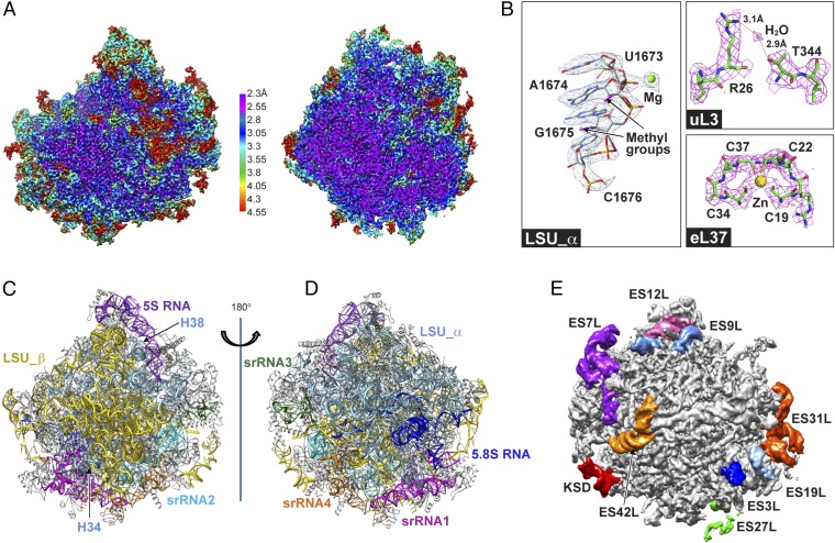
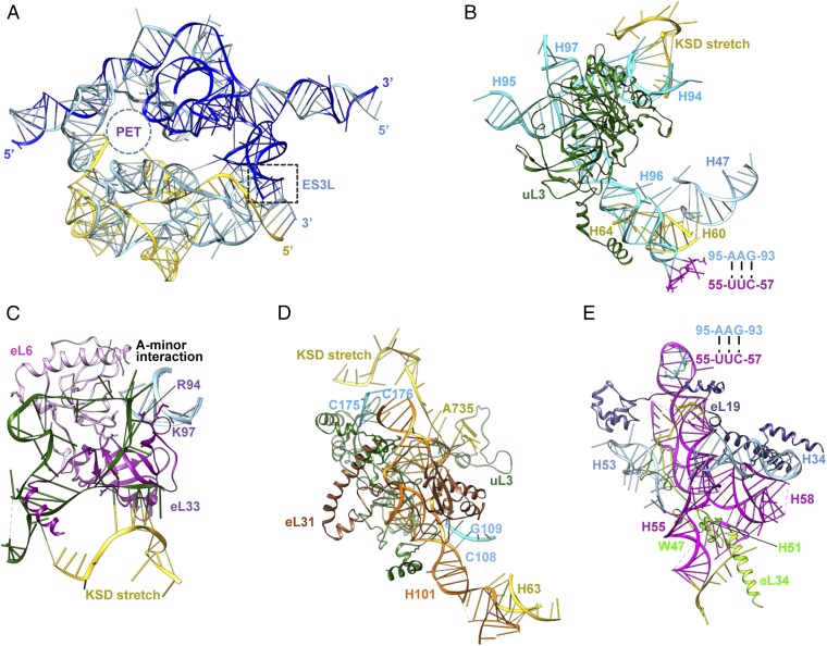
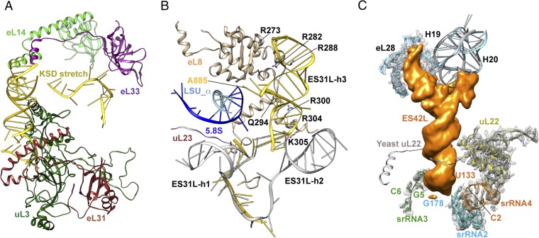
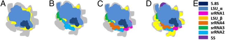
Ribosomes of trypanosomatids, a family of protozoan parasites causing debilitating human diseases, possess multiply fragmented rRNAs that together are analogous to 28S rRNA, unusually large rRNA expansion segments, and r-protein variations compared with other eukaryotic ribosomes. To investigate the architecture of the trypanosomatid ribosomes, we determined the 2.5-Å structure of the Trypanosoma cruzi ribosome large subunit by single-particle cryo-EM. Examination of this structure and comparative analysis of the yeast ribosomal assembly pathway allowed us to develop a stepwise assembly model for the eight pieces of the large subunit rRNAs and a number of ancillary "glue" proteins. This model can be applied to the characterization of Trypanosoma brucei and Leishmania spp. ribosomes as well. Together with other details, our atomic-level structure may provide a foundation for structure-based design of antitrypanosome drugs.
Keywords: Trypanosoma cruzi; antitrypanosome drug design; biogenesis; multiply fragmented rRNA; ribosome structure.
Conflict of interest statement
The authors declare no conflict of interest.
Figures
Comment in
-
Ribosome Assembly in Trypanosomatids: A Novel Therapeutic Target.Trends Parasitol. 2017 Apr;33(4):256-257. doi: 10.1016/j.pt.2016.12.003. Epub 2016 Dec 14. Trends Parasitol. 2017. PMID: 27988096 Free PMC article.
References
-
- Schmeing TM, Ramakrishnan V. What recent ribosome structures have revealed about the mechanism of translation. Nature. 2009;461(7268):1234–1242. - PubMed
-
- Yusupova G, Yusupov M. High-resolution structure of the eukaryotic 80S ribosome. Annu Rev Biochem. 2014;83:467–486. - PubMed
-
- El-Sayed NM, et al. The genome sequence of Trypanosoma cruzi, etiologic agent of Chagas disease. Science. 2005;309(5733):409–415. - PubMed
-
- El-Sayed NM, et al. Comparative genomics of trypanosomatid parasitic protozoa. Science. 2005;309(5733):404–409. - PubMed
Publication types
MeSH terms
Substances
Associated data
- Actions
Grants and funding
LinkOut - more resources
Full Text Sources
Other Literature Sources
Miscellaneous
