. 2016 Nov 1;113(44):12467-12472.
doi: 10.1073/pnas.1608110113. Epub 2016 Oct 17.

Kinetics of DNA uptake during transformation provide evidence for a translocation ratchet mechanism

Affiliations

Kinetics of DNA uptake during transformation provide evidence for a translocation ratchet mechanism

Christof Hepp et al. Proc Natl Acad Sci U S A. .

Abstract

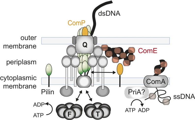
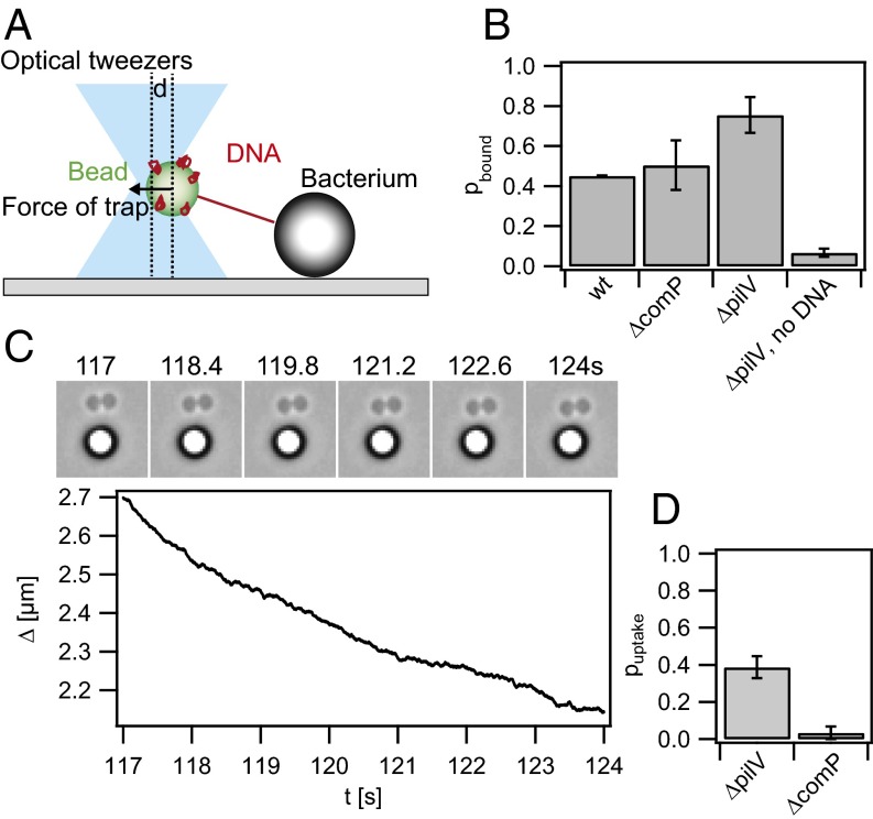
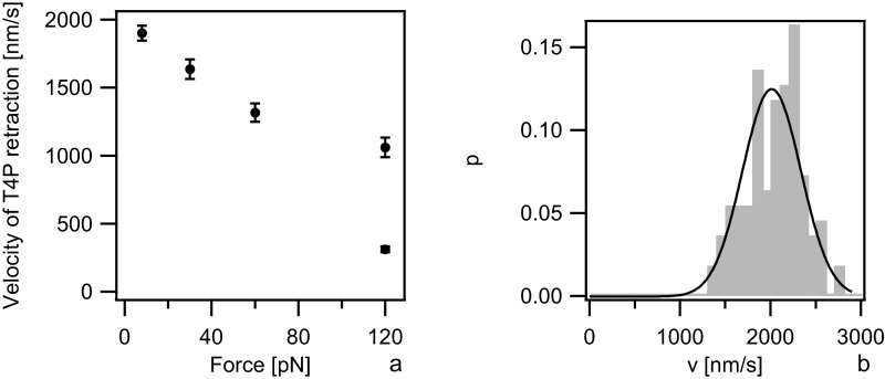
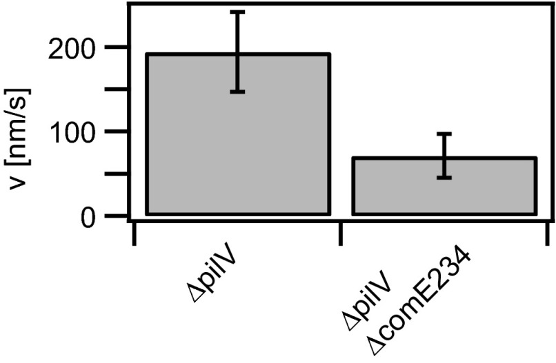
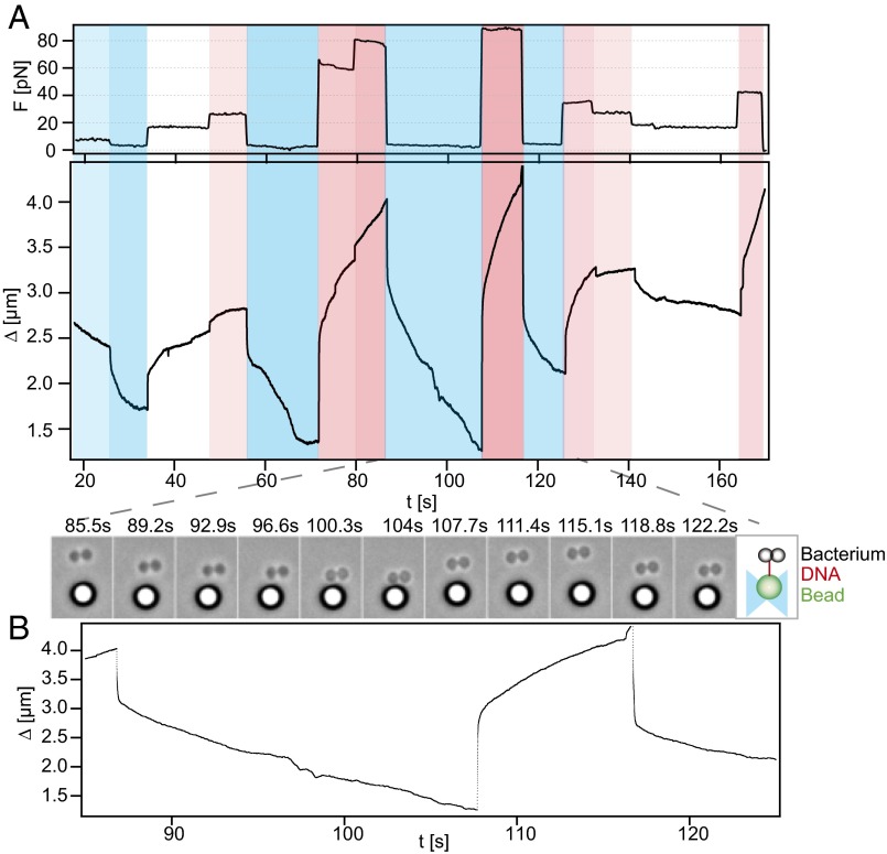
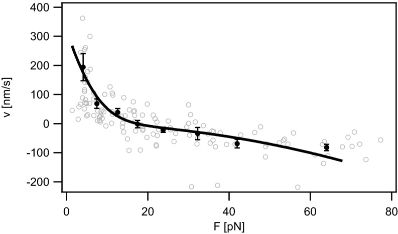
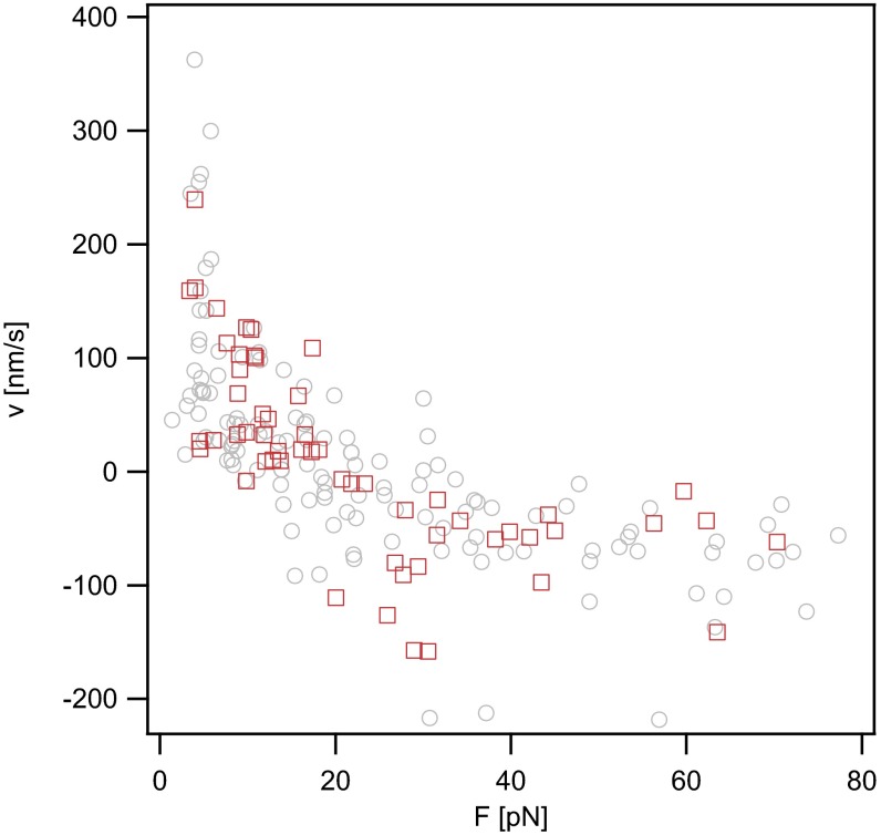
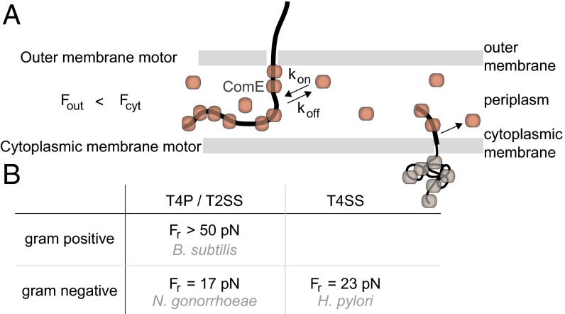
Horizontal gene transfer can speed up adaptive evolution and support chromosomal DNA repair. A particularly widespread mechanism of gene transfer is transformation. The initial step to transformation, namely the uptake of DNA from the environment, is supported by the type IV pilus system in most species. However, the molecular mechanism of DNA uptake remains elusive. Here, we used single-molecule techniques for characterizing the force-dependent velocity of DNA uptake by Neisseria gonorrhoeae We found that the DNA uptake velocity depends on the concentration of the periplasmic DNA-binding protein ComE, indicating that ComE is directly involved in the uptake process. The velocity-force relation of DNA uptake is in very good agreement with a translocation ratchet model where binding of chaperones in the periplasm biases DNA diffusion through a membrane pore in the direction of uptake. The model yields a speed of DNA uptake of 900 bp⋅s-1 and a reversal force of 17 pN. Moreover, by comparing the velocity-force relation of DNA uptake and type IV pilus retraction, we can exclude pilus retraction as a mechanism for DNA uptake. In conclusion, our data strongly support the model of a translocation ratchet with ComE acting as a ratcheting chaperone.

Keywords: bacterial transformation; gene transfer; molecular motor; translocation ratchet.


Conflict of interest statement

The authors declare no conflict of interest.

Figures

Fig. S1.
Fig. S1.
Putative molecular picture of DNA uptake machine. The proteins forming the T4P system are required for DNA uptake. They include the major pilin subunits PilE (green), the cytoplasmic ATPases PilF (F) and PilT (T), the PilQ proteins forming the outer membrane export/putative DNA import channel (Q), and various structural proteins depicted in gray. The minor pilin ComP and the periplasmic protein ComE are required for DNA uptake into the periplasm. The inner membrane pore or channel formed by ComA is essential for transport from the periplasm to the cytoplasm by an unknown mechanism. Only a single DNA strand enters the cytoplasm.
Fig. 1.
Fig. 1.
Probability of DNA binding and uptake. (A) Scheme of the experimental setup. A bead coated with DNA is trapped in an optical trap and placed close to a bacterium. (B) A binding event is defined as a deflection of the bead from the center of the trap exceeding 20 pN while the bead was moved away from the bacterium. Shown are binding probabilities of WT (Ng003), ΔcomP (Ng031), ΔpilV (Ng005), and ΔpilV (Ng005) with plain beads. (C) Typical DNA uptake event at F = 4 pN. (C, Upper) Time lapse; (C, Lower) distance Δ between bacterium and bead as a function of time. (D) Probability of DNA uptake subsequent to binding with ΔpilV and ΔcomP.
Fig. S2.
Fig. S2.
DNA construct. The DNA fragment attached to the beads is a 10-kbp PCR derivative of λ DNA. On one end three biotin residues are introduced as part of the PCR primer, one covalently attached to the 5′ end and two more covalently attached to thymines within the primer sequence. On the opposite end a DUS is introduced to face away from the bead and to be recognized for DNA uptake.
Fig. S3.
Fig. S3.
Velocity vs. force relationship of T4P retraction. The speed of T4P retraction was determined by placing uncoated beads close to a bacterium at a force clamped to different values. Pili bound to the bead and the speed was measured during retraction as described in ref. . (A) Average speed of T4P retraction at different forces of WT gonococci. Data are taken from ref. . (B) Distribution of T4P retraction speeds at F = 8 pN for ΔpilV (Ng005).
Fig. 2.
Fig. 2.
The uptake velocity depends on the concentration of ComE. Shown is average speed of DNA uptake for ΔpilV (Ng005, n = 22) and ΔpilV ΔcomE234 (Ng052, n = 10) at F = 4 pN.
Fig. 3.
Fig. 3.
DNA uptake is reversible upon application of force. Typical time series of DNA uptake and extractions (Ng005) are shown. (A, Upper) Force; (A, Lower) distance between bead and bacterium Δ as a function of time. For presentation, the data were down-sampled to 1 Hz. Colors guide the eye. Events marked in blue are retraction, and those in red are extraction. More opaque colors signify stronger forces. At Δ < 1.3 µm, tracking was not reliable because bacterium and bead were in contact. (B, Lower) Zoom-in to the distance between bead and bacterium. (B, Upper) Corresponding time-lapse microscopic images.
Fig. 4.
Fig. 4.
Velocity vs. force relationship of DNA uptake (Ng005). Shaded open circles, raw data; solid circles, data binned over 6–25 data points. Error bars: SEM. Solid line: fit to Eq. 1 with a = (1.6 ± 0.3) nm, D = (250 ± 100) nm2⋅s−1, K = 0.0012 ± 0.0008.
Fig. S4.
Fig. S4.
Velocity vs. force relationship of DNA uptake for ΔcomA. Gray open circles, ΔpilV recAind (Ng005); red open squares, ΔcomA ΔpilV recAind (Ng054).
Fig. 5.
Fig. 5.
Force generation by cytoplasmic and outer membrane motors. (A) Hypothetical model for DNA transport through the Gram-negative cell envelope. A translocation ratchet drives uptake of DNA from the environment into the periplasm by reversible ComE binding. For transport across the inner membrane to occur, ComE must unbind. B. subtilis data suggest that in agreement with this prerequisite, the force generated by the cytoplasmic motor is considerably larger. (B) Reversal forces for B. subtilis (34), N. gonorrhoeae, and H. pylori (26). T4P/T2SS, type 4 pilus/type 2 secretion system; T4SS, type 4 secretion system.

References

    1. Neupert W, Brunner M. The protein import motor of mitochondria. Nat Rev Mol Cell Biol. 2002;3(8):555–565. - PubMed
    1. Neupert W. A perspective on transport of proteins into mitochondria: A myriad of open questions. J Mol Biol. 2015;427(6 Pt A):1135–1158. - PubMed
    1. Liu S, et al. A viral packaging motor varies its DNA rotation and step size to preserve subunit coordination as the capsid fills. Cell. 2014;157(3):702–713. - PMC - PubMed
    1. Inamdar MM, Gelbart WM, Phillips R. Dynamics of DNA ejection from bacteriophage. Biophys J. 2006;91(2):411–420. - PMC - PubMed
    1. Allemand JF, Maier B. Bacterial translocation motors investigated by single molecule techniques. FEMS Microbiol Rev. 2009;33(3):593–610. - PubMed

Publication types

LinkOut - more resources