. 2016 Nov 8;113(45):12797-12802.
doi: 10.1073/pnas.1608821113. Epub 2016 Oct 24.

Synthetically derived bat influenza A-like viruses reveal a cell type- but not species-specific tropism

Affiliations

Synthetically derived bat influenza A-like viruses reveal a cell type- but not species-specific tropism

Étori Aguiar Moreira et al. Proc Natl Acad Sci U S A. .

Abstract

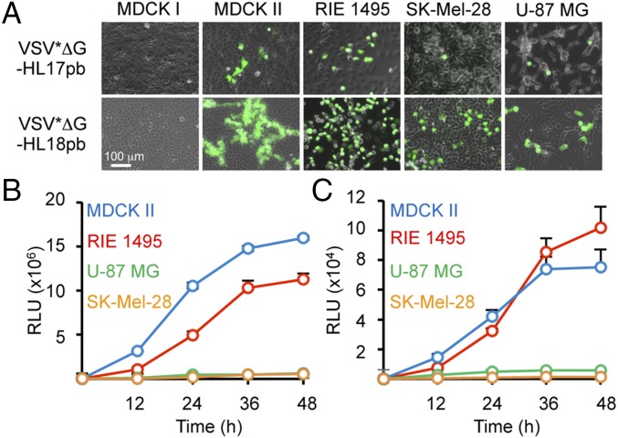
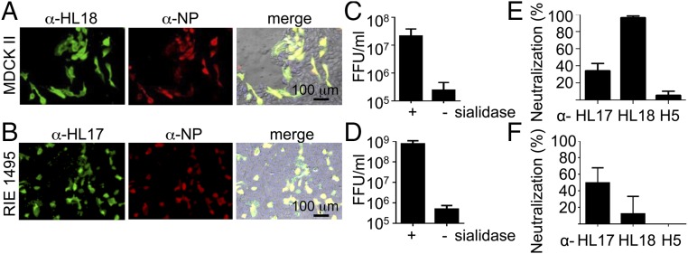
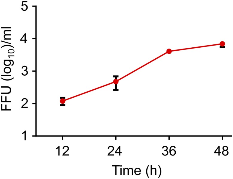
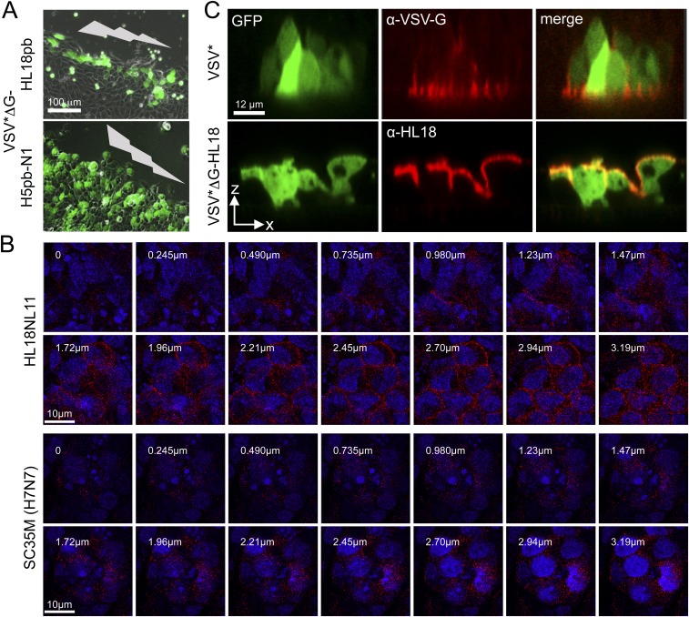
Two novel influenza A-like viral genome sequences have recently been identified in Central and South American fruit bats and provisionally designated "HL17NL10" and "HL18NL11." All efforts to isolate infectious virus from bats or to generate these viruses by reverse genetics have failed to date. Recombinant vesicular stomatitis virus (VSV) encoding the hemagglutinin-like envelope glycoproteins HL17 or HL18 in place of the VSV glycoprotein were generated to identify cell lines that are susceptible to bat influenza A-like virus entry. More than 30 cell lines derived from various species were screened but only a few cell lines were found to be susceptible, including Madin-Darby canine kidney type II (MDCK II) cells. The identification of cell lines susceptible to VSV chimeras allowed us to recover recombinant HL17NL10 and HL18NL11 viruses from synthetic DNA. Both influenza A-like viruses established a productive infection in MDCK II cells; however, HL18NL11 replicated more efficiently than HL17NL10 in this cell line. Unlike conventional influenza A viruses, bat influenza A-like viruses started the infection preferentially at the basolateral membrane of polarized MDCK II cells; however, similar to conventional influenza A viruses, bat influenza A-like viruses were released primarily from the apical site. The ability of HL18NL11 or HL17NL10 viruses to infect canine and human cells might reflect a zoonotic potential of these recently identified bat viruses.

Keywords: bat; chiroptera; emerging viruses; influenza; orthomyxoviridae.


Conflict of interest statement

The authors declare no conflict of interest.

Figures

Fig. 1.
Fig. 1.
Chimeric VSV encoding the HL proteins of bat influenza A-like viruses. (A) Genome maps of recombinant VSV vectors. sNLuc, secreted Nano luciferase. (B) Proteolytic cleavage sites of HL17, HL18, and modified versions containing a polybasic (pb) sequence motif. (C) Indicated cell lines were infected with the indicated viruses at an m.o.i. of 0.01 and maintained in serum-free medium either in the absence (-T) or presence (+T) of trypsin. Virus spreading was detected 20 h after infection by monitoring GFP-mediated fluorescence. (D) Subconfluent MDCK II cells were infected with either VSV*ΔG-HL17, VSV*ΔG-HL17pb, VSV*ΔG-HL18, or VSV*ΔG-HL18pb using an m.o.i. of 0.01. Subsequently, input virus was neutralized by incubating the cells with a monoclonal antibody directed to the VSV-G protein. The cells were maintained in serum-free medium in the absence or presence of trypsin as indicated. At the indicated times, aliquots of cell-culture supernatant were collected and titrated on MDCK II cells. Error bars indicate the mean and SD of three independent experiments. (E) Replication kinetics (m.o.i. of 0.01) of VSV*ΔG-NL11 (green circles) and VSVΔG-HL18-NL11 (violet circles) on MDCK II cells in the presence of trypsin. For comparison, replication of VSV*ΔG-HL18 (red circles) from Fig. 1D is also shown. (F) Neutralization of chimeric viruses by immune sera. VSV*ΔG-HL17 and VSV*ΔG-HL18 (100 FFU) were incubated with chicken immune sera (1:100) raised against either HL17 or HL18 or without serum (w/o) and subsequently added to MDCK II cells. Infected cells were detected 20 h after infection by GFP fluorescence.
Fig. S1.
Fig. S1.
Analysis of recombinant HL protein expression. (A) Indirect immunofluorescence analysis of Vero cells at 6 h post infection (p.i.) with either VSV*ΔG-HL17 or VSV*ΔG-HL18. Immune sera directed to either HL17 or HL18 were used for immunostaining of the cells. Bound antibodies were detected with Alexa 546-labeled secondary antibodies (red). GFP-mediated fluorescence appears in green. (B) Western blot analysis of recombinant VSV particles. Vero cells were infected with the indicated viruses (m.o.i. of 5) and maintained for 24 h in serum-free medium in the presence or absence of trypsin. The virus particles released into the cell culture supernatant were pelleted by ultracentrifugation, separated by SDS/PAGE under reducing conditions, and transferred to nitrocellulose membranes. The proteins were detected by using anti-HL17 and anti-HL18 immune sera, respectively. The positions of molecular mass markers (in kilodaltons) are shown on the left hand side; the positions of the HL subunits on the right hand side. Asterisks indicate protein bands that are not related to HL proteins.
Fig. S2.
Fig. S2.
Analysis of pH-dependent membrane fusion activity of recombinant HL proteins. (A) MDCK II and Vero cells were infected with VSV*ΔG vectors (m.o.i. of 1) expressing the indicated HL and HA proteins containing either a monobasic or polybasic (pb) cleavage site. At 6 h p.i., the cells were treated (+T) or nontreated (-T) with trypsin and shortly exposed to pH 5.2. The cells were subsequently incubated with normal growth medium for 90 min and then fixed with formalin. The nuclei were stained with DAPI (blue). Infected cells express GFP (green). The pH-dependent membrane fusion activity is indicated by the formation of polykaryons. (B) Determination of the pH threshold for induction of membrane fusion by HL17pb, HL18pb, and H5pb. MDCK II cells were infected with recombinant VSV*ΔG vectors expressing the indicated HL and HA proteins and exposed to the indicated pH values. The cells were processed as described above.
Fig. S3.
Fig. S3.
Neutralization of chimeric VSV using specific immune sera. The chimeric virus VSVΔG-HL17pb-sNLuc (A) and VSVΔG-HL18pb-sNLuc (B) were incubated with serially diluted immune sera directed to HL17, HL18, or H5 and then added to MDCK II cells. Luciferase activity was determined in cell culture supernatants at 20 h p.i. Percentage luciferase activity is shown. Activity of Nano luciferase produced by cells following infection with virus that had been treated with preimmune serum served as reference and was set to 100%.
Fig. 2.
Fig. 2.
Susceptibility of mammalian cell lines to infection with chimeric VSV. (A) The indicated cell lines were inoculated with VSV*ΔG-HL17pb or VSV*ΔG-HL18pb by using an m.o.i. of 1. At 20 h p.i., GFP expression was monitored. (B and C) The indicated cell lines were infected with either VSVΔG-HL18pb-sNLuc (B) or VSVΔG-HL17pb-sNLuc (C) by using an m.o.i. of 0.01. At the indicated times, the activity of the Nano luciferase reporter secreted into the cell-culture supernatant was determined. Emission of luminescence is expressed as relative light units (RLU). Error bars indicate the mean and SD of three independent experiments.
Fig. 3.
Fig. 3.
Generation of infectious bat influenza A-like viruses. Canine MDCK II cells (A) or RIE 1495 cells (B) were infected with either HL18NL11 or HL17NL10 both harboring an authentic proteolytic cleavage site by using an m.o.i. of 0.01. After 2 d in the presence of trypsin, the cells were fixed and stained with antibodies directed to NP and either HL18 or HL17. A merged image including phase contrast and diamidino-2-phenylindole (DAPI; Sigma; 0.1 μg/mL in ethanol) staining is shown (Right). MDCK II cells (C) or RIE 1495 cells (D) were treated or left untreated with bacterial sialidase (200 mU/mL) for 1 h at 37 °C before infection with HL18NL11 or HL17NL10 (m.o.i. of 0.01), respectively. Two days p.i., the number of infectious particles in the cell culture supernatant was determined by a focus-forming unit assay using antibodies directed against HL18 or HL17 for immunostaining of infected cell foci. HL18NL11 (2,000 FFU) (E) or HL17NL10 (2,000 FFU) (F) were incubated for 1 h with immune sera raised against HL18, HL17, or H5 (serum dilution 1:800) and then added to RIE 1495 cells. Two days after inoculation, cells were stained with antibodies directed to either HL18 (E) or HL17 (F). Error bars indicate the mean and SD of three independent experiments.
Fig. S4.
Fig. S4.
Release of HL18NL11 from infected MDCK II cells. MDCK II cells were infected with HL18NL11 (m.o.i. of 0.001). At the indicated times, infectious virus released into the cell culture supernatant was titrated on RIE 1495 cells.
Fig. 4.
Fig. 4.
Entry and egress of chimeric VSV and HL18NL11 in polarized epithelial cells. (A) MDCK II cells grown on porous filter supports (pore size 0.4 μm) were infected with the indicated viruses from either the apical or basolateral site by using an m.o.i. of 10. GFP fluorescence was detected 20 h p.i. (B) Polarized filter-grown MDCK II cells were infected with HL18NL11 (m.o.i. of 0.3) at the basolateral or apical site. The number of infectious foci in the cell layer was determined 2 d p.i. Error bars indicate the mean and SD of two independent experiments. (C) Electron-microscopic analysis of MDCK II cells infected for 50 h with HL18NL11 (m.o.i. of 0.1). Ultrathin section showing pleomorphic virus particles released from the apical cell surface. (D) Localization of HL18 at the apical plasma membrane of polarized filter-grown MDCK II cells 48 h after infection with HL18NL11 (m.o.i. of 0.01). Dashed lines indicate locations of the x and y sections. (E) Cartoon depicting the directional entry and egress of bat influenza A-like viruses in polarized epithelial cells.
Fig. S5.
Fig. S5.
Directional virus entry and egress. (A) MDCK II cells were grown on cell culture dishes until a confluent monolayer was formed. The cells were scratched with a pipette tip (indicated by the gray lightning symbol) and infected with the indicated chimeric viruses. GFP expression by infected cells was recorded at 7 h p.i. (B) Serial sections of polarized filter-grown MDCK II cells grown on Transwell filter inserts (pore size 0.4 μm) and infected with either HL18NL11 (m.o.i. of 0.3) at the basolateral site for 48 h (Upper) or the conventional influenza A virus A/SC35M (H7N7) at an m.o.i. of 5 at the apical site for 8 h (Lower). HL18 and H7 were detected at the cell surface by HL18- and H7-specific antibodies, respectively. The indicated z scans were taken by using a confocal laser scanning microscope. Numbers indicate the distance from the basal cell border. (C) Localization of newly synthesized HL18 at the apical plasma membrane of polarized MDCK II cells after infection via the basolateral membrane with VSV* or VSV*ΔG-HL18 using an m.o.i. of 10. At 8 h p.i., HL18 was detected by indirect immunofluorescence (red fluorescence). Infected cells are indicated by GFP expression (green fluorescence). XZ scans were taken by using a confocal laser scanning microscope.

Similar articles

Cited by

References

    1. Jones KE, Purvis A, MacLarnon A, Bininda-Emonds OR, Simmons NB. A phylogenetic supertree of the bats (Mammalia: Chiroptera) Biol Rev Camb Philos Soc. 2002;77(2):223–259. - PubMed
    1. Li W, et al. Bats are natural reservoirs of SARS-like coronaviruses. Science. 2005;310(5748):676–679. - PubMed
    1. Turmelle AS, Olival KJ. Correlates of viral richness in bats (order Chiroptera) EcoHealth. 2009;6(4):522–539. - PMC - PubMed
    1. Smith I, Wang LF. Bats and their virome: An important source of emerging viruses capable of infecting humans. Curr Opin Virol. 2013;3(1):84–91. - PMC - PubMed
    1. Plowright RK, et al. Ecological dynamics of emerging bat virus spillover. Proc Biol Sci. 2015;282(1798):20142124. - PMC - PubMed

LinkOut - more resources