. 2016 Nov 1;113(44):12496-12501.
doi: 10.1073/pnas.1608959113. Epub 2016 Oct 17.

Extracellular RNA is transported from one generation to the next in Caenorhabditis elegans

Affiliations

Extracellular RNA is transported from one generation to the next in Caenorhabditis elegans

Julia Marré et al. Proc Natl Acad Sci U S A. .

Abstract

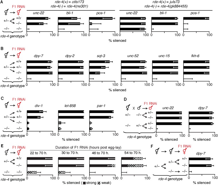
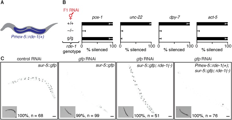
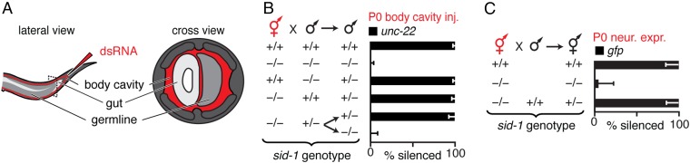
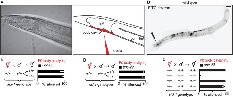
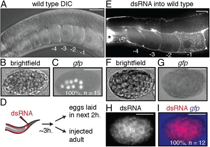
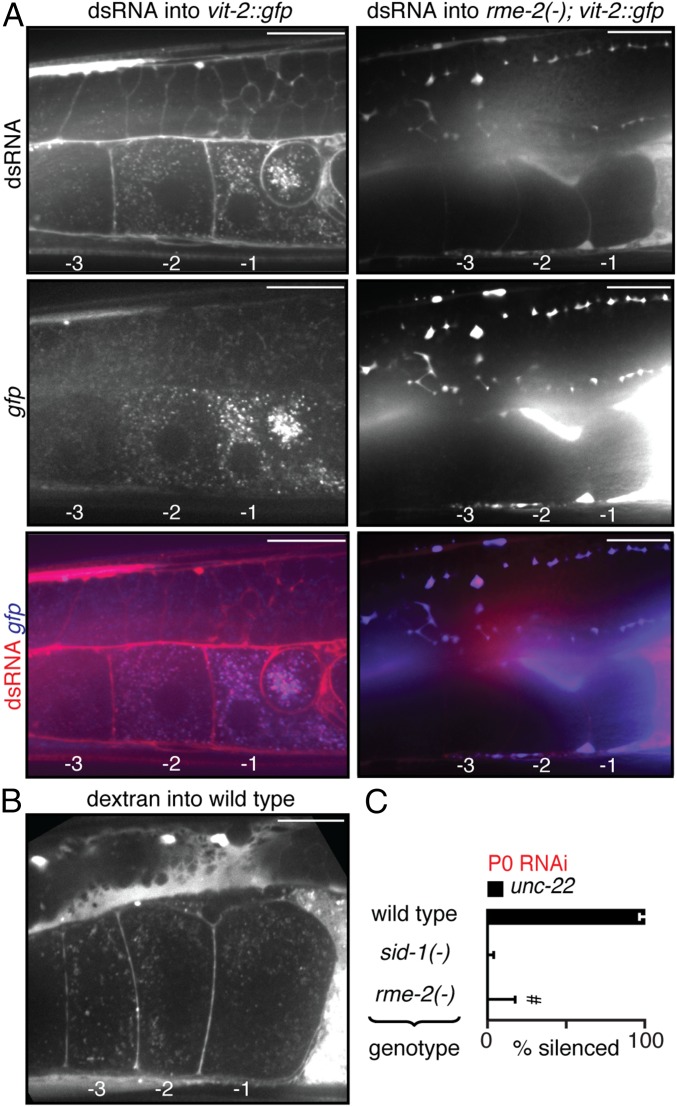
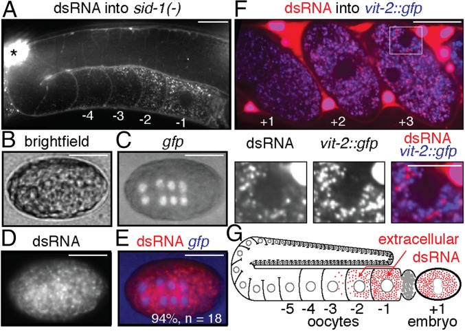
Experiences during the lifetime of an animal have been proposed to have consequences for subsequent generations. Although it is unclear how such intergenerational transfer of information occurs, RNAs found extracellularly in animals are candidate molecules that can transfer gene-specific regulatory information from one generation to the next because they can enter cells and regulate gene expression. In support of this idea, when double-stranded RNA (dsRNA) is introduced into some animals, the dsRNA can silence genes of matching sequence and the silencing can persist in progeny. Such persistent gene silencing is thought to result from sequence-specific interaction of the RNA within parents to generate chromatin modifications, DNA methylation, and/or secondary RNAs, which are then inherited by progeny. Here, we show that dsRNA can be directly transferred between generations in the worm Caenorhabditis elegans Intergenerational transfer of dsRNA occurs even in animals that lack any DNA of matching sequence, and dsRNA that reaches progeny can spread between cells to cause gene silencing. Surprisingly, extracellular dsRNA can also reach progeny without entry into the cytosol, presumably within intracellular vesicles. Fluorescently labeled dsRNA is imported from extracellular space into oocytes along with yolk and accumulates in punctate structures within embryos. Subsequent entry into the cytosol of early embryos causes gene silencing in progeny. These results demonstrate the transport of extracellular RNA from one generation to the next to regulate gene expression in an animal and thus suggest a mechanism for the transmission of experience-dependent effects between generations.

Keywords: circulating RNA; endocytosis; epigenetics; parental RNAi; transgenerational inheritance.


Conflict of interest statement

The authors declare no conflict of interest.

Figures

Fig. S1.
Fig. S1.
Published cases of silencing observed in self progeny when dsRNA against multicopy transgenes, single-copy transgenes, or endogenous genes was introduced outside the germline in hermaphrodites.
Fig. 1.
Fig. 1.
Ingested dsRNA or dsRNA-derived silencing signals can be transported to progeny through oocytes by parents that lack DNA of matching sequence. (A) Schematic of assay to assess silencing in progeny (F1) by parental ingestion of dsRNA (P0 RNAi, red). Also see Fig. S2. (B and C) Silencing of multicopy gfp transgenes in progeny by ingested dsRNA. (B) Robust silencing of Psur-5::sur-5::gfp in intestinal cells required parental ingestion of gfp-dsRNA during adulthood. L1 to L4, larval stages; yA, young adult. (C) Silencing of Pmyo-3::gfp in muscle cells after parental ingestion of gfp-dsRNA was detectable in all early progeny (0–12 h post-RNAi) but only in diminishing fractions of later progeny (12–34 h post-RNAi). (D) Males showing silencing of gfp (Pgtbp-1::gtbp-1::gfp) within the germline (sil.) did not transmit silencing to any cross-progeny (Right). Males fed control RNAi did not show any silencing. n/a, not applicable. (E) Silencing of a single-copy gfp transgene (Peft-3::gfp) in cross-progeny was detected in somatic cells even when only hermaphrodites (red) that lack gfp (none) ingested gfp-dsRNA (black bars). Error bars indicate 95% confidence interval (CI); L4-staged animals were assayed [n > 80 (B), n > 84 (C), n > 40 (D), n > 56 (E)]; and gray bars indicate silencing in progeny of animals that ingested control dsRNA (D and E).
Fig. S2.
Fig. S2.
Washing worms that ingested bacteria expressing dsRNA is sufficient to ensure that silencing in progeny is caused by inheritance of a silencing signal from parents and not by the ingestion of dsRNA by progeny. (A) Silencing (% silenced) of a multicopy gfp transgene (Psur-5::sur-5::gfp) in intestinal cells of progeny was detectable when the bacteria expressing gfp-dsRNA (P0 RNAi) were removed from parents by washing four times with buffer (4x wash) or by killing (bleaching P0 animals or placing animals on kanamycin plates). (B) A worm that was fed bacteria that express gfp-dsRNA and subsequently washed with buffer (washed P0 RNAi, black) was placed along with a worm that was not fed dsRNA but was marked with pharyngeal gfp expression (no RNAi, gray), and progeny from both worms were assessed for silencing of a multicopy gfp transgene (Pmyo-3::gfp) in muscle cells (Top). Silencing (% silenced) was detectable only in muscle cells of progeny from parents fed gfp-dsRNA (P0 gfp RNAi, black bar) (Bottom). (C) WT animals that were washed as in A, after being fed carbenicillin-resistant RNAi bacteria (washed P0 RNAi, black), were allowed to crawl on carbenicillin plates for 1 h before being cultured along with worms marked with a fluorescent marker, and the carbenicillin plates were incubated overnight to identify colonies generated by any residual bacteria that were not removed by the washes (RNAi bacteria colonies) (Left). Although washing parent worms fed P0 RNAi did not eliminate a few RNAi bacteria in some cases (5 of 15 plates had 0 colonies; 5 of 15 plates had 1 to 10 colonies; and 5 of 15 plates had 11 to 25 colonies), silencing (% silenced) of an endogenous gene (unc-22) was detectable only in progeny from parents fed unc-22-dsRNA (P0 unc-22 RNAi, black bars) and not in the cocultured fluorescently marked worms (no RNAi, gray) in all cases (Right). (D) Ingestion of bacteria that express gfp-dsRNA by animals that express a gfp transgene in intestinal cells (Pend-1::gfp, black circles) caused silencing in embryos held in utero. (E) Representative developed progeny from animals that ingested control dsRNA (Top) or from those that ingested gfp-dsRNA (Bottom) showing silencing in muscle cells (regions within square brackets) of a multicopy gfp transgene (Pmyo-3::gfp). (F) Table summarizing cases where silencing was observed in self progeny when hermaphrodites ingest dsRNA against multicopy transgenes, single-copy transgenes, or endogenous genes using the inheritance assay described above and in Materials and Methods. Error bars indicate 95% CI (AC), L4-staged animals were assayed [n > 46 (A), n > 104 (B), n > 50 (C)]. (Scale bars: D and E, 50 µm.) Gray bars are progeny from parent worms fed control RNAi (A) or no RNAi (B and C). Bleaching gravid adult worms only allows analysis of the few progeny embryos that are protected by their egg shell and held in utero. These results establish serial washing as a viable alternative.
Fig. S3.
Fig. S3.
Schematic of gamete production in Caenorhabditis elegans. Sperm is made in one batch within the gonad during the L4-stage (Left) whereas oocyte production begins after the L4-stage and continues throughout adulthood (Right).
Fig. S4.
Fig. S4.
Silencing of the male germline is dependent on SID-1 but is not detectable in all males that ingest gfp-dsRNA. (A) Ingestion of bacteria that express gfp-dsRNA (gfp RNAi) by animals with Pgtbp-1::gtbp-1::gfp caused silencing within the germline in some male animals (bottom two animals in Lower) but not in others (top animal in Lower). (B) Silencing (% silenced) of gfp fused to a genomic locus (Pgtbp-1::gtbp-1::gfp) in the somatic cells (gray bars) and in the germline (black bars) of males that ingested gfp-dsRNA was dependent on the presence of sid-1. Error bars indicate 95% CI; L4-staged animals were assayed [n > 43 (A)]. (Scale bar: A, 50 µm.)
Fig. S5.
Fig. S5.
Ingested dsRNA does not require matching DNA in parents to silence genes in progeny. (A and B) Silencing of a single-copy gfp transgene (Peft-3::gfp) in cross-progeny was detected in somatic cells even when gfp-dsRNA was ingested by hermaphrodites that lack gfp. (A) Representative images of cross-progeny for data shown in Fig. 1E. (B) Data for hermaphrodite cross-progeny. (C) Silencing (% silenced) of a single-copy gfp transgene (Peft-3::gfp) in all somatic cells in cross-progeny was not detected when males (red) that either lacked the gfp transgene (none) or that had the gfp transgene ingested gfp-dsRNA (black bars). (D) Silencing (% silenced) of a multicopy gfp transgene (Pmyo-3::gfp) in cross-progeny was detected in muscle cells even when hermaphrodites (red) that lacked gfp (none) ingested gfp-dsRNA (black bars). (Scale bar: A, 50 µm.) Error bars indicate 95% CI (BD); L4-staged animals were assayed [n > 56 (B), n > 47 (C), n > 48 (D)]; and gray bars are as in Fig. 1.
Fig. 2.
Fig. 2.
Inherited dsRNA spreads between cells in the embryo to cause silencing. (A) Model of RNA silencing in C. elegans. Extracellular long dsRNA (red) enters the cytosol of cells through SID-1 and is processed by proteins (RDE-4 and RDE-1) into primary RNA species (1° ds siRNA and 1° ss siRNA, red) that are used to find target mRNA and trigger the synthesis of secondary RNA species (2° ss siRNA, gray), which results in gene silencing. (B and C) Silencing of unc-22 in progeny of animals with rde-4 (gray bars) or rde-1 (black bars) expressed within the germline and the intestine under the mex-5 promoter [g, Pmex-5::rde(+)] progeny genotypes (−/− or g/x, where x = + or g) and type of feeding RNAi (P0 unc-22 RNAi or F1 unc-22 RNAi) are indicated. #, much weaker silencing in all animals (Movies S1–S3). (D) Presence (+) of sid-1 was necessary in late progeny (laid 72 h postinjection, black) for ∼100% silencing of unc-22 when unc-22-dsRNA was injected into the germline of hermaphrodite parents (red, P0 germline inj.). Asterisks indicate P < 0.05 (Student’s t test); error bars indicate 95% CI (BD); and L4-staged animals were assayed [n > 56 (B), n > 42 (C), and n > 22 (D)].
Fig. S6.
Fig. S6.
Requirements for SID-1, RDE-4, and RDE-1 for gene silencing in progeny upon ingestion of dsRNA by parent or by progeny. (A) Schematic of F1 RNAi. Heterozygous parents (+/−, gray) were allowed to lay progeny on a small amount of control food. One day (1 d) later, the parents were removed, and RNAi food (pink) was added to progeny (+/− or +/+, gray and −/−, white). Three days later, the animals on RNAi food were scored for silencing. (B) Presence (+) of sid-1 in parents was not sufficient for silencing (% silenced) of endogenous genes (hypodermal gene bli-1, gray bars; muscle gene unc-22, black bars) in sid-1(−) progeny when only progeny ingested matching dsRNA (F1 RNAi, red). (C and D) Presence (+) of rde-4 (C) but not of rde-1 (D) in parents was sufficient for silencing (% silenced) of endogenous genes (hypodermal gene bli-1, gray bars; muscle gene unc-22, black bars) in mutant progeny that lack the corresponding rde gene when only progeny ingest dsRNA (F1 RNAi, red). (E) Presence (+) of sid-1 in parents was sufficient for silencing (% silenced) of a single-copy gfp transgene (Peft-3::gfp) in progeny when parents ingested gfp-dsRNA. (F and G) Presence (+) of rde-4 (F) and rde-1 (G) in parents was sufficient for silencing (% silenced) of a single-copy gfp transgene (Peft-3::gfp) in the soma of progeny when only parents ingested gfp-dsRNA. Error bars indicate 95% CI (BG); x = + or − (BG); L4-staged animals were assayed [n > 13 (B), n > 31 (E), n > 25 (C), n > 14 (D), n > 9 (F), n > 13 (G)]; and gray bars in E–G are as in Fig. 1. Entry of dsRNA into cytosol through SID-1, processing by RDE-4, and processing by RDE-1 can all occur in parents or during early development in progeny and be sufficient for silencing in progeny when parents ingest dsRNA. Parental RDE-4 can enable silencing in rde-4(−) progeny when progeny ingest dsRNA as larvae.
Fig. S7.
Fig. S7.
Presence of rde-4 in parents is sufficient for silencing somatic but not germline genes in rde-4(−) progeny when only progeny ingest dsRNA. (A) Presence (+) of rde-4 in parents was sufficient for silencing (% silenced) of the somatic genes unc-22 and bli-1 but not the germline gene pos-1 when only progeny (−/− or +/x, where x = + or −) ingested the corresponding dsRNA (F1 RNAi, red). Similar results were observed for both the strong ne301 mutant allele (Left, balanced with the fluorescent transgene otIs173) and the weak gk884455 mutant allele (Right, balanced with the fluorescent transgene juIs73) of rde-4. (B) Presence (+) of rde-4 in parents was sufficient for silencing (% silenced) of the somatic genes dpy-7, dpy-2, sqt-3, unc-52, unc-15, and fkh-6 in response to F1 RNAi. (C) Presence (+) of rde-4 in parents was not sufficient for silencing (% silenced) of the germline genes div-1, let-858, and par-1 in response to F1 RNAi. (D) Presence (+) of rde-4 in hermaphrodite but not male parents was sufficient for silencing (% silenced) of the endogenous genes unc-22 (Left) and dpy-7 (Right) when only cross-progeny ingested the corresponding dsRNA (F1 RNAi, red). (E) Presence (+) of rde-4 in parents was sufficient for silencing (% silenced) of unc-22 when progeny ingested unc-22-dsRNA even as late as 54 h post-egg lay. (F) Presence (+) of rde-4 in hermaphrodite grandparents was not sufficient for silencing (% silenced) of dpy-7 when only mutant progeny of mutant progeny (i.e., mutant grandprogeny) ingested dpy-7 dsRNA (F1 RNAi, red). Error bars indicate 95% CI, and L4-staged animals were assayed [n > 11 (A), n > 13 (B), n > 10 (C), n > 26 (D), n > 13 (E), n > 28 (F)]. Sufficient parental RDE-4 is present in progeny to enable silencing of genes expressed in somatic tissues—even when feeding RNAi is initiated beyond the fourth larval stage of progeny (unc-22).
Fig. S8.
Fig. S8.
Expression of RDE-1 under the control of the mex-5 promoter enables silencing within the germline and intestinal cells but not within hypodermal or muscle cells. (A) Schematic of worm showing the expression of rde-1 restricted to the germline (blue) with a promoter reported to be specific to the germline [Pmex-5::rde-1(+)]. (B) Expression of rde-1 under the control of Pmex-5 (g) was sufficient for silencing (% silenced) the germline gene pos-1 and the intestinal gene act-5, but not the muscle gene unc-22 or the hypodermal gene dpy-7 when animals ingested the corresponding dsRNA (F1 RNAi, red). Error bars indicate 95% CI, and L4-staged animals were assayed (n > 42). (C) Expression of rde-1 under the control of Pmex-5 (g) also supports silencing of gfp expression within the intestine. Silencing of gfp by F1 RNAi in animals that express sur-5::gfp in a WT, rde-1(−), or rde-1(−); Pmex-5::rde-1(+) background was scored, and representative worms were imaged (the percentage indicates animals with similar phenotype and n indicates number of L4 animals scored). Insets are bright-field images. (Scale bars: 50 µm.) Silencing of gfp was not observed upon F1 RNAi in ∼25% of progeny from sur-5::gfp; rde-1(−); Pmex-5::rde-1(+)/+ parents (n > 25 F1s each from five P0 animals; total n = 206 L4 animals), consistent with lack of silencing in sur-5::gfp; rde-1(−) progeny and with lack of rescue from rde-1(+) expression within the parental germline. These results suggest that our strain, with expression of rde-1(+) under the control of a mex-5 promoter, supports gene silencing by feeding RNAi in germline and intestinal cells but not in in muscle or hypodermal cells.
Fig. 3.
Fig. 3.
Extracellular dsRNA does not need to enter the cytosol of any cell in parents to cause silencing in progeny. (A) Schematic showing injection of dsRNA into the body cavity. (B) Presence (+) of sid-1 in progeny was sufficient for silencing of an endogenous gene (unc-22) in the muscles of progeny when dsRNA matching the gene (unc-22-dsRNA, red) was injected into the body cavity of hermaphrodite parents. Also see Fig. S9. (C) Presence (+) of sid-1 in hermaphrodite parents was not required for silencing of a single-copy gfp transgene (Pmex-5::gfp) in the germline of progeny when gfp-dsRNA was expressed within parental neurons (P0 neur. expr., red) of hermaphrodite parents. Error bars indicate 95% CI (B and C), and L4-staged animals were assayed [n > 49 (B), n > 38 (C)].
Fig. S9.
Fig. S9.
Presence of SID-1 in parents or progeny is sufficient for silencing in progeny when dsRNA is injected into the body cavity of parents. (A) Injection of dsRNA into the body cavity (Figs. 3–6) was performed by inserting a microinjection needle containing dsRNA (needle) past the bend of the posterior gonad arm. Representative image of set up (Left) and schematic (Right) for injection are shown. (B) Representative image of fluorescence (black) in a WT adult animal from fluorescein-labeled 10-kDa dextran injected into the body cavity (black needle indicates site of injection). (C and D) Presence (+) of sid-1 in parents was sufficient for silencing (% silenced) of an endogenous gene (unc-22) in the muscles of hermaphrodite (C) and male (D) progeny when dsRNA matching the gene (unc-22-dsRNA, red) was injected into the body cavity of hermaphrodite parents. (E) Injections and crosses were performed as described in Fig. 3 A and B, and hermaphrodite cross-progeny were assayed for silencing. (Scale bar: B, 50 µm.) Error bars indicate 95% CI (CE); x = + or − (DE); and L4-staged animals were assayed [n > 31 (C), n > 35 (D), n > 139 (E)].
Fig. 4.
Fig. 4.
Extracellular dsRNA accumulates in proximal oocytes and subsequently within embryos where it can silence genes of matching sequence. (AC) Images of adult germline showing proximal oocytes (−1 through −4 with respect to the spermatheca) (A) and embryos (B) expressing GFP in intestinal cells (Pend-1::gfp) (C). (D) Strategy to visualize silencing by fluorescently labeled gfp-dsRNA. (E) In injected WT animals, dsRNA concentrated in proximal oocytes (−1 and −2). Slices from Z-series (Movies S5 and S6) were spliced. Asterisk indicates brightly fluorescent intestinal cell. (FI) WT embryos inherited the gfp-dsRNA, and silencing of Pend-1::gfp occurred. (Scale bars: 20 µm.) Multiple adults (n = 4 in E) and embryos (n = 15 in B and C, and n = 12 in FI) were imaged for each experiment.
Fig. S10.
Fig. S10.
Annealing sense RNA and Atto 565-labeled antisense RNA generates fluorescent dsRNA. (A) Schematic of fluorescent gfp-dsRNA. The Atto 565 label is attached to the 5′ end of the antisense strand of dsRNA. (B) RNA was run in a 12% polyacrylamide gel and imaged for fluorescence (Bottom, Atto 565) and stained with ethidium bromide (Top, EtBr).
Fig. 5.
Fig. 5.
Import of dsRNA into oocytes relies on RME-2–mediated endocytosis. (A) Import of dsRNA and of the Vitellogenin VIT-2 into oocytes both require RME-2. Fluorescently labeled dsRNA (Top) and VIT-2::GFP (Middle, gfp) accumulate similarly in proximal oocytes (merge, Bottom) in WT (Left) but not in rme-2(−) (Right) animals. Also, see Movie S7. (B) Fluorescent dextran from the body cavity also accumulates in punctate structures within oocytes. (Scale bars: 20 µm.) Proximal oocytes are numbered as in Fig. 4. Multiple adults (n = 4 in A, Left; n = 5 in A, Right; n = 3 in B) were imaged for each experiment. (C) Ingestion of dsRNA by animals that lack rme-2 [rme-2(−)] does not result in detectable silencing in progeny. #, much weaker silencing in 2 of 23 animals. Error bars and gray bars are as in Fig. 1, and n > 22 L4-staged animals.
Fig. 6.
Fig. 6.
Extracellular dsRNA can accumulate without cytosolic entry in proximal oocytes and subsequently within embryos. (A) In injected sid-1(−) animals, dsRNA concentrated in proximal oocytes (−1 and −2). Slices from Z-series (Movies S8 and S9) were spliced. Asterisk indicates brightly fluorescent coelomocyte. (B–E) The sid-1(−) embryos inherited the gfp-dsRNA, but no silencing of Pend-1::gfp occurred. (Scale bars: 20 µm.) Proximal oocytes are numbered as in Fig. 4. Multiple adults (n = 4 in A) and embryos (n = 18 in B–E) were imaged for each experiment. (F) Colocalization of fluorescently labeled dsRNA and VIT-2::GFP is reduced as the embryo develops. Single confocal slice of an adult animal showing accumulation of fluorescently labeled dsRNA that was injected into the body cavity of a strain with vit-2::gfp in embryos held in utero (n = 6 <4-cell embryos and n = 10 ≥4-cell embryos). (Top) Merged image showing +1, +2, and +3 embryos after fertilization. (Scale bar: 20 µm.) (Bottom) Zoomed image of highlighted region (white box) in Top image for individual channels of dsRNA and VIT-2::GFP fluorescence and merge. (Scale bar: 10 µm.) (G) Model illustrating that extracellular dsRNA can be transported through oocytes to progeny with or without entry into the cytosol.

Similar articles

Cited by

References

    1. Heard E, Martienssen RA. Transgenerational epigenetic inheritance: Myths and mechanisms. Cell. 2014;157(1):95–109. - PMC - PubMed
    1. Bale TL. Epigenetic and transgenerational reprogramming of brain development. Nat Rev Neurosci. 2015;16(6):332–344. - PMC - PubMed
    1. Nestler EJ. Transgenerational epigenetic contributions to stress responses: Fact or fiction. PLoS Biol. 2016;14(3):e1002426. - PMC - PubMed
    1. Rando OJ. Intergenerational transfer of epigenetic information in sperm. Cold Spring Harb Perspect Med. 2016;6(5):a022988. - PMC - PubMed
    1. Pembrey ME, et al. ALSPAC Study Team Sex-specific, male-line transgenerational responses in humans. Eur J Hum Genet. 2006;14(2):159–166. - PubMed

Publication types

MeSH terms

Substances

LinkOut - more resources