. 2016 Nov 22;113(47):E7490-E7499.
doi: 10.1073/pnas.1615455113. Epub 2016 Oct 17.

p62- and ubiquitin-dependent stress-induced autophagy of the mammalian 26S proteasome

Affiliations

p62- and ubiquitin-dependent stress-induced autophagy of the mammalian 26S proteasome

Victoria Cohen-Kaplan et al. Proc Natl Acad Sci U S A. .

Abstract

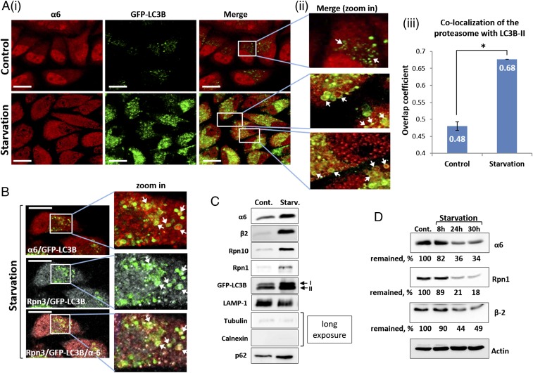
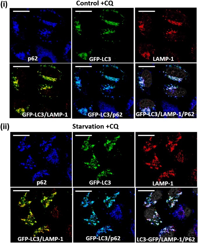
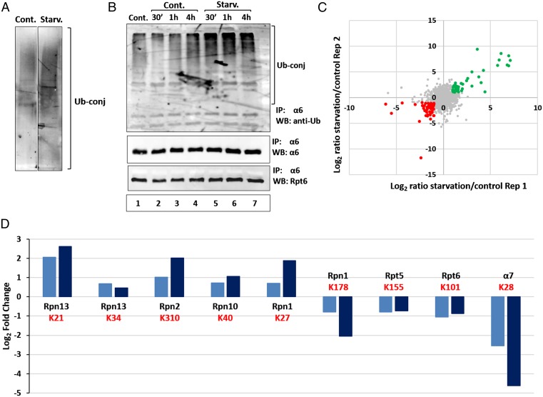
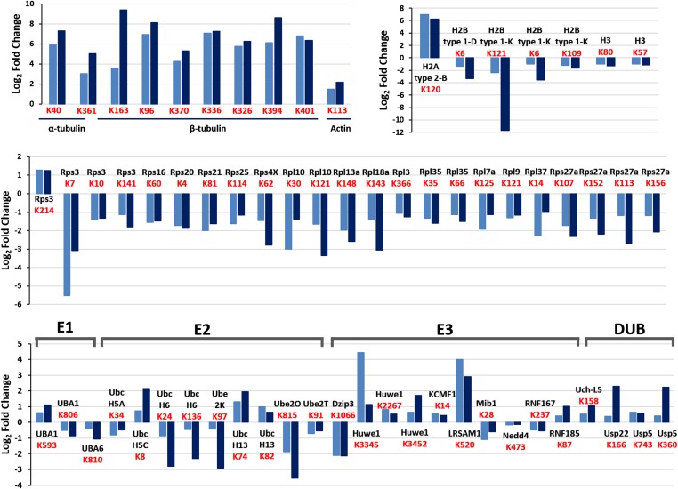
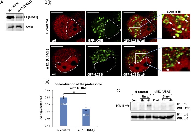
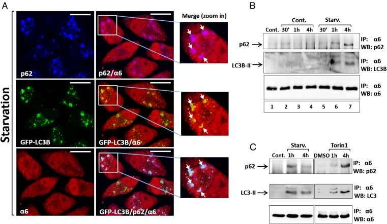
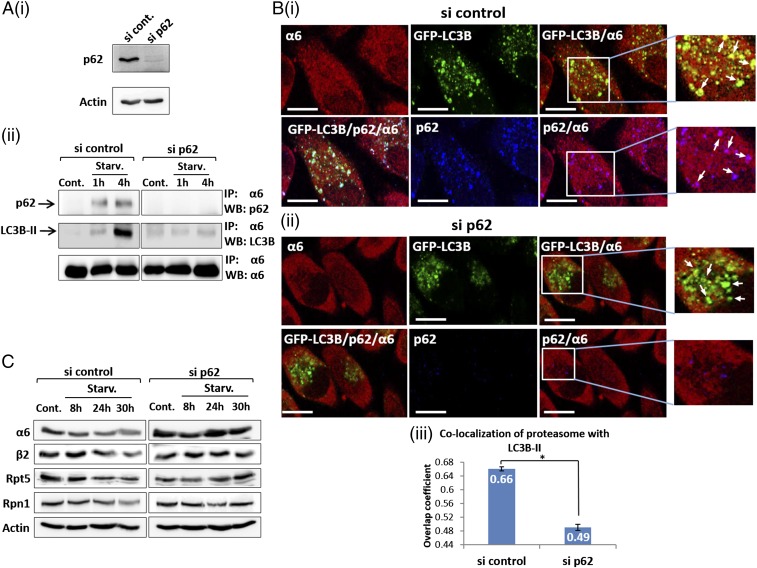
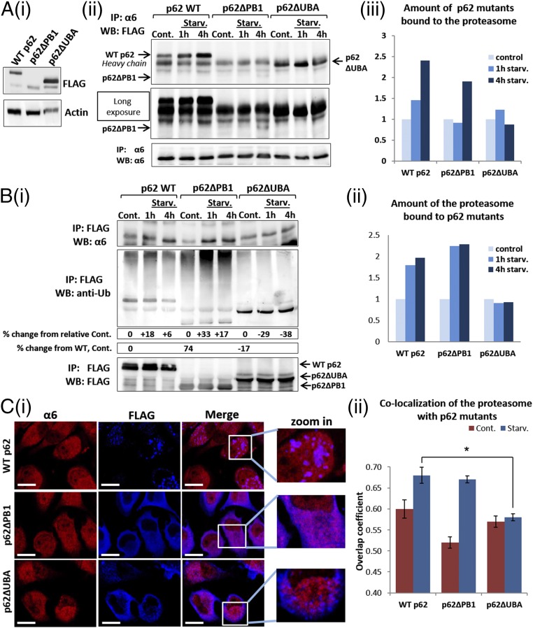
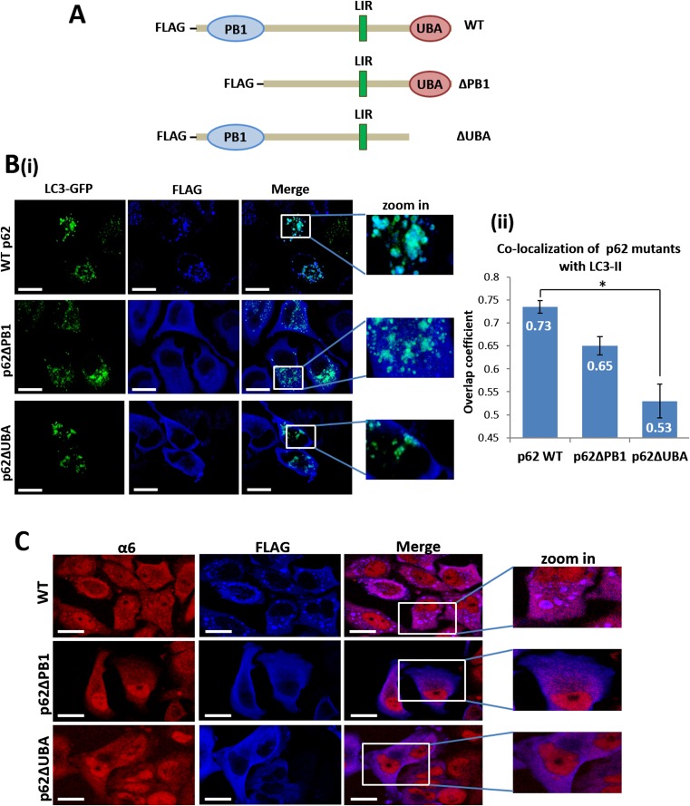
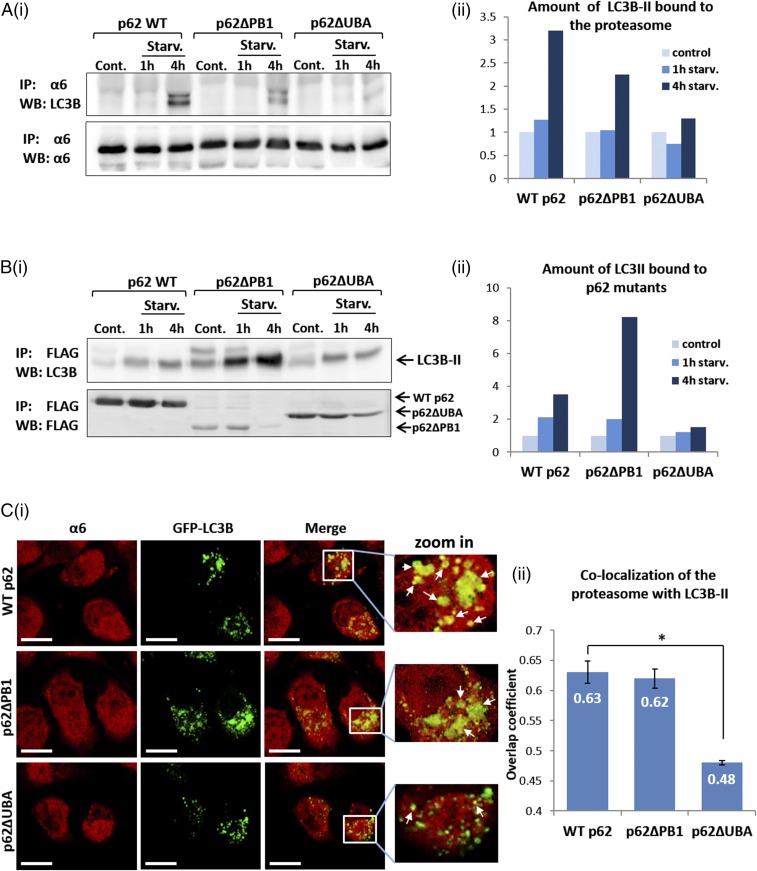
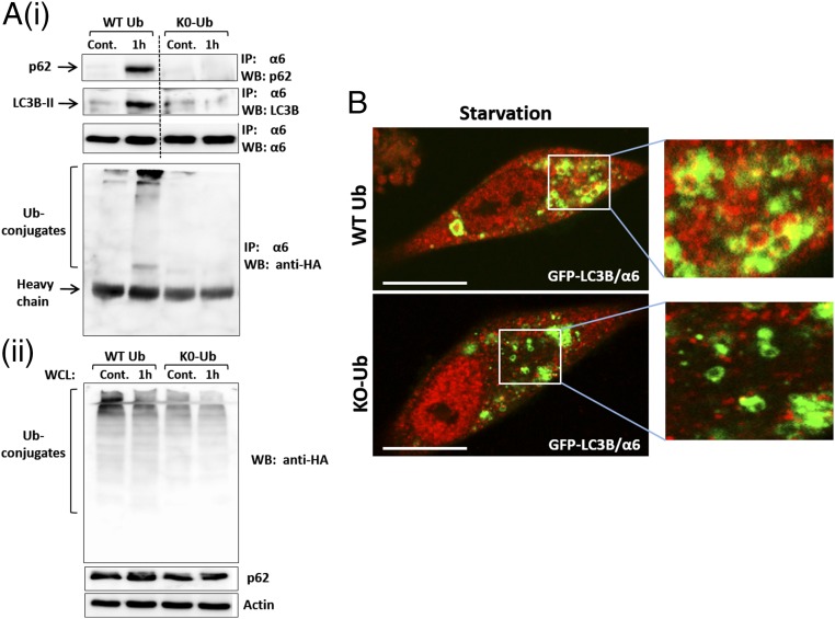
The ubiquitin-proteasome system and autophagy are the two main proteolytic systems involved in, among other functions, the maintenance of cell integrity by eliminating misfolded and damaged proteins and organelles. Both systems remove their targets after their conjugation with ubiquitin. An interesting, yet incompletely understood problem relates to the fate of the components of the two systems. Here we provide evidence that amino acid starvation enhances polyubiquitination on specific sites of the proteasome, a modification essential for its targeting to the autophagic machinery. The uptake of the ubiquitinated proteasome is mediated by its interaction with the ubiquitin-associated domain of p62/SQSTM1, a process that also requires interaction with LC3. Importantly, deletion of the PB1 domain of p62, which is important for the targeting of ubiquitinated substrates to the proteasome, has no effect on stress-induced autophagy of this proteolytic machinery, suggesting that the domain of p62 that binds to the proteasome determines the function of p62 in either targeting substrates to the proteasome or targeting the proteasome to autophagy.

Keywords: autophagy; degradation; proteasome; ubiquitin.


Conflict of interest statement

The authors declare no conflict of interest.

Figures

Fig. 1.
Fig. 1.
Autophagic uptake of the proteasome and its subsequent degradation are induced by amino acid starvation. (A) (i) HeLa cells stably expressing GFP-LC3B were incubated for 4 h in the presence of complete (Upper) or amino acid-depleted medium (Lower). Fixed cells were stained with anti-α6 antibody (red). (Scale bars: 20 μm.) (ii) Highly magnified fields. White arrows point to proteasome-containing autophagosomes. (iii) Manders overlap coefficients of LC3B-II (GFP) and α6 (red) colocalization were calculated. *P < 0.0000012. (B) Immunofluorescent staining of GFP-LC3B–transfected HeLa cells with anti-α6 (red; Upper) and Rpn3 (gray; Middle) antibodies following 4 h of amino acid starvation. (Scale bars: 20 μm.) White arrows point to the 20S CP (merge, zoom in; Upper) and 19S RP (merge, zoom in; Middle) within autophagosomal vesicles. (Lower) Merged image of α6, Rpn3, and LC3B-II. (C) Autophagosome-lysosome vesicles were purified from control (Cont.) and 4 h starved cells (Starv.). Isolated vesicles were lysed, resolved via SDS/PAGE, and subjected to immunoblotting with anti-α6 and anti-β2 (20S CP); with anti-Rpn10 and anti-Rpn1 (19S RP); and with anti-LAMP1 (lysosome), anti-tubulin, anti-calnexin and anti-p62 (autophagic machinery). (D) GFP-LC3B–expressing HeLa cells were left untreated (Cont.) or starved for the indicated times. Cell lysates were subjected to immunoblotting after electrophoresis with anti-α6, anti-Rpn1, anti-β2, and anti-actin.
Fig. S1.
Fig. S1.
Colocalization of autophagosomes with lysosomes following addition of CQ. Fed (i) and 4-h amino acid-starved (ii) GFP-LC3B–expressing HeLa cells were stained with anti-p62 (blue) and anti-LAMP-1 (red). (Scale bars: 20 μm.)
Fig. 2.
Fig. 2.
Amino acid deprivation stimulates proteasome ubiquitination. (A) Autophagosomes isolated from control and starved cells (Fig. 1C) were resolved via SDS/PAGE and blotted with anti-Ub conjugates antibody. (B) HeLa cells were incubated for the indicated times in the presence of complete (lanes 1–4; Cont.) or amino acid-depleted (lanes 5–7; Starv.) medium, in the absence (lane 1; Cont.) or presence (lanes 2–7) of CQ. Cell lysates were immunoprecipitated (IP) with anti-α6, and the immunoprecipitates were resolved and subjected to immunoblotting (WB) with anti-Ub, anti-Rpt6, and anti-α6. (C) Log2 ratios of two biological replicates of Ub-anchoring sites (K-ε-GG) enriched from HeLa cells incubated for 1 h under control or starved conditions. Green dots represent ubiquitinated peptides, the level of which increased during starvation, whereas red dots represent those whose level was decreased. (D) Log2 ratios of ubiquitinated sites (K-ε-GG) of specific proteasome subunits obtained in two replicated biological experiments. The Ub-modified lysine (K) is denoted in red.
Fig. S2.
Fig. S2.
Amino acid deprivation stimulates the ubiquitination of specific UPS and cellular proteins. Shown are log2 ratios of the ubiquitination sites (K-ε-GG) of components of the UPS and of α- and β-tubulin, actin, histones, and ribosomal proteins that were up-regulated (ratio >1) or down-regulated (ratio <−1) in the two independent biological replicates. Ubiquitination sites are depicted in red.
Fig. 3.
Fig. 3.
Autophagosomal uptake of the proteasome depends on Ub conjugation (A) GFP-LC3B–overexpressing HeLa cells were transfected with control or anti-E1 (UBA1) siRNA oligonucleotides. After 2 d, cells were lysed, resolved via SDS/PAGE, and examined for E1 expression. (B) (i) After silencing of E1, cells were starved for amino acids for 4 h and then stained with anti-α6. (Scale bars: 10 μm.) White arrows point to proteasome-containing autophagosomes. (ii) Overlap coefficients of colocalization of the proteasome with LC3B-II were measured according to the method of Manders. *P < 0.000027. (C) HeLa cells were transfected with control or anti-E1 (UBA1) siRNAs. Cells were left untreated (Cont.) or starved to amino acids (Starv.) for the indicated times. Cell lysates were immunoprecipitated (IP) with anti-α6, and the precipitates were resolved, followed by immunoblotting (WB) with anti-LC3B and anti-α6.
Fig. 4.
Fig. 4.
Amino acid starvation stimulates the interaction of the proteasome with p62 and LC3B. (A) After 4 h of amino acid starvation, GFP-LC3B–expressing HeLa cells were stained with anti-p62 (blue; Upper Left) and anti-α6 (red; Lower Left). (Scale bars: 20 μm.) White arrows point to colocalized proteasome and p62 (zoom in; Upper), LC3B-II (zoom in; Middle), and colocalization of all three (zoom in; Lower). (B) GFP-LC3B–expressing HeLa cells were incubated for the indicated times in the presence of complete (lanes 1–4) or amino acid-depleted (lanes 5–7; Starv.) medium, in the absence (lane 1; Cont.) or presence (lanes 2–7) of CQ. Cell lysates were immunoprecipitated (IP) with anti-α6, and the immunoprecipitates were resolved and blotted (WB) with anti-p62, anti-LC3B, and anti-α6. (C) GFP-LC3B–expressing HeLa cells were left untreated (Cont.) or starved for amino acids (Starv.; Left), or were supplemented with DMSO (DMSO) or Torin1 (0.5 μΜ) for the indicated times (Right). CQ was present in the starved and the Torin1-treated cells. Cell lysates were subjected to immunoprecipitation (IP) and, following resolution, were immunoblotted (WB) with anti-p62, anti-LC3B, and anti-α6.
Fig. 5.
Fig. 5.
p62/SQSTM1 mediates autophagosomal uptake of the proteasome. (A) (i) GFP-LC3B–expressing HeLa cells were transfected with control or anti-p62 siRNAs. At 2 d after transfection, cells were lysed and p62 expression was detected by Western blot analysis. (ii) si control-transfected and si p62-transfected cells were left untreated (Cont.) or starved for amino acids (Starv.) for the indicated times. Cell lysates were subjected to immunoprecipitation (IP) with anti-α6, followed by electrophoresis and immunoblotting (WB) with anti-p62, anti-LC3B, and anti-α6. (B) After treatment with si control (i) and si p62 (ii), GFP-LC3B–expressing HeLa cells were subjected to amino acid starvation, then stained with anti-p62 and anti-α6. (Scale bars: 20 μm.) (iii) Overlap coefficients of colocalization of the proteasome with LC3B-II in control and p62-silenced cells were measured according to the method of Manders. *P < 0.0000211. (C) Cells were treated as described in A and B, but for different starvation times, and lysates were resolved and blotted with anti-α6, anti-β2, anti-Rpt5, anti-Rpn1, and anti-actin.
Fig. 6.
Fig. 6.
The UBA domain of p62 is required for its interaction with the ubiquitinated proteasome following amino acid starvation. (A) (i) Whole-cell lysates of FLAG-tagged WT p62-, p62∆PB1-, and p62∆UBA-expressing HeLa cells were analyzed for expression of the different p62 proteins by immunoblotting with anti-FLAG. (ii) HeLa cells expressing the different p62 species (as described in A, i) were left untreated (Cont.) or starved for amino acids for the indicated times. Cell lysates were immunoprecipitated (IP) with anti-α6, resolved, and immunoblotted (WB) with anti-FLAG and anti-α6. (iii) Densitometric analysis of the p62 proteins depicted in A, ii. The value of p62 in the control lane of each construct was arbitrarily set to 1. (B) (i) The cells and the experimental setup were as described in A, ii. Cell lysates were subjected to immunoprecipitation (IP) with immobilized anti-FLAG, followed by immunoblotting (WB) with anti-α6, anti-FLAG, and anti-Ub conjugates. (ii) Densitometric analysis of the amount of the proteasome coprecipitated with the p62 species after amino acid deprivation. The value of the α6 subunit in the control lane of each p62 construct was arbitrarily set to 1. (C) (i) HeLa cells transiently transfected with FLAG-tagged WT p62, p62∆PB1, and p62∆UBA were starved for amino acids for 4 h and then stained with anti-FLAG and anti-α6. (Scale bars: 20 μm.) (ii) Manders overlap coefficients of colocalization of the different p62 proteins with the proteasome. *P < 0.000313.
Fig. S3.
Fig. S3.
Deletion of the UBA domain of p62 decreases its colocalization with both the proteasome and LC3B-II. (A) Schematic representation of the p62 constructs. (B) (i) GFP-LC3B–expressing HeLa cells were transiently transfected with FLAG-tagged WT (Upper), ∆PB1 (Middle), and ∆UBA (Lower) p62s. Cells were starved for amino acids for 4 h and then stained with anti-FLAG. (Scale bars: 20 μm.) (ii) Overlap coefficients of LC3B-II colocalization with the p62 constructs were calculated according to the method of Manders. *P < 0.0002. (C) Same as in Fig. 6B, except that the cells were not starved. These cells served as a control for the experiment shown in Fig. 6C, ii. (Scale bars: 20 μm.)
Fig. 7.
Fig. 7.
The interaction of the ubiquitinated proteasome with the UBA domain of p62 promotes its autophagosomal uptake. (A) (i) FLAG-tagged WT, ∆PB1, and ∆UBA p62s were transiently transfected to GFP-LC3B–expressing HeLa cells and either left untreated or starved to amino acids for the indicated times. Cell lysates were subjected to immunoprecipitation (IP) with anti-α6, resolved, and immunoblotted (WB) with anti-LC3B and anti-α6. (ii) Densitometric analysis of the amount of LC3B-II precipitated with the proteasome. The value of LC3B-II in the control lane of each p62 construct was arbitrarily set to 1. (B) (i) Same as under A, i, except that anti-FLAG was used to immunoprecipitate the different p62 species. (ii) Densitometric analysis of the amount of LC3B-II precipitated with the different p62 proteins. The value of LC3B-II in the control lane of each p62 construct was arbitrarily set to 1. (C) (i) WT, ∆PB1, and ∆UBA p62s were overexpressed in GFP-LC3B–expressing HeLa cells and stained with anti-α6 (red). (Scale bars: 20 μm.) White arrows indicate points of colocalization of the proteasome with LC3B-II. (ii) Coefficients of colocalization of the proteasome with LC3B-II in the different p62 species-expressing cells were calculated according to the method of Manders. *P < 0.000015.
Fig. 8.
Fig. 8.
Inhibition of polyubiquitination decreases uptake of the proteasome by autophagosomes. (A) (i) GFP-LC3B–expressing HeLa cells were transiently transfected with p62. At 24 h later, the cells were infected with adenoviruses coding for either WT HA-Ub or HA-K0-Ub. Cells were split and were either left untreated or starved for amino acids for 1 h. The proteasome was immunoprecipitated from cell lysates, and the precipitates were resolved and blotted with anti-p62, anti-LC3B, anti-α6, and anti-HA. (ii) Expression of WT Ub and K0-Ub (Upper) and p62 (Middle) was detected in whole-cell lysates (WCL). (B) Immunofluorescent staining with anti-α6 of GFP-LC3B–expressing HeLa cells infected with WT Ub (Upper) or K0-Ub (Lower) was performed after 4 h of amino acid starvation. (Scale bars: 20 μm.)

Comment in

  • How the proteasome is degraded.
    Hoeller D, Dikic I. Hoeller D, et al. Proc Natl Acad Sci U S A. 2016 Nov 22;113(47):13266-13268. doi: 10.1073/pnas.1616535113. Epub 2016 Nov 11. Proc Natl Acad Sci U S A. 2016. PMID: 27837026 Free PMC article. No abstract available.
  • p62 at the crossroad of the ubiquitin-proteasome system and autophagy.
    Cohen-Kaplan V, Ciechanover A, Livneh I. Cohen-Kaplan V, et al. Oncotarget. 2016 Dec 20;7(51):83833-83834. doi: 10.18632/oncotarget.13805. Oncotarget. 2016. PMID: 27974671 Free PMC article. No abstract available.

References

    1. Finley D. Recognition and processing of ubiquitin-protein conjugates by the proteasome. Annu Rev Biochem. 2009;78(1):477–513. - PMC - PubMed
    1. Cohen-Kaplan V, Livneh I, Avni N, Cohen-Rosenzweig C, Ciechanover A. The ubiquitin-proteasome system and autophagy: Coordinated and independent activities. Int J Biochem Cell Biol. 2016 doi: 10.1016/j.biocel.2016.07.019. - DOI - PubMed
    1. Ruggiano A, Foresti O, Carvalho P. Quality control. ER-associated degradation: Protein quality control and beyond. J Cell Biol. 2014;204(6):869–879. - PMC - PubMed
    1. Glickman MH, Ciechanover A. The ubiquitin-proteasome proteolytic pathway: Destruction for the sake of construction. Physiol Rev. 2002;82(2):373–428. - PubMed
    1. Hartmann-Petersen R, Gordon C. Protein degradation: Recognition of ubiquitinylated substrates. Curr Biol. 2004;14(18):R754–R756. - PubMed

Publication types

LinkOut - more resources