. 2016 Nov 8;113(45):12709-12714.
doi: 10.1073/pnas.1615902113. Epub 2016 Oct 24.

Structure of the mitochondrial ATP synthase from Pichia angusta determined by electron cryo-microscopy

Affiliations

Structure of the mitochondrial ATP synthase from Pichia angusta determined by electron cryo-microscopy

Kutti R Vinothkumar et al. Proc Natl Acad Sci U S A. .

Abstract

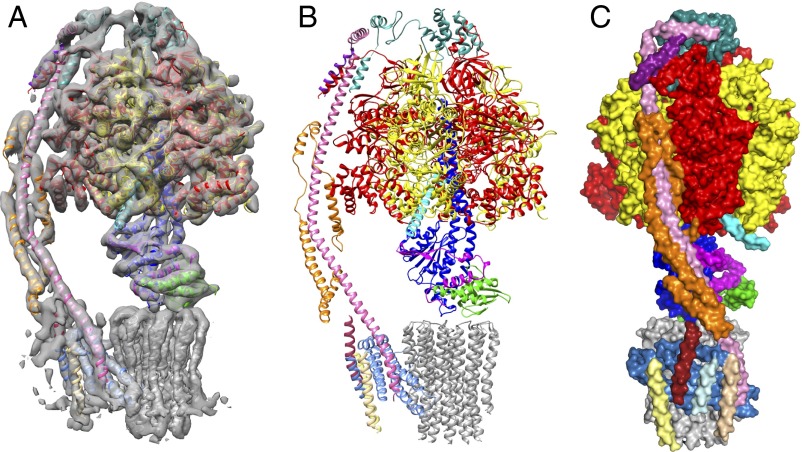
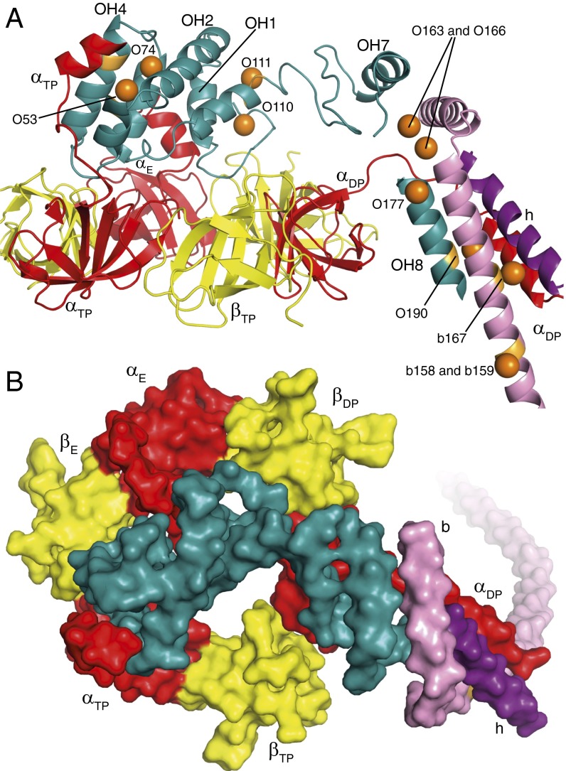
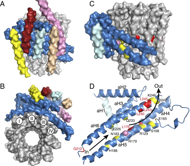
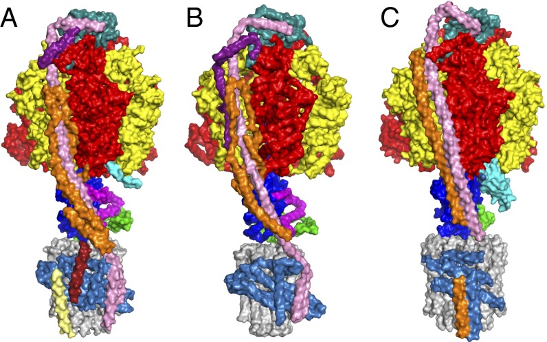
The structure of the intact monomeric ATP synthase from the fungus, Pichia angusta, has been solved by electron cryo-microscopy. The structure provides insights into the mechanical coupling of the transmembrane proton motive force across mitochondrial membranes in the synthesis of ATP. This mechanism requires a strong and integral stator, consisting of the catalytic α3β3-domain, peripheral stalk, and, in the membrane domain, subunit a and associated supernumerary subunits, kept in contact with the rotor turning at speeds up to 350 Hz. The stator's integrity is ensured by robust attachment of both the oligomycin sensitivity conferral protein (OSCP) to the catalytic domain and the membrane domain of subunit b to subunit a. The ATP8 subunit provides an additional brace between the peripheral stalk and subunit a. At the junction between the OSCP and the apparently stiff, elongated α-helical b-subunit and associated d- and h-subunits, an elbow or joint allows the stator to bend to accommodate lateral movements during the activity of the catalytic domain. The stator may also apply lateral force to help keep the static a-subunit and rotating c10-ring together. The interface between the c10-ring and the a-subunit contains the transmembrane pathway for protons, and their passage across the membrane generates the turning of the rotor. The pathway has two half-channels containing conserved polar residues provided by a bundle of four α-helices inclined at ∼30° to the plane of the membrane, similar to those described in other species. The structure provides more insights into the workings of this amazing machine.

Keywords: ATP synthase; Pichia angusta; proton translocation; structure.


Conflict of interest statement

The authors declare no conflict of interest.

Figures

Fig. 1.
Fig. 1.
The structure of the F-ATPase from P. angusta. The enzyme was inhibited with residues 1–60 of the bovine inhibitor protein IF1. (A) The cryo-EM map of state 1 and structural model viewed from the side, with the peripheral stalk on the left, the catalytic domain at the top, attached by the central and peripheral stalks to the membrane domain below. (B and C) Side views of the enzyme-inhibitor complex in cartoon and surface representation, respectively. C is rotated to the right by 90° relative to A and B. The α-, β-, γ-, δ-, and ε-subunits forming the membrane extrinsic catalytic domain are red, yellow, royal blue, green, and magenta, respectively; the inhibitor protein is cyan; and the peripheral stalk subunits OSCP, b, d, and h are sea-green, pink, orange, and purple, respectively. In the membrane domain, the c10-rotor is gray, the resolved region of the associated subunit a is corn-flower blue. Chains Ch1–Ch4 are pale yellow, brick-red, pale cyan, and beige, respectively, and have been assigned as transmembrane α-helices in subunit f and, in ATP8, as aH1 and bH1, respectively. In SI Appendix, Fig. S5, the identities of subunits are placed directly on an enlarged version of C.
Fig. 2.
Fig. 2.
Attachment of the peripheral stalk to the crown of the F1-catalytic domain of of the F-ATPase from P. angusta. The diagrams are based on state 1. (A and B) Views from the side and from above the N-terminal crown of the F1-domain in cartoon and surface representation, respectively. The OSCP is sea-green, the three α-subunits are red, the three β-subunits are yellow, the b-subunit is pink, and subunit h is purple. In A, the N-terminal α-helical regions of the α-subunits are labeled αE, αDP, and αTP. In the OSCP, the positions of α-helices OH1, OH2, OH4, OH7, and OH8 are indicated. The orange spheres represent the positions of selenium atoms in the structure of bovine F1-ATPase with the truncated peripheral stalk (bF1-tPS), and orange patches indicate the positions of corresponding amino acids in the fungal OSCP. They are labeled according to the subunit where they reside, with the prefix O for OSCP and b for subunit b, followed by the residue number in the respective subunits. O53, O74, O110, O111, O190, b167, and b158–159 correspond to residues in resolved regions of the structure of the P. angusta F-ATPase. O163, O166, and O177 indicate the positions of the selenomethionine residues in bF1-tPS and indicate the positions of the equivalent P. angusta residues in unresolved regions of the current structure.
Fig. 3.
Fig. 3.
The membrane domain of the F-ATPase from P. angusta. In AD, the a-subunit is corn-flower blue. (A and B) Views in solid representation from the side and below the membrane domain. The c10-ring is gray, the b-subunit (upper part not shown) is pink, and the pale yellow, brick-red, light cyan, and beige segments are transmembrane α-helices, Ch1–Ch4 assigned to subunit f, ATP8, aH1, and bH1, respectively. In the c-ring, I–IV indicate the four transmembrane C-terminal α-helices in contact with subunit a. (C and D) Views of the a-subunit in solid and cartoon representation viewed from outside and looking out from the interface with the c-ring, respectively, with aH1 in pale cyan. Conserved polar residues are yellow; the positions of human mutations associated with pathologies (SI Appendix, Table S1) are red. The pink sphere denotes the conserved Arg-179 in aH5 that is essential for proton translocation. The lower arrow indicates the inlet pathway for protons that transfer to Glu-59 in the C-terminal α–helix-II of the c-ring. They are carried around the ring by anticlockwise rotation, as viewed from above, until they arrive at Arg-179 where they enter the exit pathway, denoted by the upper arrow.
Fig. 4.
Fig. 4.
Comparison of the current structures of the F-ATPases. The structures of the three complexes are viewed from the side toward the peripheral stalk with the F1-catalytic domain above and the membrane domain beneath. Enzymes (A and B) from P. angusta and bovine mitochondria (7) and (C) from P. denitrificans (9). The α-, β-, and γ-subunits are red, yellow, and royal blue, respectively. The c-rings (made of 10, 8, and 12 subunits in AC, respectively) are gray, and the adjacent a-subunits are corn-flower blue. In A and B, the OSCP subunits (Top), and in C, the orthologous δ-subunit, are sea-green; the δ-subunits (A and B) and orthologous ε-subunit in C are green. In A and B only, the ε-subunit (next to the green δ-subunit) is magenta. In A and B, in addition to the OSCP, the peripheral stalks contain the b-subunits (pink), the d-subunits (orange), and the orthologous h and F6-subunits (purple), respectively. In C, in addition to the δ-subunit, the peripheral stalk contains a b-subunit (pink) and a b′-subunit (orange). In A, the pale yellow and brick-red α-helices packed against the c-rings have been assigned tentatively to the f and ATP8 subunits, respectively.

References

    1. Watanabe R, Noji H. Chemomechanical coupling mechanism of F1-ATPase: Catalysis and torque generation. FEBS Lett. 2013;587(8):1030–1035. - PubMed
    1. Walker JE. The ATP synthase: The understood, the uncertain and the unknown. Biochem Soc Trans. 2013;41(1):1–16. - PubMed
    1. Robinson GC, et al. The structure of F₁-ATPase from Saccharomyces cerevisiae inhibited by its regulatory protein IF₁. Open Biol. 2013;3(2):120164. - PMC - PubMed
    1. Bason JV, Montgomery MG, Leslie AGW, Walker JE. Pathway of binding of the intrinsically disordered mitochondrial inhibitor protein to F1-ATPase. Proc Natl Acad Sci USA. 2014;111(31):11305–11310. - PMC - PubMed
    1. Bason JV, Montgomery MG, Leslie AGW, Walker JE. How release of phosphate from mammalian F1-ATPase generates a rotary substep. Proc Natl Acad Sci USA. 2015;112(19):6009–6014. - PMC - PubMed

LinkOut - more resources