. 2016 Oct 25;17(11):1777.
doi: 10.3390/ijms17111777.

Exogenous Melatonin Improves Plant Iron Deficiency Tolerance via Increased Accumulation of Polyamine-Mediated Nitric Oxide

Affiliations

Exogenous Melatonin Improves Plant Iron Deficiency Tolerance via Increased Accumulation of Polyamine-Mediated Nitric Oxide

Cheng Zhou et al. Int J Mol Sci. .

Abstract

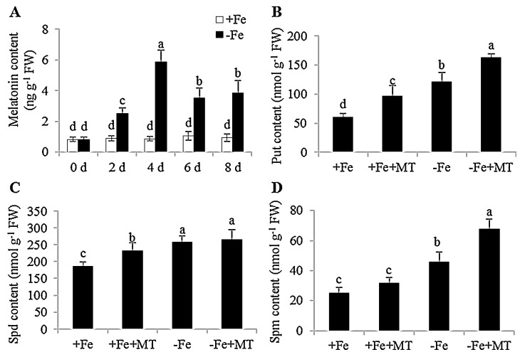
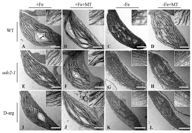
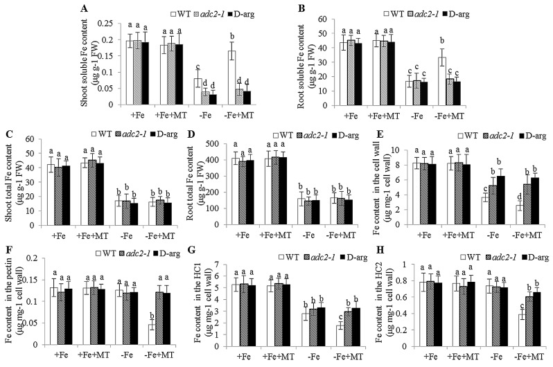
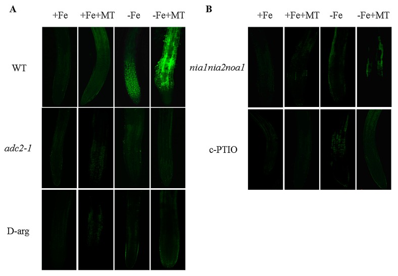
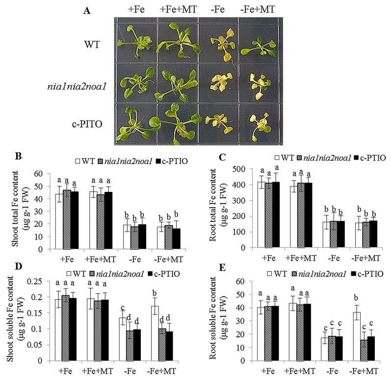
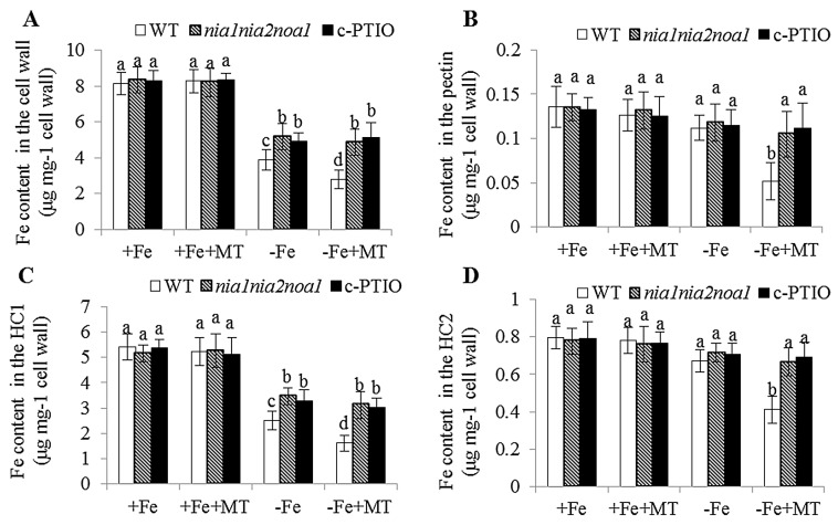
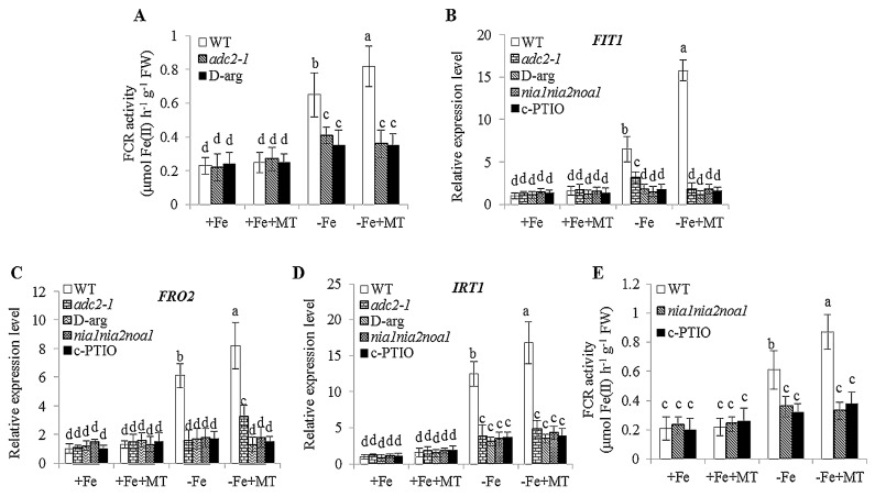
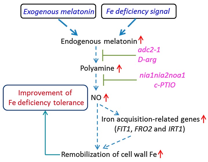
Melatonin has recently been demonstrated to play important roles in the regulation of plant growth, development, and abiotic and biotic stress responses. However, the possible involvement of melatonin in Fe deficiency responses and the underlying mechanisms remained elusive in Arabidopsis thaliana. In this study, Fe deficiency quickly induced melatonin synthesis in Arabidopsis plants. Exogenous melatonin significantly increased the soluble Fe content of shoots and roots, and decreased the levels of root cell wall Fe bound to pectin and hemicellulose, thus alleviating Fe deficiency-induced chlorosis. Intriguingly, melatonin treatments induced a significant increase of nitric oxide (NO) accumulation in roots of Fe-deficient plants, but not in those of polyamine-deficient (adc2-1 and d-arginine-treated) plants. Moreover, the melatonin-alleviated leaf chlorosis was blocked in the polyamine- and NO-deficient (nia1nia2noa1 and c-PTIO-treated) plants, and the melatonin-induced Fe remobilization was largely inhibited. In addition, the expression of some Fe acquisition-related genes, including FIT1, FRO2, and IRT1 were significantly up-regulated by melatonin treatments, whereas the enhanced expression of these genes was obviously suppressed in the polyamine- and NO-deficient plants. Collectively, our results provide evidence to support the view that melatonin can increase the tolerance of plants to Fe deficiency in a process dependent on the polyamine-induced NO production under Fe-deficient conditions.

Keywords: iron deficiency; iron remobilization; melatonin; nitric oxide (NO); polyamine.


Conflict of interest statement

The authors declare no conflict of interest. The funding sponsors had no role in the design of the study; in the collection, analyses, or interpretation of data; in the writing of the manuscript, and in the decision to publish the results.

Figures

Figure 1
Figure 1
Changes of endogenous melatonin and polyamine content in response to Fe deficiency, and the effects of exogenous melatonin on cellular polyamine levels in Arabidopsis plants. Seven-day-old seedlings were placed in 1/2 MS liquid medium with or without the presence of 50 μM Fe for the indicated time, then these plants were taken to measure melatonin content (A). In addition, seve-day-old seedlings were treated with or without 5 μM melatonin under −Fe and +Fe conditions for 8 days. These plants were then used to determine cellular polyamine, including Put (B), Spd (C), and Spm (D). Each bar is the mean ± SE of at least three replicates, and different lowercase letters above the bars indicating significant differences using a Tukey’s test at p < 0.05.
Figure 2
Figure 2
The effect of exogenous melatonin on Fe deficiency symptoms in the WT, adc2-1, and d-arg-treated Arabidopsis plants. Seven-day-old seedlings were grown under +Fe (50 μM Fe) and −Fe (0 μM Fe) conditions for 8 days, then these plants were sampled to measure the content of endogenous polyamine, including Put (A), Spd (B), and Spm (C). Additionally, seven-day-old seedlings were grown under +Fe and −Fe conditions with or without the presence of 5 μM melatonin for 8 days, respectively. Then, these plants were used to analyze the growth phenotype (D) and chlorophyll content (E). Each bar is the mean ± SE of at least three replicates, and different lowercase letters above the bars indicate significant differences using Tukey’s test at p < 0.05.
Figure 3
Figure 3
Transmission electron micrographic (TEM) analyses of the effect of exogenous melatonin on the chloroplast ultrastructure in the WT (AD), adc2-1 (EH), and d-arg-treated (IL) Arabidopsis plants. Seven-day-old seedlings were treated with or without 5 μM melatonin in the presence or absence of 50 μM Fe for 8 days. Then the leaves from these plants were sampled to analyze the chloroplast ultrastructure: (A,E,I), +Fe; (B,F,J), +Fe + MT; (C,G,K), −Fe; (D,H,L), −Fe + MT; Scale bar = 1 μm.
Figure 4
Figure 4
The effect of exogenous melatonin on Fe content in the WT, adc2-1 and d-arg-treated Arabidopsis plants. Seven-day-old seedlings were treated with or without 5 μM melatonin in the presence or absence of 50 μM Fe for 8 days. Then these plants were taken to measure the soluble Fe content of shoots (A) and roots (B), the total Fe content of shoots (C) and roots (D), and to measure the Fe content in root cell wall components: total cell wall (E), pectin (F), HC1 (G), and HC2 (H) fractions. Each bar is the mean ± SE of at least three replicates, and different lowercase letters above the bars indicate significant difference using Tukey’s test at p < 0.05.
Figure 5
Figure 5
The effect of melatonin on the NO accumulation in roots of polyamine- and NO-deficient Arabidopsis plants. Seven-day-old seedlings were treated with or without 5 μM melatonin in the presence or absence of 50 μM Fe for 8 days. Then, NO-associated fluorescence was detected in the roots of polyamine-deficient (adc2-1 and d-arg-treated) (A) and NO-deficient (nia1nia2noa1 and c-PTIO-treated) plants (B).
Figure 6
Figure 6
The effect of exogenous melatonin on Fe deficiency symptoms in the WT, nia1nia2noa1, and c-PTIO-treated Arabidopsis plants. Seven-day-old seedlings were treated with or without 5 μM melatonin in the presence or absence of 50 μM Fe for 8 days. Then, these plants were used to analyze the growth phenotype (A), total Fe content of shoots (B) and roots (C), and soluble Fe content of shoots (D) and roots (E). Each bar is the mean ± SE of at least three replicates, and different lowercase letters above the bars indicate significant differences using Tukey’s test at p < 0.05.
Figure 7
Figure 7
The effect of exogenous melatonin on Fe content in root cell wall components in the WT, nia1nia2noa1 and c-PTIO-treated Arabidopsis plants. Seven-day-old seedlings were grown in −Fe (0 μM Fe) or +Fe (50 μM Fe) medium without or with the presence of 5 μM melatonin (+MT) for 8 days. Then, these plants were taken to measure the Fe content in the root total cell wall (A), pectin (B), HC1 (C), and HC2 (D) fractions. Each bar is the mean ± SE of at least three replicates, and different lowercase letters above the bars indicate significant difference using Tukey’s test at p < 0.05.
Figure 8
Figure 8
The effect of exogenous melatonin on chloroplast ultrastructure in the WT (AD), nia1nia2noa1 (EH), and c-PTIO-treated (IL) Arabidopsis plants. Seven-day-old seedlings were treated with or without 5 μM melatonin in the presence or absence of 50 μM Fe for 8 days. Then the leaves from these plants were sampled for transmission electron micrographic analyses of the chloroplast ultrastructure: (A,E,I), +Fe; (B,F,J), +Fe + MT; (C,G,K), −Fe; (D,H,L), −Fe + MT; Scale bar = 1 μm.
Figure 9
Figure 9
The effect of exogenous melatonin on the pH values of Arabidopsis plants. Seven-day-old seedlings were grown in −Fe (0 μM Fe) or +Fe (50 μM Fe) medium (pH = 5.6) without or with the presence of 5 μM melatonin (−MT or +MT) for the indicated times for measuring the pH values (A). In addition, soluble Fe content of shoots (B) and roots (C) were determining after seven-day-old WT and aha2 were placed in −Fe or +Fe medium with or without the presence of 5 μM melatonin for 8 days. Each bar is the mean ± SE of at least three replicates, and different lowercase letters above the bars indicate significant differences using a Tukey’s test at p < 0.05.
Figure 10
Figure 10
The effect of exogenous melatonin on the FCR activities and the expression of Fe-acquisition-related genes in polyamine- (adc2-1 and d-arg-treated) and NO-deficient (nia1nia2noa1 and c-PTIO-treated) Arabidopsis plants. Seven-day-old seedlings were treated with or without 5 μM melatonin in the presence or absence of 50 μM Fe for 8 days. Roots from adc2-1 and d-arg-treated plants were sampled for measuring the FCR activities (A). The expression levels of Fe acquisition-related genes, including FIT1 (B), FRO2 (C), and IRT1 (D), were examined after seven-day-old seedlings were placed in the −Fe or +Fe medium with or without the presence of 5 μM melatonin (−MT or +MT) for 8 days. In addition, roots from nia1nia2noa1 and c-PTIO-treated plants were used to determine the FCR activities (E). Each bar is the mean ± SE of at least three replicates, and different lowercase letters above the bars indicate significant differences using Tukey’s test at p < 0.05.
Figure 11
Figure 11
A proposed model illustrating the link between melatonin and improved tolerance of plants to Fe deficiency. The increased melatonin level induces the increment of the polyamine content, with the subsequent induction of NO accumulation. The enhancement of the NO signal then activates Fe deficiency responses and increases the remobilization cell wall Fe, which results in the increased soluble Fe in response to the low Fe supply. Dashed arrows indicate regulatory pathways. Red upright arrows denote increases in content or effects.

Similar articles

Cited by

References

    1. Lerner A.B., Case J.D., Takahashi Y., Lee T.H., Mori W. Isolation of melatonin, a pineal factor that lightens melanocytes. J. Am. Chem. Soc. 1958;80 doi: 10.1021/ja01543a060. - DOI
    1. Zawilska J.B., Skene D.J., Arendt J. Physiology and pharmacology of melatonin in relation to biological rhythms. Pharmacol. Rep. 2009;61:383–410. doi: 10.1016/S1734-1140(09)70081-7. - DOI - PubMed
    1. Reiter R.J., Mayo J.C., Tan D.X., Sainz R.M., Alatorre-Jimenez M., Qin L. Melatonin as an antioxidant: Under promises but over delivers. J. Pineal Res. 2016 doi: 10.1111/jpi.12360. - DOI - PubMed
    1. Calvo J.R., González-Yanes C., Maldonado M.D. The role of melatonin in the cells of the innate immunity: A review. J. Pineal Res. 2013;55:103–120. doi: 10.1111/jpi.12075. - DOI - PubMed
    1. Zhang N., Zhang H.J., Zhao B., Sun Q.Q., Cao Y.Y., Li R., Wu X.X., Weeda S., Li L., Ren S., et al. The RNA-seq approach to discriminate gene expression profiles in response to melatonin on cucumber lateral root formation. J. Pineal Res. 2014;56:39–50. doi: 10.1111/jpi.12095. - DOI - PubMed

MeSH terms

LinkOut - more resources