. 2016 Dec;54(12):613-625.
doi: 10.1002/dvg.22990. Epub 2016 Nov 12.

Spatiotemporal differences in otoconial gene expression

Affiliations

Spatiotemporal differences in otoconial gene expression

Yinfang Xu et al. Genesis. 2016 Dec.

Abstract

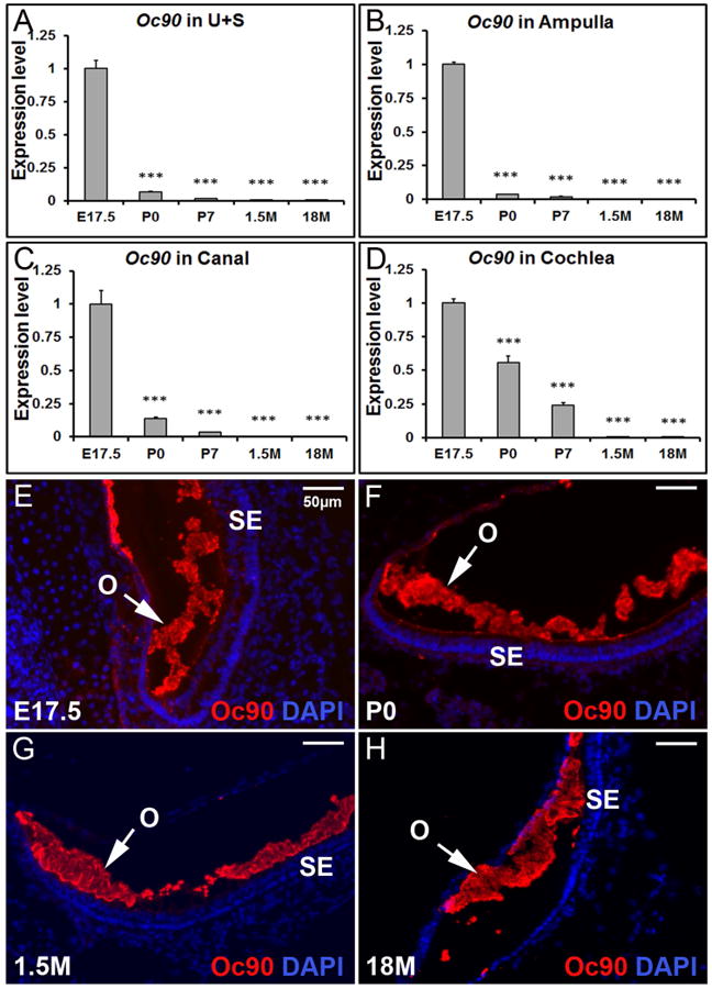
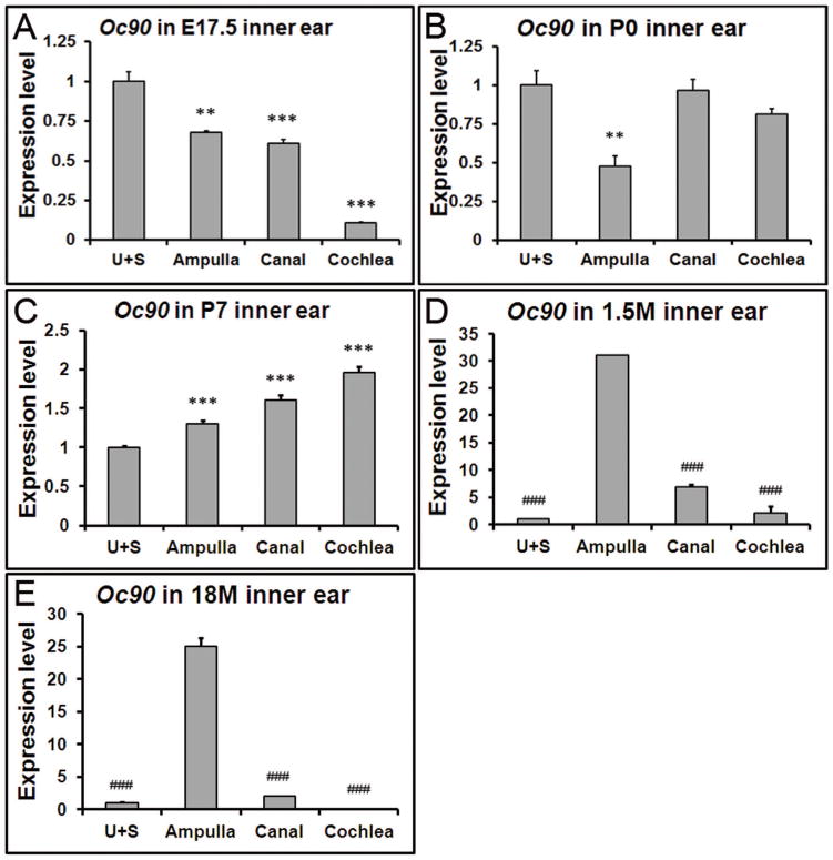
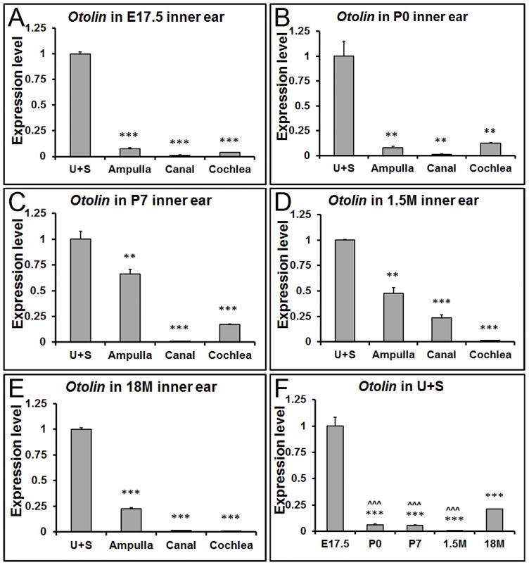
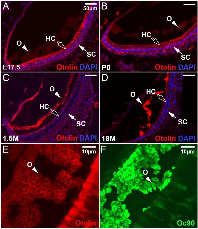
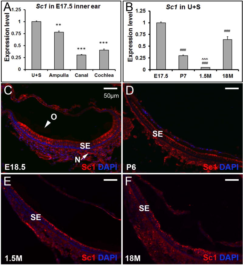
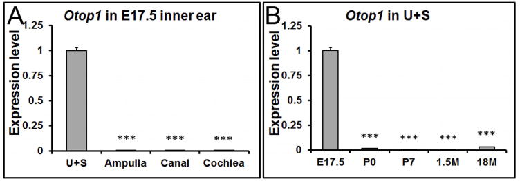
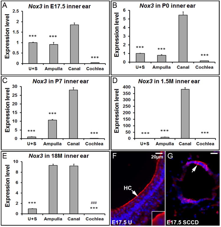
Otoconia are minute biocrystals composed of glycoproteins, proteoglycans, and CaCO3 , and are indispensable for sensory processing in the utricle and saccule. Otoconia abnormalities and degeneration can cause or facilitate crystal dislocation to the ampulla, leading to vertigo and imbalance in humans. In order to better understand the molecular mechanism controlling otoconia formation and maintenance, we have examined the spatial and temporal expression differences of otoconial genes in the mouse inner ear at developmental, mature and aging stages using whole transcriptome sequencing (RNA-Seq) and quantitative RT-PCR. We show that the expression levels of most otoconial genes are much higher in the utricle and saccule compared with other inner ear tissues before postnatal stages in C57Bl/6J mice, and the expression of a few of these genes is restricted to the embryonic utricle and saccule. After the early postnatal stages, expression of all otoconial genes in the utricle and saccule is drastically reduced, while a few genes gain expression dominance in the aging ampulla, indicating a potential for ectopic debris formation in the latter tissue at old ages. The data suggest that the expression of otoconial genes is tightly regulated spatially and temporally during developmental stages and can become unregulated at aging stages. Birth Defects Research (Part A) 106:613-625, 2016. © 2016 Wiley Periodicals, Inc.

Keywords: RNA-Seq; aging; development; otoconial genes; spatial; temporal.


Figures

Figure 1
Figure 1. Temporal changes in Oc90 transcript (A-D) and protein (E-H) in mouse inner ear tissues
Based on qRT-PCR, Oc90 is expressed mostly before birth in the utricle and saccule (A), ampulla (B), canal (C) and cochlea (D). *** denote p<0.001 when compared with E17.5 (n=6 for each age group). U+S, utricle and saccule. Based on fluorescent immunostaining, Oc90 protein is abundant in the developing (E & F), mature (G) and aging (H) otoconia (utricle shown). O, otoconia; SE, sensory epithelia.
Figure 2
Figure 2. Spatial changes in Oc90 expression at different ages
Data presented in each panel here are not just re-analysis of Figure 1, but were independently obtained in separate qRT-PCR plates for accurate comparison. (A) Oc90 shows the highest expression level in the utricle and saccule (U+S) at E17.5, but such a pattern is reversed at postnatal (C), adult (D) and aging stages (E). (D, E) In the 1.5 and 18 month-old inner ears, Oc90 shows the highest expression level in the ampulla. ** and *** denote p<0.01, <0.001 when compared with the U+S and ### indicates p<0.001 compared with the ampulla (n=6 for each group).
Figure 3
Figure 3. The spatiotemporal expression pattern of Otolin transcript in the inner ear
(A-E) qRT-PCR shows that Otolin has the highest expression level in the utricle and saccule at all ages, especially at developmental stages. (F) In the utricle and saccule, Otolin is mainly expressed at embryonic stages. **, *** denote p<0.01 and p<0.001 compared to the utricle and saccule (U+S), respectively (n=6 for each tissue and age group). ˆˆˆ indicates p < 0.001 when compared with 18M.
Figure 4
Figure 4. Localization of Otolin protein in the utricle at different ages
(A) Otolin is present in hair cells (HC), supporting cells (SC), and transitional epithelia at E17.5, but the signal was faint in otoconia at this age. (B) At P0, Otolin signal is slightly stronger in the otoconia (O), but less intense in the underlying epithelia when compared to the corresponding tissues at E17.5. (C) At 1.5 months old, Otolin accumulates further in otoconia and in the apical extracellular matrix of the sensory epithelium of the utricle. (D) In the aging utricle, Otolin is only detectable in otoconia and otoconial membrane, but not in the epithelia. (E) Otolin accumulates mostly in the matrix rim of otoconial crystallites at E17.5, whereas Oc90 signals (F) are more diffuse. Bars = 50μm in A-D and 20 μm in E, F.
Figure 5
Figure 5. The expression pattern of Sc1 in the inner ear
(A) At E17.5, expression of Sc1 is significantly higher in the utricle and saccule by qRT-PCR, but the spatial specification is not as striking as that of Otolin. ** and *** denote p<0.01 and <0.001 when compared with the utricle and saccule (n=6 in each group). (B) Sc1 is re-expressed in the utricle and saccule at 18 months old by qRT-PCR. ### indicates p < 0.001 when compared with E17.5, and ˆˆˆ denote p < 0.001 when compared with 18M (n=6 for each age group). (C-F) Sc1 protein is hardly detectable in otoconia (O), but is present in the sensory epithelium (SE) and nerve fibers (N) (utricle shown).
Figure 6
Figure 6. The expression of Otop1 is restricted to the embryonic utricle and saccule by qRT-PCR
(A) At E17.5, the expression of Otop1 is confined to the utricle (U) and saccule (S). *** denote p<0.001 compared with the utricle and saccule (n=6 in each tissue group). (B) In the utricle and saccule (U+S), Otop1 shows the highest expression level at embryonic stages. ### denote p<0.001 compared with E17.5 (n=6 in each age group).
Figure 7
Figure 7. Nox3 shows the highest expression level in the semi-circular canals and aging ampulla
(A-E) qRT-PCR shows that Nox3 has a significantly higher expression level in the canals compared to other inner ear tissues at different stages. *** denote p<0.001 when compared with the canals (n=6 each tissue group). In the 18 month-old mice, the ampullar expression level of Nox3 is also much higher than the utricle/saccule, and cochlea. ### denote p<0.001 when compared with the ampulla (n=6 each tissue group). (F) The E17.5 utricle (U) was stained with a Nox3 antibody (red) and DAPI (blue). (G) Immunostaining of the semi-circular canal duct (SCCD) at E17.5 with Nox3 and DAPI. HC, hair cell; SCCD, semi-circular canal duct.

Similar articles

Cited by

References

    1. Andrade LR, Lins U, Farina M, Kachar B, Thalmann R. Immunogold TEM of otoconin 90 and otolin - relevance to mineralization of otoconia, and pathogenesis of benign positional vertigo. Hear Res. 2012;292:14–25. - PMC - PubMed
    1. Andrade LR. Evidence for changes in beta- and gamma-actin proportions during inner ear hair cell life. Cytoskeleton (Hoboken) 2015;72:282–291. - PMC - PubMed
    1. Anniko M. Development of otoconia. Am J Otolaryngol. 1980;1:400–410. - PubMed
    1. Anniko M, Wenngren BI, Wroblewski R. Aberrant elemental composition of otoconia in the dancer mouse mutant with a semidominant gene causing a morphogenetic type of inner ear defect. Acta Otolaryngol. 1988;106:208–212. - PubMed
    1. Anniko M, Ylikoski J, Wroblewski R. Microprobe analysis of human otoconia. Acta Otolaryngol. 1984;97:283–289. - PubMed

MeSH terms

LinkOut - more resources