Review
. 2017 Mar;27(2):123-137.
doi: 10.1111/bpa.12454. Epub 2017 Jan 11.

Multiple sclerosis animal models: a clinical and histopathological perspective

Affiliations
Review

Multiple sclerosis animal models: a clinical and histopathological perspective

Markus Kipp et al. Brain Pathol. 2017 Mar.

Abstract

There is a broad consensus that multiple sclerosis (MS) represents more than an inflammatory disease: it harbors several characteristic aspects of a classical neurodegenerative disorder, that is, damage to axons, synapses and nerve cell bodies. While we are equipped with appropriate therapeutic options to prevent immune-cell driven relapses, effective therapeutic options to prevent the progressing neurodegeneration are still missing. In this review article, we will discuss to what extent pathology of the progressive disease stage can be modeled in MS animal models. While acute and relapsing-remitting forms of experimental autoimmune encephalomyelitis (EAE), which are T cell dependent, are aptly suited to model relapsing-remitting phases of MS, other EAE models, especially the secondary progressive EAE stage in Biozzi ABH mice is better representing the secondary progressive phase of MS, which is refractory to many immune therapies. Besides EAE, the cuprizone model is rapidly gaining popularity to study the formation and progression of demyelinating CNS lesions without T cell involvement. Here, we discuss these two non-popular MS models. It is our aim to point out the pathological hallmarks of MS, and discuss which pathological aspects of the disease can be best studied in the various animal models available.

Keywords: EAE; PPMS; SPMS; cuprizone; disability; disease progression; multiple sclerosis; neurodegeneration; treatment.


Conflict of interest statement

The authors report no conflict of interest.

Figures

Figure 1
Figure 1
(A) Histopathologic characteristics of an acute, inflammatory MS lesion within the white matter. Demyelination is indicated by a focal, well demarcated loss of anti‐PLP (proteolipid protein) immunoreactivity. The entire lesion is interspersed with activated monocytes and microglia, expressing MHC‐II protein. In (B) the correlate in EAE is shown. Even in HE‐stained sections, inflammatory infiltrates are clearly visible. In EAE, such lesions are mainly found within the spinal cord and cerebellum. (C) The three pathological hallmarks of MS pathology, namely focal inflammation, diffuse white matter injury and gray matter injury are illustrated. While focal inflammation causes acute relapses (see red arrows), diffuse white and gray matter pathology induce neurodegeneration and in consequence accumulation of irreversible clinical disability (red arrowhead). Blue arrow indicates clinical remission.
Figure 2
Figure 2
EAE clinical disease score in C57BL/6 mice immunized with MOG35–55 peptide is shown. After 9–10 days post‐immunization, inflammatory demyelination of the spinal cord results in overt clinical disability of experimental animals. After the peak of the disease (around day 15 post‐immunization) animals slightly recover, but from then on do not progress any more. Figure adapted from J Mol Neurosci. 2016 Sep;60(1):102‐14. Arrows highlight inflammatory lesions within the spinal cord.
Figure 3
Figure 3
Histopathology of secondary progressive EAE in Biozzi ABH mice. Compared to aged‐matched controls in which myelin integrity is intact and in which few if any Iba‐1+ cells are present (A), in SP‐EAE demyelination is demarcated by the loss of PLP associated with highly immunereactive Iba‐1+ microglia/macrophages (B). In white matter, SMI32 depicting damaged axons is prominent in regions of highly reactive Iba‐1+ cells (C). In addition to Iba‐1 positive cells in the white matter, large infiltrates are also observed in gray matter in close association with motor neurons (D). Like MS, clusters of activated microglia are observed in normal appearing white matter in mice with SP‐EAE (E). In comparison to acute EAE in Biozzi ABH mice, a sparse number of adaptive immune cells are observed during SP‐EAE, and when observed are generally restricted to the leptomeninges (F).
Figure 4
Figure 4
(A) Histopathological characteristics of early (1 week treatment) cuprizone‐induced lesions. While the myelination appears still normal in an anti‐PLP stain, microglia activation and oligodendrocyte apoptosis is clearly evident. Left column control, right column 1 week cuprizone‐intoxication. (B) The diffuse pathology after prolonged (5 weeks treatment) cuprizone intoxication is illustrated. Besides the midline of the corpus callosum, the cortex, hippocampus and diverse subcortical areas are affected. Demyelination is paralleled by intense microglia (Iba1) and astrocyte (GFAP) activation. Furthermore, acute axonal damage can be seen in brain sections processed for amyloid precursor protein (APP)‐immunohistochemistry. APP is a membrane‐spanning glycoprotein which is transported from neuron cell bodies to axon terminals by fast anterograde axonal transport. In healthy axons, APP is below the immunohistochemical detection limit. Under pathological conditions, for example cytoskeletal disruption, anterograde axonal transport is disturbed and APP accumulates as ovoid spheroids 70. Accumulation of such axonal spheroids can be directly demonstrated by electron microscopy. In yellow, the intact part of the axon is highlighted, red shows the axonal spheroid. Blue shows severe axonal enlargement.
Figure 5
Figure 5
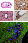
Distribution and characteristics of inflammatory forebrain lesions in the Cup/EAE model is shown 115. Infiltrates were found widespread within the forebrain, including the cortex, corpus callosum and subcortical regions (upper‐left part). Anti‐GFAP stains show the breakdown of the glia limitans perivascularis. Anti‐CD3 stains demonstrate that a significant number of perivascular cells are lymphocytes. Double staining for GFAP and the water channel protein AQP4 (aquaporin‐4) clearly demonstrate functional impairment of astrocytes around such inflammatory lesions.

Similar articles

Cited by

References

    1. Acs P, Kipp M, Norkute A, Johann S, Clarner T, Braun A et al (2009) 17Beta‐estradiol and progesterone prevent cuprizone provoked demyelination of corpus callosum in male mice. Glia 57:807–814. - PubMed
    1. Al‐Izki S, Pryce G, Hankey DJ, Lidster K, von Kutzleben SM, Browne L et al (2014) Lesional‐targeting of neuroprotection to the inflammatory penumbra in experimental multiple sclerosis. Brain 137:92–108. - PubMed
    1. Al‐Izki S, Pryce G, O'Neill JK, Butter C, Giovannoni G, Amor S, Baker D (2012) Practical guide to the induction of relapsing progressive experimental autoimmune encephalomyelitis in the Biozzi ABH mouse. Mult Scler Relat Disord 1:29–38. - PubMed
    1. Baker D, Amor S (2014) Experimental autoimmune encephalomyelitis is a good model of multiple sclerosis if used wisely. Mult Scler Relat Disord 3:555–564. - PubMed
    1. Bakker DA, Ludwin SK (1987) Blood–brain barrier permeability during Cuprizone‐induced demyelination. Implications for the pathogenesis of immune‐mediated demyelinating diseases . J Neurol Sci 78:125–137. - PubMed

MeSH terms