. 2016 Nov 22;7(47):78009-78028.
doi: 10.18632/oncotarget.12870.

Methylation-associated silencing of microRNA-129-3p promotes epithelial-mesenchymal transition, invasion and metastasis of hepatocelluar cancer by targeting Aurora-A

Affiliations

Methylation-associated silencing of microRNA-129-3p promotes epithelial-mesenchymal transition, invasion and metastasis of hepatocelluar cancer by targeting Aurora-A

Shiyun Cui et al. Oncotarget. .

Abstract

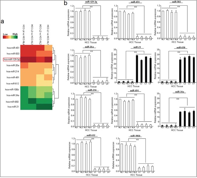
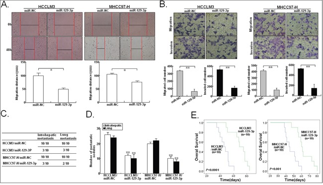
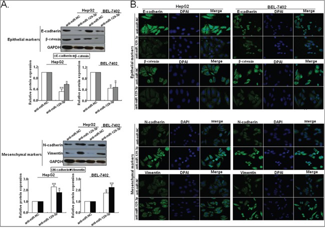
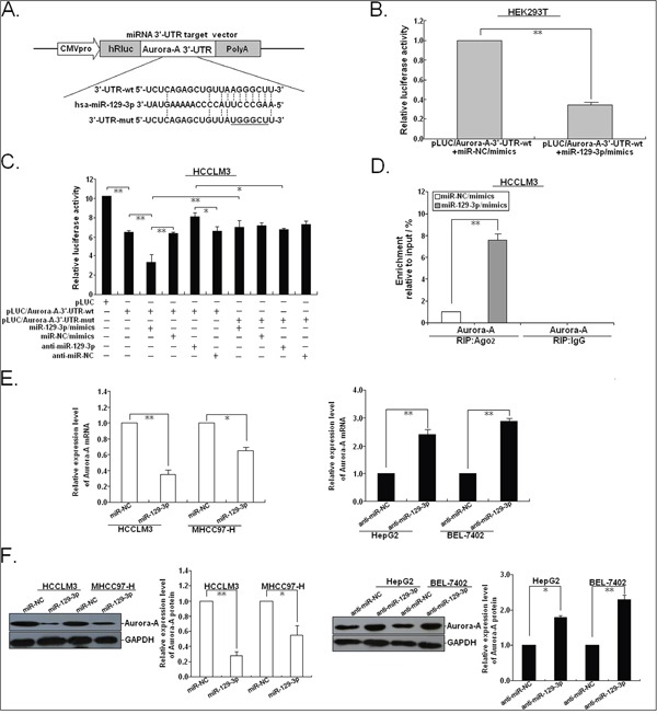
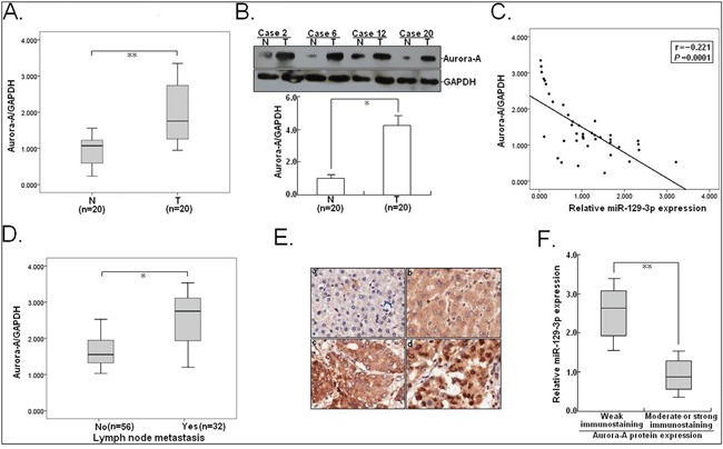
Metastasis and recurrence has become one major obstacle for further improving the survival of hepatocelluar cancer (HCC) patients. Therefore, it is critical to elucidate the mechanisms involved in HCC metastasis. This study aimed to investigate the roles of microRNA (miR)-129-3p in HCC metastasis and its possible molecular mechanisms. By using microarray analysis to compare levels of different miRNAs in HCC tissues with or without lymph node metastasis (LNM), we showed that HCC tissues with LNM had reduced levels of miR-129-3p, which was related to its promoter hypermethylation and correlated with tumor metastasis, recurrence and poor prognosis. Gain - and loss - of - function assays indicated that re-expression of miR-129-3p could reverse epithelial-mesenchymal transition (EMT), and reduce in vitro invasion and in vivo metastasis of HCC cells. Aurora-A, a serine/threonine protein kinase, was identified as a direct target of miR-129-3p. Knockdown of Aurora-A phenocopied the effect of miR-129-3p overexpression on HCC metastasis. In addition, Aurora-A upregulation could partially rescue the effect of miR-129-3p. We further demonstrated that activation of PI3K/Akt and p38-MAPK signalings were involved in miR-129-3p-mediated HCC metastasis. These findings suggest that methylation-mediated miR-129-3p downregulation promotes EMT, in vitro invasion and in vivo metastasis of HCC cells via activation of PI3K/Akt and p38-MAPK signalings partially by targeting Aurora-A. Therefore, miR-129-3p may be a novel prognostic biomarker and potential therapeutic target for HCC.

Keywords: Aurora-A; epithelial-mesenchymal transition; hepatocelluar cancer; metastasis; miR-129-3p.


Conflict of interest statement

CONFLICTS OF INTEREST

The authors declare no conflicts of interest.

Figures

Figure 1
Figure 1. miR-129-3p is significantly downregulated in metastatic HCC cell lines or tissues
A. MiRNA array analysis showed differentially expressed miRNAs in HCC tissues with LNM (HCC-L, n=4) and HCC tissues without LNM (HCC-NL, n=4). Overexpression is indicated in green, whereas underexpression is indicated in red. B. Validation of microarray analysis data by qRT-PCR. The relative expression levels of 11 dysregulated miRNAs (miR-129-3p, 451, 503, 26a, 21, 650, 214, 491, 34a, 612 and 106b) were determined by qRT-PCR. U6 was used as an internal control. Each qRT-PCR experiment was performed in triplicate.*P<0.05; **P<0.01. HCC-NL: HCC without LNM; HCC-L: HCC with LNM.
Figure 2
Figure 2. Reduced miR-129-3p correlates with HCC progression
Expression of miR-129-3p was determined by qRT-PCR. U6 was used as an internal control. A. Relative miR-129-3p expression in paired HCC and adjacent nontumor liver tissues. T: HCC tissues; N: nontumor liver tisssues. B. Relative miR-129-3p expression in HCC tissues with or without LNM. C. Relative miR-129-3p expression in HCC tissues with or without vascular invasion. D. Relative miR-129-3p expression in HCC tissues with different TNM stage. E. Relative miR-129-3p expression in HCC tissues with or without recurrence. F. Kaplan-Meier analysis of DFS or OS of HCC patients. The survival data were compared with the log-rank test. Each experiment was performed in triplicate. *P<0.05; **P<0.01.
Figure 3
Figure 3. miR-129-3p inhibits in vitro migration or invasion and in vivo metastasis in HCC cells
A. Wound healing assay. A confluent monolayer of miR-NC/mimics or miR-129-3p/mimics-transfected HCCLM3 or MHCC97-H cells was wounded. Photographs were taken immediately (0 h) and at 48 h after wounding, quantification of wound closure was done. The data present the mean distance of cell migration to the wound area at 48 h after wounding in three independent wound sites per group. B. Transwell migration and invasion assay of miR-NC/mimics or miR-129-3p/mimics-transfected HCCLM3 or MHCC97-H cells. Cells in six random fields of view at 100× magnification were counted and expressed as the average number of cells per field of view. C. Incidence of intrahepatic or lung metastasis in different groups of nude mice transplanted with miR-NC/mimics or miR-129-3p/mimics-transfected HCCLM3 or MHCC97-H cells (n=10/group). D. Hematoxylin and eosin staining of intrahepatic and lung metastatic tumor nodules formed from miR-NC/mimics or miR-129-3p/mimics-transfected HCCLM3 or MHCC97-H cells (n=10/group). The numbers of metastatic nodules in each nude mice were counted and statistically analyzed. E. The OS time of different groups of nude mice transplanted with miR-NC/mimics or miR-129-3p/mimics-transfected HCCLM3 or MHCC97-H cells (n=10/group). The survival data were compared with the log-rank test. *P<0.05; **P<0.01.
Figure 4
Figure 4. Upregulation of miR-129-3p reverses EMT in HCC cells (high initial metastatic potential)
A. Western blotting and B. immunofluorescence staining assays indicated that the increased expression of epithelial markers (E-cadherin and β-catenin) and the decreased expression of mesenchymal markers (N-cadherin and Vimentin) could be obviously observed in miR-129-3p/mimics-transfected HCCLM3 or MHCC97-H cells, compared with miR-NC/mimics-transfected cells. Each experiment was performed in triplicate.*P<0.05; **P<0.01.
Figure 5
Figure 5. MiR-129-3p binds to the 3′-UTR of human Aurora-A mRNA
A. Mutation was generated on the Aurora-A 3′-UTR sequence in the complementary site for the seed region of miR-129-3p, as shown. A human Aurora-A 3′-UTR fragment containing wild-type or mutant miR-129-3p-binding sequence was cloned into downstream of the luciferase reporter gene in pLUC-luc vector. B. pLUC vector contains Aurora-A mRNA 3′-UTR and miR-129-3p/mimics or control miR-NC/miR-mimics were co-transfected into HEK293T cells, Cells lysates were prepared after 48 h for measuring luciferase activity, which was normalized to normalized to Renilla luciferase activity. C. Relative luciferase activity was analyzed after wildtype or mutant 3′-UTR reporter plasmids were co-transfected with miR-129-3p/mimics or anti-miR-129-3p in HCCLM3 cells. The histogram shows the mean±SEM of the normalized luciferase activity from three independent experiments. D. The RIP analysis revealed recruitment of Aurora-A mRNAs to miRNP complex in miR-129-3p/mimics (or miRNA-NC/mimics)-transfected HCCLM3 cells following immunoprecipitation against Ago2. The IgG immunoprecipitation was used as a negative control. E. qRT-PCR detection of Aurora-A mRNA expression in miR-129-3p/mimics (or miR-NC/mimics)-transfected HCCLM3 or MHCC97-H cells and anti-miR-129-3p (or anti-miR-NC)-transfected HepG2 or BEL-7402 cells. F. Western blotting detection of Aurora-A protein expression in miR-129-3p/mimics (or miR-NC/mimics)-transfected HCCLM3 or MHCC97-H cells and anti-miR-129-3p (or anti-miR-NC)-transfected HepG2 or BEL-7402 cells. GAPDH was used as an internal control. *P<0.05; **P<0.01.
Figure 6
Figure 6. Silencing of Aurora-A inhibits in vitro invasion and in vivo metastatic capacity, and reverses EMT in HCC cells
A. qRT-PCR detection of Aurora-A mRNA expression in pSil/shcontrol or pSil/shAurora-A-transfected HCCLM3 or MHCC97-H cells. B. Western blotting detection of Aurora-A protein expression in pSil/shcontrol or pSil/shAurora-A-transfected HCCLM3 or MHCC97-H cells. C. Wound healing assay. A confluent monolayer of pSil/shcontrol or pSil/shAurora-A-transfected HCCLM3 or MHCC97-H cells was wounded. D. Transwell migration and invasion assay of pSil/shcontrol or pSil/shAurora-A-transfected HCCLM3 or MHCC97-H cells. E. Incidence of intrahepatic or lung metastasis in different groups of nude mice transplanted with pSil/shcontrol or pSil/shAurora-A-transfected HCCLM3 or MHCC97-H cells (n=10/group). F. Hematoxylin and eosin staining of intrahepatic and lung metastatic tumor nodules formed from pSil/shcontrol or pSil/shAurora-A-transfected HCCLM3 or MHCC97-H cells (n=10/group). The numbers of metastatic nodules in each nude mice were counted and analyzed using Student t test. G. The OS time of different groups of nude mice transplanted with pSil/shcontrol or pSil/shAurora-A-transfected HCCLM3 or MHCC97-H cells (n=10/group). H. Western blotting and immunofluorescence staining detection of epithelial markers and mesenchymal markersin pSil/shcontrol or pSil/shAurora-A-transfected HCCLM3 or MHCC97-H cells. Each experiment was performed in triplicate. *P<0.05; **P<0.01.
Figure 7
Figure 7. Overexpression of Aurora-A reverses the effects of miR-129-3p upregulation on HCC cells
48h after HCCLM3 cells were co-transfected with miR-129-3p/mimics and pMD-Auro (or pMD-NC), A. qRT-PCR and B. Western blotting detection of Aurora-A mRNA and protein expression. GAPDH was used as an internal control. C. Wound healing assay. A confluent monolayer of HCCLM3 cells co-transfected with miR-129-3p/mimics and pMD-Auro (or pMD-NC) was wounded. D. Transwell migration and invasion assay of HCCLM3 cells co-transfected with miR-129-3p/mimics and pMD-Auro (or pMD-NC). E. Incidence of intrahepatic or lung metastasis in different groups of nude mice transplanted with HCCLM3 cells co-transfected with miR-129-3p/mimics and pMD-Auro (or pMD-NC) (n=10/group). F. Hematoxylin and eosin staining of intrahepatic and lung metastatic tumor nodules formed from HCCLM3 cells co-transfected with miR-129-3p/mimics and pMD-Auro (or pMD-NC) (n=10/group). The numbers of metastatic nodules in each nude mice were counted and analyzed. G. The OS time of different groups of nude mice transplanted with HCCLM3 cells co-transfected with miR-129-3p/mimics and pMD-Auro (or pMD-NC) (n=10/group). The survival data were compared with the log-rank test. H. Western blotting and immunofluorescence staining detection of expression of epithelial markers (E-cadherin and β-catenin) and mesenchymal markers (N-cadherin and Vimentin) in HCCLM3 cells co-transfected with miR-129-3p/mimics and pMD-Auro or pMD-NC. Each experiment was performed in triplicate. *P<0.05; **P<0.01.
Figure 8
Figure 8. miR-129-3p and Aurora-A are involved in PI3K/Akt and p38-MAPK signalings in HCC cells
A. Western blotting detection of Aurora-A, pAkt, p-p38-MAPK, total Akt, total p38-MAPK and MMP-2 proteins in miR-NC/mimics or miR-129-3p/mimics-transfected HCCLM3 cells or HCCLM3 cells co-transfected with miR-129-3p/mimics and pMD-Auro. B. Western blotting detection of Aurora-A, p-Akt, p-p38-MAPK, total Akt, total p38-MAPK proteins in anti-miR-NC or anti-miR-129-3p-transfected HepG2 cells, followed by the treatment of the inhibitor of p38-MAPK (SB202190) or Akt (LY294002). C. Western blotting detection of EMT-related proteins in anti-miR-NC or anti-miR-129-3p-transfected HepG2 cells, followed by the treatment of SB202190 or LY294002. D. Wound healing assay. A confluent monolayer of HCCLM3 or MHCC97-H cells treated with SB202190 or LY294002 was wounded. The data present the mean distance of cell migration to the wound area at 48 h after wounding in three independent wound sites per group. E. Transwell migration and invasion assay of HCCLM3 or MHCC97-H cells treated with SB202190 or LY294002. F. Wound healing and G. transwell migration and invasion assays of HepG2 cells transfected with anti-miR-NC or anti-miR-129-3p, followed by the treatment of SB202190 or LY294002. H. Schematic overview of miR-129-3p regulatory signaling. GAPDH was used as an internal control. *P<0.05; **P<0.01.
Figure 9
Figure 9. Aurora-A is significantly upregulated in HCC tissues and negatively correlates with miR-129-3p
A. qRT-PCR detection of Aurora-A mRNA expression in HCC tissues and corresponding nontumor liver tissues (n=20). T: HCC tissues; N: nontumor liver tissues. B. Western blotting detection of Aurora-A protein expression in HCC tissues and corresponding nontumor liver tissues (n=20). C. Statistically significant inverse correlation between miR-129-3p and Aurora-A protein expression in 20 cases of HCC tissues (r = −0.221; P=0.0001). D. qRT-PCR detection of Aurora-A mRNA expression in HCC tissues without LNM (n=56) and HCC tissues with LNM (n=32). E. Immunostaining of Aurora-A protein was negatively or very weakly positive in corresponding nontumor liver tissues (a and b), but was moderately or strongly positive in HCC tissues (c and d). Origninal magnification, ×100. F. The immunoreactivity of Aurora-A protein in HCC tissues showed a statistically significant inverse correlation the relative level of miR-129-3p expression (P<0.01). Each experiment was performed in triplicate. GAPDH was used as an internal control. Data were presented as mean±SEM (n=3). *P<0.05; **P<0.01.

Similar articles

Cited by

References

    1. Jemal A, Siegel R, Xu J, Ward E. Cancer statistics, 2010. CA Cancer J Clin. 2010;60:277–300. - PubMed
    1. Zhou XD. Recurrence and metastasis of hepatocellular carcinoma: progress and prospects. Hepatobiliary Pancreat Dis Int. 2002;1:35–41. - PubMed
    1. Chen PY, Meister G. microRNA-guided posttranscriptional gene regulation. Biol Chem. 2005;386:1205–1218. - PubMed
    1. Jeker LT, Bluestone JA. MicroRNA regulation of T-cell differentiation and function. Immunol Rev. 2013;253:65–81. - PMC - PubMed
    1. Li C, Hashimi SM, Good DA, Cao S, Duan W, Plummer PN, Mellick AS, Wei MQ. Apoptosis and microRNA aberrations in cancer. Clin Exp Pharmacol Physiol. 2012;39:739–746. - PubMed

MeSH terms