. 2016 Dec 13;7(50):82305-82323.
doi: 10.18632/oncotarget.12929.

Cranial irradiation induces transient microglia accumulation, followed by long-lasting inflammation and loss of microglia

Affiliations

Cranial irradiation induces transient microglia accumulation, followed by long-lasting inflammation and loss of microglia

Wei Han et al. Oncotarget. .

Abstract

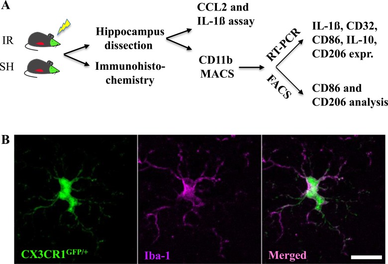
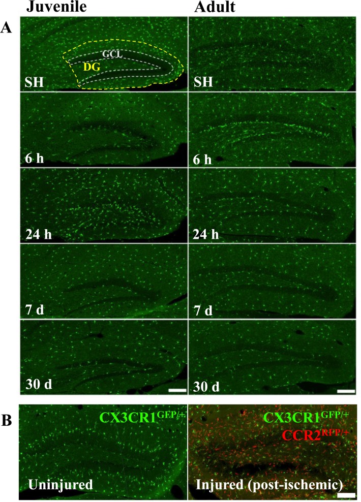
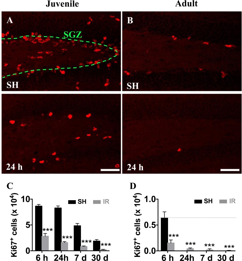
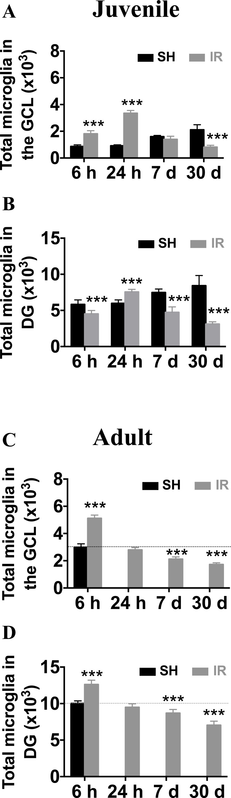
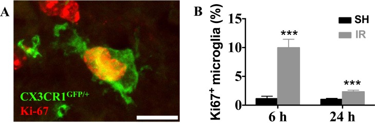
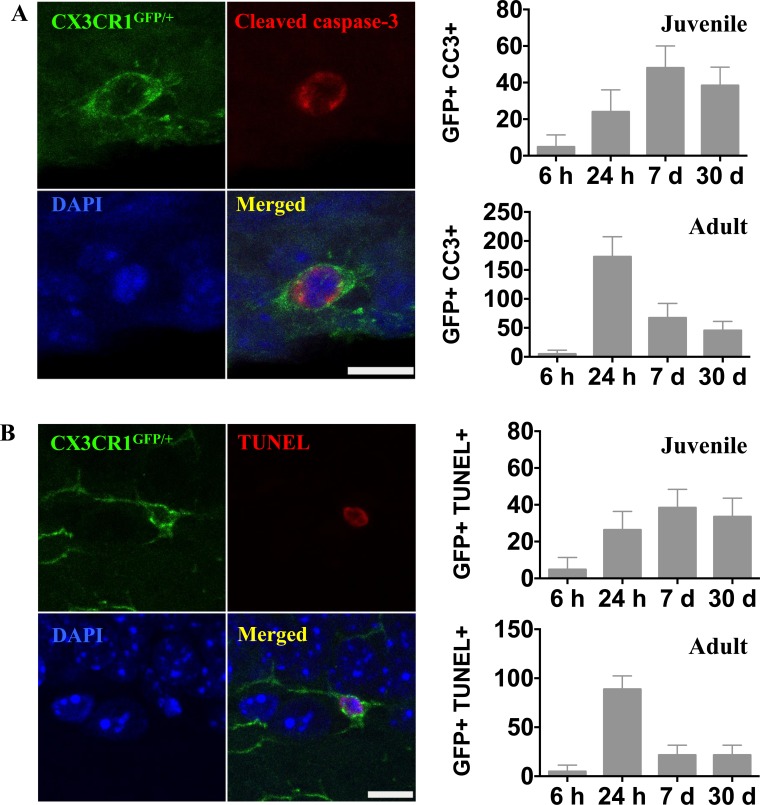
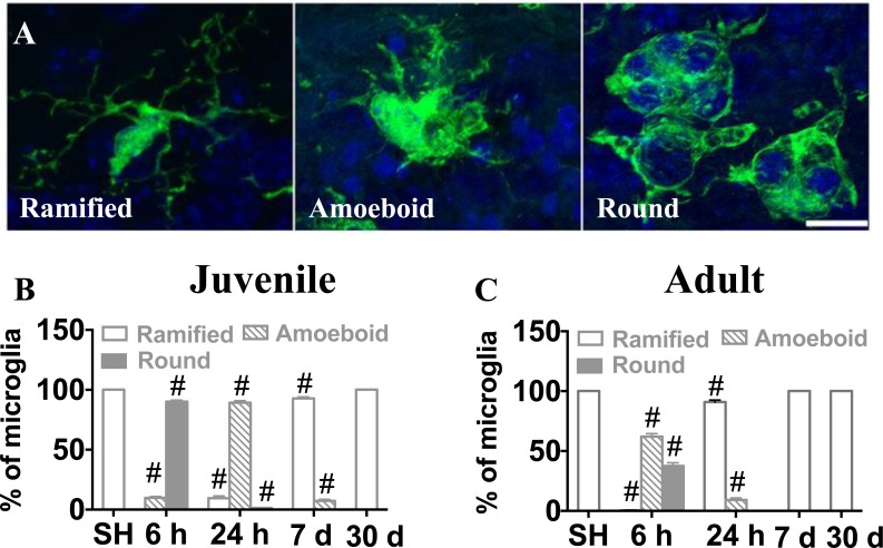
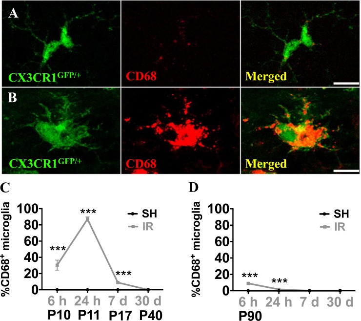
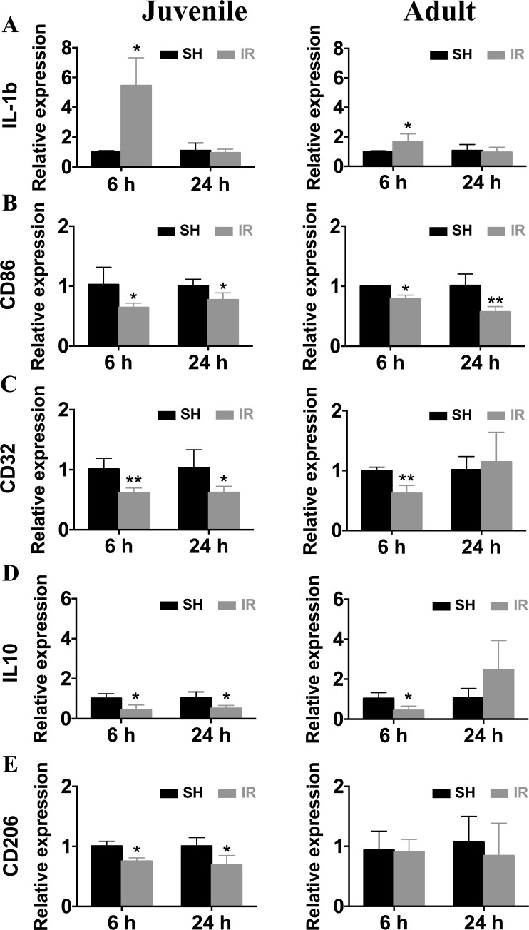
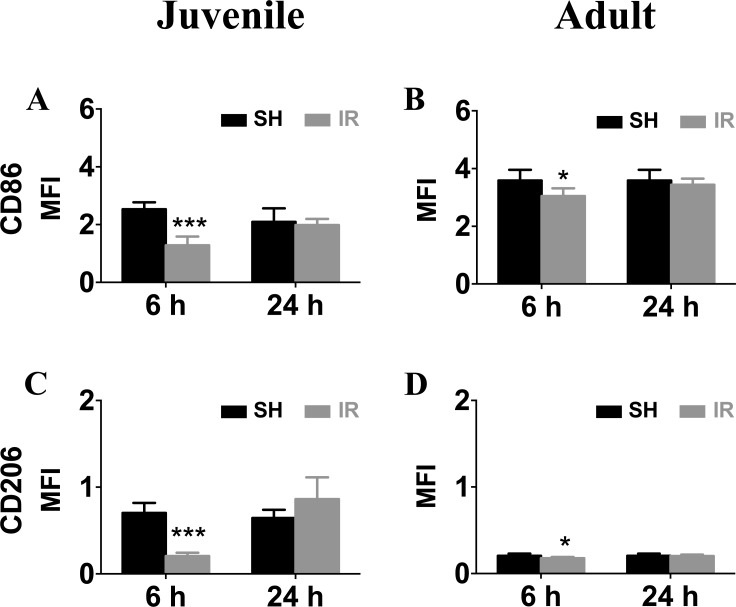
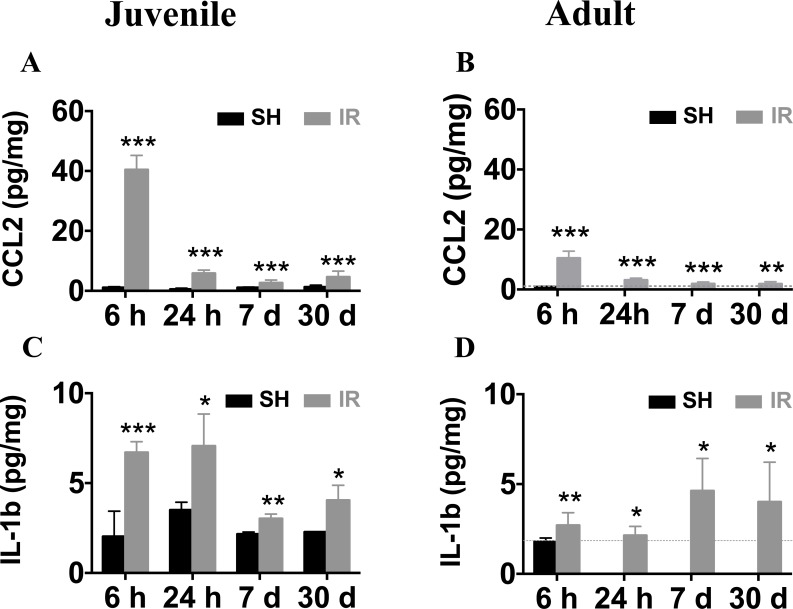
The relative contribution of resident microglia and peripheral monocyte-derived macrophages in neuroinflammation after cranial irradiation is not known. A single dose of 8 Gy was administered to postnatal day 10 (juvenile) or 90 (adult) CX3CR1GFP/+ CCR2RFP/+ mouse brains. Microglia accumulated in the subgranular zone of the hippocampal granule cell layer, where progenitor cell death was prominent. The peak was earlier (6 h vs. 24 h) but less pronounced in adult brains. The increase in juvenile, but not adult, brains was partly attributed to proliferation. Microglia numbers then decreased over time to 39% (juvenile) and 58% (adult) of controls 30 days after irradiation, largely as a result of cell death. CD68 was expressed in 90% of amoeboid microglia in juvenile hippocampi but only in 9% of adult ones. Isolated hippocampal microglia revealed reduced CD206 and increased IL1-beta expression after irradiation, more pronounced in juvenile brains. CCL2 and IL-1 beta increased after irradiation, more in juvenile hippocampi, and remained elevated at all time points. In summary, microglia activation after irradiation was more pronounced, protracted and pro-inflammatory by nature in juvenile than in adult hippocampi. Common to both ages was long-lasting inflammation and the absence of monocyte-derived macrophages.

Keywords: irradiation; macrophage; microglia; monocyte; neurogenesis; neuroinflammation.


Conflict of interest statement

CONFLICT OF INTEREST

The author(s) declare that they have no competing interests.

Figures

Figure 1
Figure 1. Experimental design
Reporter mice, CX3CR1GFP/+ and CCR2RFP/+, expressing GFP in resident microglia and RFP in monocyte-derived macrophages, were subjected to irradiation (IR) or sham procedures (SH). Hippocampal tissue was subjected to immunohistochemistry and stereological quantification, or analyzed for CCL2 and IL-1ß protein using ELISA. Microglia were isolated from the hippocampus using MACS and further analyzed using either RT-PCR or FACS (A). All CX3CR1GFP/+-labeled cells expressed Iba-1 in sham control brains, and all Iba-1-positive cells were positive for CX3CR1GFP/+. Scale bar, 10 μm (B).
Figure 2
Figure 2. Irradiation did not recruit peripheral monocytes into the hippocampus
No CCR2RFP/+-labeled cells were detected in the hippocampus at any of the time points after IR, neither in juvenile nor in adult brains. Scale bar, 150 μm (A). Unlike after IR, infiltrating monocyte-derived macrophages (CCR2RFP/+-labled cells, red) appeared in the injured hippocampus already 1 day after an ischemic insult (B., right panel), in contrast to the uninjured hippocampus where only CX3CR1GFP/+-labeled cells (green) were detected (B, left panel). Scale bar, 100 μm. DG = dentate gyrus, GCL = granule cell layer.
Figure 3
Figure 3. Irradiation reduced the number of hippocampal proliferating cells in both juvenile and adult brains
Representative pictures showing cell proliferation (predominantly neural progenitor cells), as demonstrated by the numbers of Ki67-positive (red) cells, in the subgraular zone of the GCL in juvenile (A) and adult (B) hippocampi in sham (SH) controls and 24 h after IR. Ki67-positive cells decreased progressively after IR in both juvenile (C) and adult (D) brains. Scale bars, 50 μm. ***P < 0.001.
Figure 4
Figure 4. Irradiation induced a more pronounced and protracted microglia increase in the juvenile hippocampus
The numbers of microglia were quantified in the GCL (including the SGZ) and the entire DG in sham controls (SH) or 6 h, 24 h, 7 days and 30 days after irradiation (IR). Data are shown as mean ± S.D., n = 6 for the sham groups, n = 10-11 for the irradiated groups. Asterisks indicate comparison between irradiated groups and the corresponding sham controls. For the juvenile brains, each time point is compared with a separate control group, taking into account possible developmental changes. ***P < 0.001.
Figure 5
Figure 5. Irradiation induced microglial proliferation only in the juvenile brain
A representative microphotograph showing co-localization of microglia (CX3CR1GFP/+, green) and Ki67 (red) in the juvenile DG after IR, indicating proliferating microglia (A) In juvenile brains, microglial proliferation was increased 6 h and 1 d after IR (B) No Ki67-positive microglia could be detected in the DG of adult brains.
Figure 6
Figure 6. Irradiation induced microglial cell death
A. Representative microphotographs showing co-localization of microglia (CX3CR1GFP/+, green) and activated caspase-3 (red) in the juvenile DG after IR. The numbers of activated caspase-3-positive microglia were quantified in the entire DG in sham controls (SH) or 6 h, 24 h, 7 days and 30 days after irradiation (IR). Data are shown as mean ± S.D., n = 6 for the sham groups, n = 10-11 for the irradiated groups. Scale bar, 10 μm. B. Representative microphotographs showing triple staining of CX3CR1GFP/+, TUNEL (red) and DAPI (blue). Only microglia with a clearly TUNEL-positive nucleus were counted, distinguishing them from microglia with engulfed chromatin fragments from phagocytosed cells. The numbers of TUNEL-positive microglia were quantified in the entire DG in sham controls (SH) or 6 h, 24 h, 7 days and 30 days after irradiation (IR). Data are shown as mean ± S.D., n = 6 for the sham groups, n = 10-11 for the irradiated groups. Scale bar, 10 μm.
Figure 7
Figure 7. Microglia in the SGZ underwent morphological changes after irradiation
Microglia were classified into ramified, amoeboid and round phenotypes based on their morphological characteristics (A) The percentages of microglia with the different phenotypes of the total number of microglia in sham-irradiated (SH) or 6 h, 24 h, 7 days and 30 days after IR in juvenile (B) and adult (C) brains were assessed. Sham controls for the juvenile brains collectively represent P10, P11, P17 and P40, since the results were identical. Data are shown as mean ± S.D., n = 6 for the sham groups, 10-11 for the irradiated groups.Single hash tags indicate comparison between irradiated groups and the corresponding sham controls. #P < 0.001.
Figure 8
Figure 8. Microglia in the SGZ expressed CD68 after irradiation
CD68 expression was very low in ramified microglia in sham controls, but increased strongly in some microglia after IR (A, B) The numbers of microglia with high CD68 staining (as in B) were counted. Percentages of microglia with high CD68 expression over the total number of microglia in the SGZ after sham irradiation (SH) or 6 h, 1 day, 1 week and 1 month after irradiation (IR) in juvenile (C)and adult (D) brains. Data are shown as mean ± S.D., n = 6 for the sham groups, 10-11 for the irradiated groups. Asterisks indicate comparison between irradiated groups and the corresponding sham controls. ***P < 0.001. Scale bars, 10 μm.
Figure 9
Figure 9. Irradiation induced changes in the gene expression of microglial markers
Microglia from juvenile and adult hippocampi expressed a significantly higher IL-1β mRNA levels 6 h after IR compared to sham controls (SH) (A) The mRNA levels of CD86 (B), CD32 (C), IL-10 (D) and CD206 (E) were significantly reduced after irradiation (IR), and the reduction was more pronounced and protracted in microglia from juvenile brains compared with their adult counterparts. Data are shown as mean ± S.D., n = 3 for the sham groups, 5-6 for the irradiated groups. Asterisks indicate comparison between irradiated groups and the corresponding sham controls. *P < 0.05, **P < 0.01.
Figure 10
Figure 10. Irradiation downregulated microglia surface expression of CD86 and CD206
Representative histograms of CD86 and CD206 surface levels on microglia isolated from sham control (SH) or irradiated (IR) hippocampi from juvenile (left panel) and adult brains (right panel). Surface mean fluorescence intensity (MFI) for CD86 (A) and CD206 (B) Data are shown as mean ± S.D., n = 3 for the sham groups, 5-6 for the irradiated groups. Asterisks indicate comparison between irradiated groups and the corresponding sham controls. *P < 0.05, ***P < 0.001.
Figure 11
Figure 11. Irradiation induced persistent elevation of CCL2 and IL-1β expression in the hippocampus
Very low levels of CCL2 were detected in sham controls (SH). Irradiation (IR) increased CCL2 expression in both juvenile (A) and adult (B) hippocampi, displaying a peak 6 h after IR, and still being elevated 30 days after IR. The expression levels of IL-1β were also persistently elevated for at least 30 days both in juvenile (C) and adult (D) brains. Data are shown as mean ± S.D., n = 6 for the sham groups, 10-11 for the irradiated groups. Asterisks indicate comparison between irradiated groups and the corresponding sham controls. For the juvenile brains, each time point is compared with a separate control group, taking into account possible developmental changes. *P < 0.05, **P < 0.01, ***P < 0.001.

Similar articles

Cited by

References

    1. Mulhern RK, Merchant TE, Gajjar A, Reddick WE, Kun LE. Late neurocognitive sequelae in survivors of brain tumours in childhood. Lancet Oncol. 2004;5:399–408. - PubMed
    1. Packer RJ, Sutton LN, Atkins TE, Radcliffe J, Bunin GR, D'Angio G, Siegel KR, Schut L. A prospective study of cognitive function in children receiving whole-brain radiotherapy and chemotherapy: 2-year results. J Neurosurg. 1989;70:707–13. - PubMed
    1. Crossen JR, Garwood D, Glatstein E, Neuwelt EA. Neurobehavioral sequelae of cranial irradiation in adults: a review of radiation-induced encephalopathy. J Clin Oncol. 1994;12:627–42. - PubMed
    1. Lee PW, Hung BK, Woo EK, Tai PT, Choi DT. Effects of radiation therapy on neuropsychological functioning in patients with nasopharyngeal carcinoma. J Neurol Neurosurg Psychiatry. 1989;52:488–92. - PMC - PubMed
    1. Raber J, Rola R, LeFevour A, Morhardt D, Curley J, Mizumatsu S, VandenBerg SR, Fike JR. Radiation-induced cognitive impairments are associated with changes in indicators of hippocampal neurogenesis. Radiat Res. 2004;162:39–47. - PubMed

MeSH terms