Multicenter Study
. 2016 Sep 27;13(1):280.
doi: 10.1186/s12974-016-0718-0.

MOG-IgG in NMO and related disorders: a multicenter study of 50 patients. Part 2: Epidemiology, clinical presentation, radiological and laboratory features, treatment responses, and long-term outcome

Affiliations
Multicenter Study

MOG-IgG in NMO and related disorders: a multicenter study of 50 patients. Part 2: Epidemiology, clinical presentation, radiological and laboratory features, treatment responses, and long-term outcome

Sven Jarius et al. J Neuroinflammation. .

Abstract

Background: A subset of patients with neuromyelitis optica spectrum disorders (NMOSD) has been shown to be seropositive for myelin oligodendrocyte glycoprotein antibodies (MOG-IgG).

Objective: To describe the epidemiological, clinical, radiological, cerebrospinal fluid (CSF), and electrophysiological features of a large cohort of MOG-IgG-positive patients with optic neuritis (ON) and/or myelitis (n = 50) as well as attack and long-term treatment outcomes.

Methods: Retrospective multicenter study.

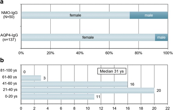
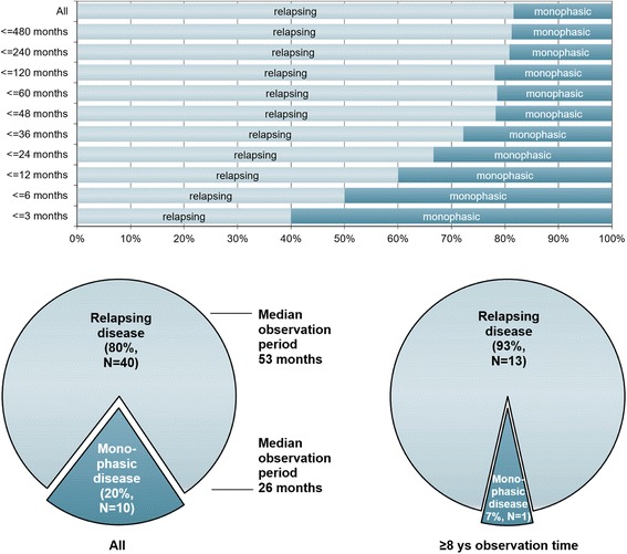
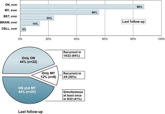
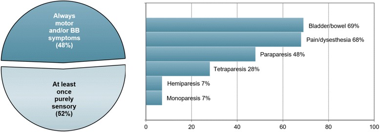
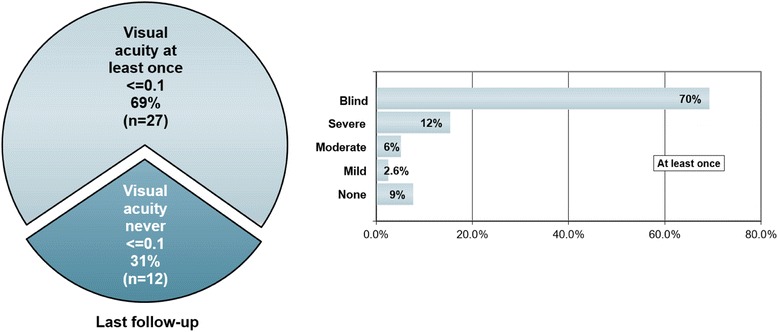
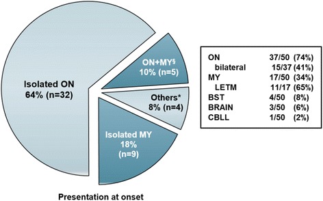
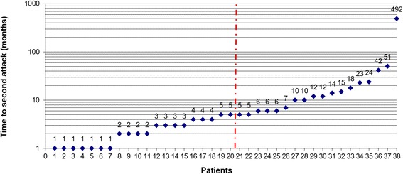
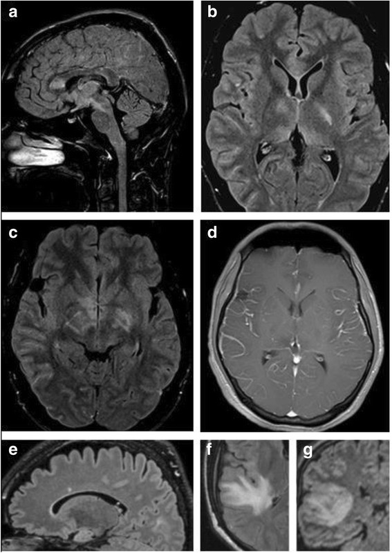
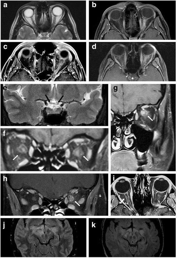
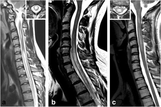
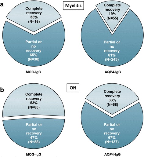
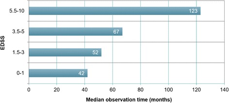
Results: The sex ratio was 1:2.8 (m:f). Median age at onset was 31 years (range 6-70). The disease followed a multiphasic course in 80 % (median time-to-first-relapse 5 months; annualized relapse rate 0.92) and resulted in significant disability in 40 % (mean follow-up 75 ± 46.5 months), with severe visual impairment or functional blindness (36 %) and markedly impaired ambulation due to paresis or ataxia (25 %) as the most common long-term sequelae. Functional blindess in one or both eyes was noted during at least one ON attack in around 70 %. Perioptic enhancement was present in several patients. Besides acute tetra-/paraparesis, dysesthesia and pain were common in acute myelitis (70 %). Longitudinally extensive spinal cord lesions were frequent, but short lesions occurred at least once in 44 %. Fourty-one percent had a history of simultaneous ON and myelitis. Clinical or radiological involvement of the brain, brainstem, or cerebellum was present in 50 %; extra-opticospinal symptoms included intractable nausea and vomiting and respiratory insufficiency (fatal in one). CSF pleocytosis (partly neutrophilic) was present in 70 %, oligoclonal bands in only 13 %, and blood-CSF-barrier dysfunction in 32 %. Intravenous methylprednisolone (IVMP) and long-term immunosuppression were often effective; however, treatment failure leading to rapid accumulation of disability was noted in many patients as well as flare-ups after steroid withdrawal. Full recovery was achieved by plasma exchange in some cases, including after IVMP failure. Breakthrough attacks under azathioprine were linked to the drug-specific latency period and a lack of cotreatment with oral steroids. Methotrexate was effective in 5/6 patients. Interferon-beta was associated with ongoing or increasing disease activity. Rituximab and ofatumumab were effective in some patients. However, treatment with rituximab was followed by early relapses in several cases; end-of-dose relapses occurred 9-12 months after the first infusion. Coexisting autoimmunity was rare (9 %). Wingerchuk's 2006 and 2015 criteria for NMO(SD) and Barkhof and McDonald criteria for multiple sclerosis (MS) were met by 28 %, 32 %, 15 %, 33 %, respectively; MS had been suspected in 36 %. Disease onset or relapses were preceded by infection, vaccination, or pregnancy/delivery in several cases.

Conclusion: Our findings from a predominantly Caucasian cohort strongly argue against the concept of MOG-IgG denoting a mild and usually monophasic variant of NMOSD. The predominantly relapsing and often severe disease course and the short median time to second attack support the use of prophylactic long-term treatments in patients with MOG-IgG-positive ON and/or myelitis.

Keywords: Aquaporin-4 antibodies (AQP4-IgG, NMO-IgG); Autoantibodies; Azathioprine; Barkhof criteria; Cerebrospinal fluid; Electrophysiology; Evoked potentials; Glatiramer acetate; IPND criteria; Infections; Interferon beta; International consensus diagnostic criteria for neuromyelitis optica spectrum disorders; Longitudinally extensive transverse myelitis; Magnetic resonance imaging; McDonald criteria; Methotrexate; Multiple sclerosis; Myelin oligodendrocyte glycoprotein antibodies (MOG-IgG); Natalizumab; Neuromyelitis optica spectrum disorders (NMOSD); Ofatumumab; Oligoclonal bands; Optic neuritis; Outcome; Pregnancy; Rituximab; Therapy; Transverse myelitis; Treatment; Vaccination; Wingerchuk criteria 2006 and 2015.


Figures

Fig. 1
Fig. 1
Sex ratio and age distribution. a Sex ratio in MOG-IgG-positive patients with ON and/or LETM compared with AQP4-IgG-positive ON and/or LETM (the latter data are taken from ref. [34]). b Age distribution at disease onset in 50 MOG-IgG-positive patients with ON and/or myelitis
Fig. 2
Fig. 2
Disease course in relation to observation time in 50 MOG-IgG-positive patients with ON and/or myelitis. Upper panel: Note the decrease in the proportion of monophasic cases with increasing observation time; however, in some patients no relapse has occurred more than 10 years after the initial attack. Lower panel: Note the shorter observation time in the ‘monophasic’ group (left lower panel) and the lower percentage of non-relapsing cases among patients with a long observation period (≥8 years; right lower panel)
Fig. 3
Fig. 3
Attack history at last follow-up. Upper panel: Frequencies of MOG-IgG-positive patients (N = 50) with a history of clinically manifest acute optic neuritis (ON), myelitis (MY), brainstem encephalitis (BST), supratentorial encephalitis (BRAIN), and cerebellitis (CBLL) at last follow-up. Lower panel: Frequencies of MOG-IgG patients with a history of optic neuritis (ON) and myelitis, ON but not myelitis, and myelitis (LETM in all cases) but not ON, respectively, at last follow-up (n = 50)
Fig. 4
Fig. 4
Symptoms present during attacks involving acute myelitis (N = 28 patients). BB = bladder and/or bowel.
Fig. 5
Fig. 5
High-contrast visual acuity (VA) loss during acute ON (N = 39 patients). Blind: complete or functional blindness (VA ≤0.1) in one or both eyes at least once; severe: VA ≤0.5; moderate: VA ≤0.75; mild: ≤1.0; none: high-contrast VA not affected, but low-contrast visual loss, color desaturation, and/or scotoma present
Fig. 6
Fig. 6
Presentation at onset. ON = optic neuritis, MY = myelitis, LETM = longitudinally extensive transverse myelitis, BST = brainstem encephalitis, BRAIN = supratentorial encephalitis, CBLL = cerebellitis. § Includes two cases of simultaneous ON, myelitis and brainstem encephalitis at onset. *Other presentations included simultaneous myelitis, rhombencephalitis and supratentorial encephalitis; simultaneous myelitis and supratentorial encephalitis (2 ×); and isolated brainstem encephalitis. No data on spinal cord lesion length at disease onset were available from 1 patient
Fig. 7
Fig. 7
Time to first relapse in months. The red line indicates the median. The first relapse was defined as a new clinical attack occurring more than 30 days after onset of the initial attack. No exact data was available in two cases
Fig. 8
Fig. 8
Examples of brain lesions detected by MRI. a Sagittal FLAIR image showing callosal lesions as well as lesions extending from the diencephalon to the pons (see case 8 in part 3 of this article series [31] for details). b Axial FLAIR MRI demonstrating lesions in the basal ganglia, juxtacortically on the right side, und in the genu corporis callosi in the same patient. c Axial FLAIR image at the diencephalic level revealing periependymal lesions (in addition to basal ganglia lesions). d Axial T1-weighted image with Gd demonstrating leptomeningeal enhancement (see case 8 in part 3 [31]). E: Sagittal MRI showing a callosal lesion (see case 10 in the Appendix for details). f, g Axial T2-weighted (f) and coronal FLAIR (g) images showing large, confluent T2 hyperintense lesions in the right temporal lobe (see case 7 in part 3 [31])
Fig. 9
Fig. 9
Examples of optic nerve lesions detected by MRI. a, b T2-weighted (a) and T1-weighted (B, with Gd) MRI reveals swelling and Gd enhancement of the left optic nerve. c, d (fat-suppressed): Longitudinal extensive Gd enhancement of the optic nerve (see cases 9 and 12 in part 3 [31] for details). e Longitudinally extensive bilateral optic neuritis extending from the chiasm (E, black arrows) into the orbits, affecting the left more than the right optic nerve. f-h Coronal T1-weighted MRIs display marked contrast enhancement of the intraorbital optic nerve as well as concurrent enhancement of the perioptic nerve sheath, partly extending in the surrounding orbital fat, in patients with acute ON (cases 11, 29 and 19). I: Axial T1-weighted MRI shows Gd enhancement along the right optic nerve in another patient (see case 13 in part 3 of this article series [31]). j, k Axial FLAIR imaging demonstrates bilateral lesions in the optic tract (see case 8 in part 3 [31] for details) (j MRI at attack onset; k follow-up MRI 1 month later)
Fig. 10
Fig. 10
Examples of spinal cord MRI findings. a Sagittal T2-weighted spinal MRI performed at disease onset revealed a large longitudinal centrally located lesion extending over the entire spinal cord as well as swelling of the cord. b Longitudinal extensive central spinal cord T2 lesion in another patient. c T2-hyperintense lesions extending from the pontomedullary junction throughout the cervical cord to C5 in a third patient. The insets in A and C show axial sections of the thoracic cord at lesion level
Fig. 11
Fig. 11
Outcome after acute attacks in MOG-IgG-positive patients compared with a previously published AQP4-IgG-positive cohort. a Outcome after acute myelitis in MOG-IgG-positive (46 evaluable attacks) and in AQP4-IgG-positive patients (298 evaluable attacks [34]). b Outcome after acute ON in MOG-IgG-positive (134 evaluable attacks) and in AQP4-IgG-positive patients (205 evaluable attacks; see ref. [34]). Note that ‘complete recovery’ includes ‘almost complete recovery’ in the left graph (no such distinction was made in the AQP4-IgG-positive cohort)
Fig. 12
Fig. 12
Increase in median EDSS scores with observation time in 47 MOG-IgG-positive patients

References

    1. Jarius S, Wildemann B. The history of neuromyelitis optica. J. Neuroinflammation. 2013;10:8. - PMC - PubMed
    1. Lennon VA, Wingerchuk DM, Kryzer TJ, Pittock SJ, Lucchinetti CF, Fujihara K, Nakashima I, Weinshenker BG. A serum autoantibody marker of neuromyelitis optica: distinction from multiple sclerosis. Lancet. 2004;364:2106–12. doi: 10.1016/S0140-6736(04)17551-X. - DOI - PubMed
    1. Jarius S, Wildemann B. AQP4 antibodies in neuromyelitis optica: diagnostic and pathogenetic relevance. Nat. Rev. Neurol. 2010;6:383–92. doi: 10.1038/nrneurol.2010.72. - DOI - PubMed
    1. Jarius S, Paul F, Franciotta D, Waters P, Zipp F, Hohlfeld R, Vincent A, Wildemann B. Mechanisms of disease: aquaporin-4 antibodies in neuromyelitis optica. Nat. Clin. Pract. Neurol. 2008;4:202–14. - PubMed
    1. Lennon VA, Kryzer TJ, Pittock SJ, Verkman AS, Hinson SR. IgG marker of optic-spinal multiple sclerosis binds to the aquaporin-4 water channel. J. Exp. Med. 2005;202:473–7. doi: 10.1084/jem.20050304. - DOI - PMC - PubMed

Publication types

MeSH terms