. 2016:581:517-539.
doi: 10.1016/bs.mie.2016.08.016. Epub 2016 Oct 10.

Interferometric Scattering Microscopy for the Study of Molecular Motors

Affiliations

Interferometric Scattering Microscopy for the Study of Molecular Motors

J Andrecka et al. Methods Enzymol. 2016.

Abstract

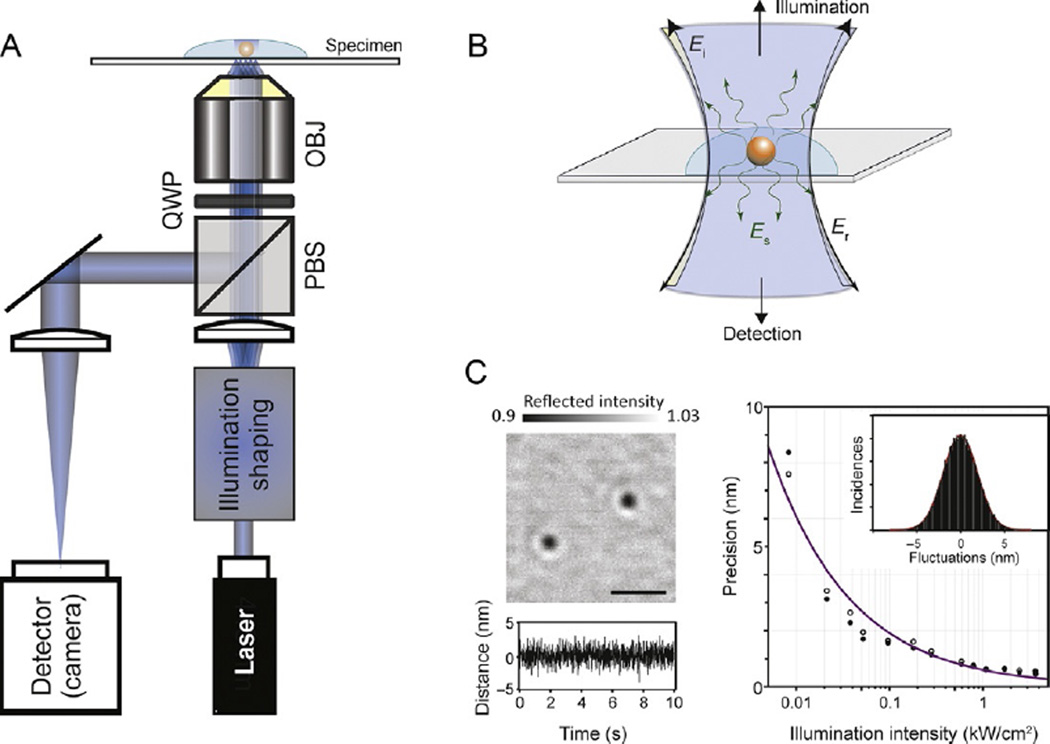
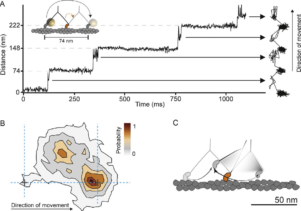
Our understanding of molecular motor function has been greatly improved by the development of imaging modalities, which enable real-time observation of their motion at the single-molecule level. Here, we describe the use of a new method, interferometric scattering microscopy, for the investigation of motor protein dynamics by attaching and tracking the motion of metallic nanoparticle labels as small as 20nm diameter. Using myosin-5, kinesin-1, and dynein as examples, we describe the basic assays, labeling strategies, and principles of data analysis. Our approach is relevant not only for motor protein dynamics but also provides a general tool for single-particle tracking with high spatiotemporal precision, which overcomes the limitations of single-molecule fluorescence methods.

Keywords: Dynein; High speed; Interferometric scattering microscopy; Kinesin; Molecular motors; Myosin; Single molecule; Single-particle tracking.


Figures

Fig. 1
Fig. 1
Principles and performance of iSCAT for single-particle tracking. (A) Experimental setup. OBJ, microscope objective; PBS, polarizing beam splitter; QWP, quarter-wave plate. Illumination shaping refers to different schemes for illuminating the sample without generating excessive speckle such as slim field (Piliarik & Sandoghdar, 2014) or rapid beam scanning (Kukura et al., 2009). (B) Operating principle of iSCAT detection. Light reflected at the sample/glass interface is collected along with any scattered light from the object of interest. Ei, incident electric field; Er, reflected electric field; Es, scattered electric field. (C) Localization of 20 nm gold particles in the shot-noise-limited regime. Left: flat-field-corrected iSCAT image of 20 nm gold nanoparticles immobilized on a glass coverslip in water. Scale bar: 1 µm. The bottom trace shows the fluctuation in the distance between two nanoparticles as a function of time sampled at 1000 frames per second, caused solely by shot noise in the measurement. Right: localization precision as a function of illumination intensity for 20 nm gold nanoparticles. An increase in illumination intensity improves the localization precision for a given exposure time. Open and filled circles: localization precision in x and y, respectively. The line depicts the expected behavior for shot-noise-limited imaging.
Fig. 2
Fig. 2
iSCAT assay for gold-labeled myosin-5a tracking. Flat-field-corrected image showing actin filaments and some 20 nm gold-labeled myosin-5a molecules bound to actin. Inset: zoom of the indicated region showing 20 nm gold-labeled myosin-5a bound to actin (left) and the same region after background subtraction, which removes all static features including the actin filament and any immobile particles (right). Scale bars: 1 µm.
Fig. 3
Fig. 3
iSCAT imaging of gold-labeled dynein. 30 nm gold-labeled dynein bound to a microtubule. In this assay, a gold label produces a positive iSCAT contrast when placed into focus (bright) due to the increased distance between the label and the coverglass surface, in contrast to myosin-5a head-labeled gold, which invariably appeared dark (Fig. 2). Scale bars: 1 µm.
Fig. 4
Fig. 4
Myosin-5a head motion during processive stepping. (A) iSCAT tracking results: distance traveled as a function of time for a single-motor domain labeled with a 20-nm gold particle at its N-terminus and the corresponding 2D-trajectory, which is shown to the right of the time trace. The arrows indicate ~40 nm off-axis side position of the unbound motor domain (transient state). Inset: schematic of myosin-5a labeling and movement. Imaging speed: 1000 frames per second. (B) Contour map of a two-dimensional histogram with a 10 × 10-nm2 bin width obtained from the transient state of the unbound head x–y trajectories (number of steps=486). All contributing steps were aligned and those to the right of the filament when viewed in the direction of motion were mirrored. (C) The data lead to a hand-over-hand spinning model (shown schematically), in which each step proceeds via a single, spatially constrained transient state of the detached head (Andrecka et al., 2015).
Fig. 5
Fig. 5
Example results of kinesin tracking with iSCAT. (A) Schematic showing detectable transitions in the two-step cycle of kinesin with only one motor domain tagged. The entire step (including both one- and two-head-bound durations) of the unlabeled head as well as the two-heads-bound duration of the labeled head precedes the first translocation of the bead (black), and the one-head-bound duration of the labeled head precedes the second translocation of the bead (red). (B) Example XY trajectory for kinesin-1 at saturating ATP. The amount of time that the motor spends on the microtubule and off the microtubule is easily determined. Black and red coloring corresponds to (A). Vertical lines show available microtubule-binding sites. Imaging speed: 1000 frames per second (Mickolajczyk et al., 2015).
Fig. 6
Fig. 6
iSCAT tracking of dynein. Example x–y plot of cytoplasmic dynein position along a microtubule exhibiting side deviations from the track (Δy). The schematic presents one possible explanation of the observed trace where the dynein molecule switches microtubule protofilaments. Dynein was tracked at 200 frames per second using a 20-nm gold nanoparticle bound to AAA domains 5 and 6 (indicated by orange and red circles, respectively).

References

    1. Allen RD, Metuzals J, Tasaki I, Brady ST, Gilbert SP. Fast axonal-transport in squid giant-axon. Science. 1982;218(4577):1127–1129. - PubMed
    1. Andreasson JOL, Milic B, Chen GY, Guydosh NR, Hancock WO, Block SM. Examining kinesin processivity within a general gating framework. eLife. 2015;4:e07403. - PMC - PubMed
    1. Andrecka J, Arroyo JO, Lewis K, Cross RA, Kukura P. Label-free imaging of microtubules with sub-nm precision using interferometric scattering microscopy. Biophysical Journal. 2016;110(1):214–217. - PMC - PubMed
    1. Andrecka J, Ortega Arroyo J, Takagi Y, de Wit G, Fineberg A, MacKinnon L, Kukura P. Structural dynamics of myosin 5 during processive motion revealed by interferometric scattering microscopy. eLife. 2015;4:e05413. - PMC - PubMed
    1. Arroyo JO, Cole D, Kukura P. Interferometric scattering microscopy and its combination with single-molecule fluorescence imaging. Nature Protocols. 2016;11(4):617–633. - PubMed

MeSH terms

LinkOut - more resources