. 2017 Jan;97(Pt A):11-23.
doi: 10.1016/j.nbd.2016.10.005. Epub 2016 Oct 26.

Morphometric alterations of Golgi apparatus in Alzheimer's disease are related to tau hyperphosphorylation

Affiliations

Morphometric alterations of Golgi apparatus in Alzheimer's disease are related to tau hyperphosphorylation

Alejandro Antón-Fernández et al. Neurobiol Dis. 2017 Jan.

Abstract

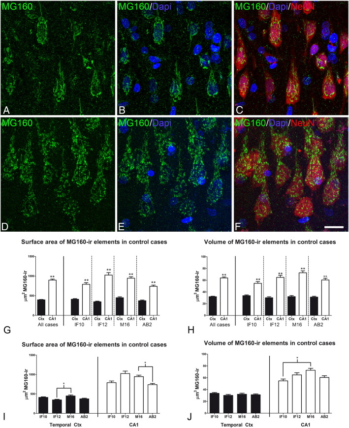
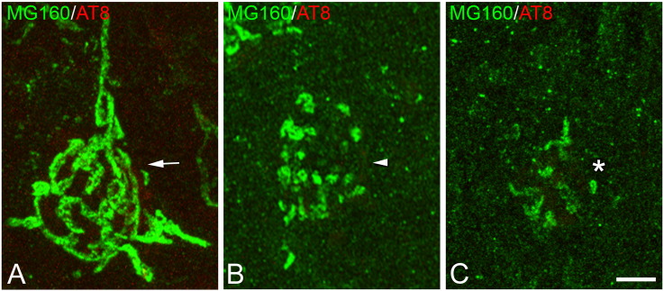
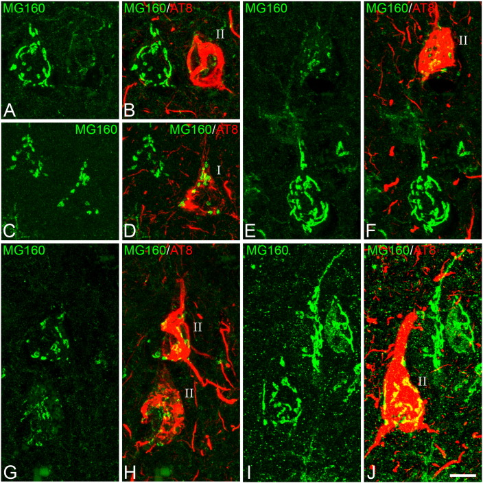
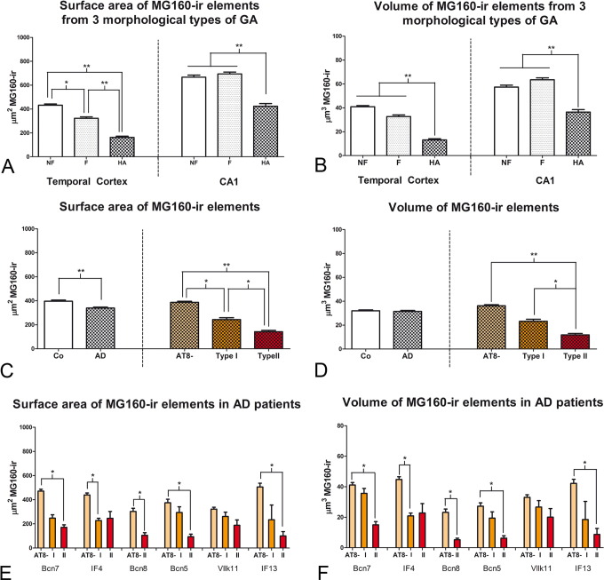
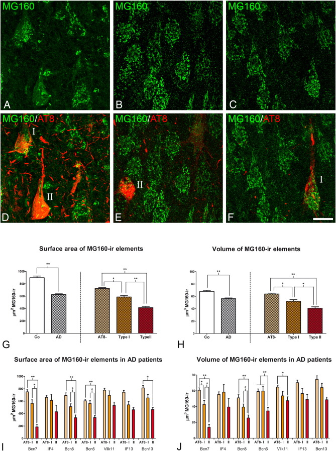
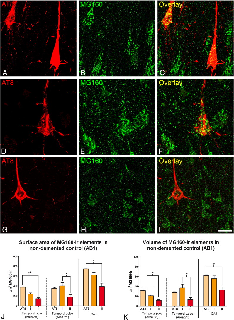
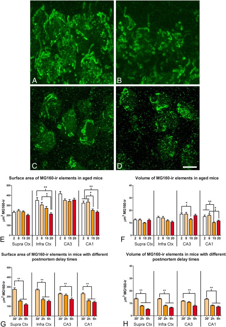
The Golgi apparatus (GA) is a highly dynamic organelle, which is mainly involved in the post-translational processing and targeting of cellular proteins and which undergoes significant morphological changes in response to different physiological and pathological conditions. In the present study, we have analyzed the possible alterations of GA in neurons from the temporal neocortex and hippocampus of Alzheimer's disease (AD) patients, using double immunofluorescence techniques, confocal microscopy and 3D quantification techniques. We found that in AD patients, the percentage of temporal neocortical and CA1 hippocampal pyramidal neurons with a highly altered GA is much higher (approximately 65%) in neurons with neurofibrillary tangles (NFT) than in NFT-free neurons (approximately 6%). Quantitative analysis of the surface area and volume of GA elements in neurons revealed that, compared with NFT-free neurons, NFT-bearing neurons had a reduction of approximately one half in neocortical neurons and one third in CA1 neurons. In both regions, neurons with a pre-tangle stage of phospho-tau accumulation had surface area and GA volume values that were intermediate, that is, between those of NFT-free and NFT-bearing neurons. These findings support the idea that the progressive accumulation of phospho-tau is associated with structural alterations of the GA including fragmentation and a decrease in the surface area and volume of GA elements. These alterations likely impact the processing and trafficking of proteins, which might contribute to neuronal dysfunction in AD.

Keywords: Dementia; Human hippocampus; Human neocortex; Microtubules; Neurofibrillary tangles; Taupathy.


Figures

Fig. 1
Fig. 1
Distribution of MG160 in the GA of human pyramidal neurons A–F: Trios of confocal stack projection images taken from the temporal neocortex (A–C) and the CA1 hippocampal region (D–F) of sections double-immunostained for MG160/NeuN and counterstained with DAPI showing the distribution of MG160 in the GA. Scale bar in F indicates 18 μm in A–C and 16 μm in D–F. G–J: Histograms showing surface area (G, I) and volume (H, J) values (mean ± SE) of GA elements immunoreactive for MG160, obtained from cropped confocal image stacks including complete single pyramidal neurons from temporal neocortex (black bars; total n = 323) and CA1 (white bars; total n = 189). G and H show the statistical comparisons of mean values (surface area and volume, respectively) between neocortical and hippocampal neurons. I and J show the comparisons of the values obtained across the different cases in each region (Friedman test, *p ≤ 0.01; **p ≤ 0.001). Note that in all cases the surface area and volume values of MG160-ir elements in CA1 are higher than in neocortical pyramidal cells (G, H) despite the inter-individual differences (I, J).
Fig. 2
Fig. 2
Morphological types of Golgi apparatus Confocal stack projection mages taken from the temporal neocortex of sections from control cases, double-immunostained for MG160/AT8 showing examples of neurons with a non-fragmented (A), fragmented (B) or highly altered (C) GA. Scale bar indicates 4,8 μm.
Fig. 3
Fig. 3
Distribution of MG160 in the GA of AT8 − and AT8 + human neocortical pyramidal neurons A, B; C, D; E, F; G, H; I, J: pairs of confocal stack projection images taken from the temporal neocortex of sections double-immunostained for MG160/AT8 showing the distribution of MG160 in the GA of AT8 −, and AT8 + (type I and type II) pyramidal neurons. Scale bar shown in J indicates 6.4 μm in A and B, 9.1 μm in C and D, 8.1 μm in E and F, 9.6 μm in G and H and 8.8 μm in I and J.
Fig. 4
Fig. 4
Measurements of the GA in neocortical pyramidal neurons from AD patients A and B: histograms show comparisons in surface area (A) and volume (B) values of MG160-ir elements between neurons with non-fragmented, fragmented or highly altered GA in AD cases (Friedman test, *p ≤ 0.01; **p ≤ 0.001). C–D: histograms showing, on the left side, surface area (C) and volume (D) values (mean ± SE) of MG160-ir GA elements, in control (white bars, n = 323) and AD (patterned bars, n = 733) pyramidal neurons from temporal neocortex. Note that Golgi elements had a higher surface area (but not volume) in control than in neurons from AD patients. Also note (in the histograms on the right) that for AD neurons (taking all cases together) both surface area and volume values were reduced in AT8 + (especially in type II) neurons, compared to AT8 − neurons. Note that in E and F there was a tendency for this reduction in all AD patients analyzed, with statistically significant differences between the different cell types of each patient in five out of the six cases analyzed (Friedman test, *p ≤ 0.01; **p ≤ 0.001).
Fig. 5
Fig. 5
Distribution of MG160 in the GA of AT8 − and AT8 + human CA1 pyramidal neurons A, D; B, E; C, F; Pairs of confocal stack projection images taken from sections from the hippocampal CA1 region double-immunostained for MG160/AT8 showing the distribution of MG160 in the GA of AT8 −, and AT8 + (type I and type II) pyramidal neurons. Scale bar in F indicates 20 μm in A–D and C–F, and 19 μm in B–E. Histograms showing, on the left side, higher surface area (G) and volume (H) values (mean ± SE) of MG160-ir GA elements, in control (white bars, n = 189) compared with AD (patterned bars, n = 713) CA1 pyramidal neurons. Note on the right side that for AD neurons (taking all cases together) both surface area and volume values were reduced in AT8 + (especially in type II) neurons, compared to AT8 − neurons. Note that in I and J there was a tendency for this reduction in all AD patients analyzed, with statistically significant differences between the different cell types of each patient in four out of the seven cases analyzed.
Fig. 6
Fig. 6
Distribution of MG160 in the GA of AT8 − and AT8 + human CA1 and neocortical pyramidal neurons in a non-demented control case Pairs of confocal stack projection images taken from CA1 (A–C) and areas 21 (D–F) and 38 (G–I) of the temporal neocortex from sections double-immunostained for MG160/AT8 showing the distribution of MG160 in the GA of AT8 −, and AT8 + (type I and type II) pyramidal neurons from a human in a non-demented control case. Scale bar in I indicates 24 μm in A–C, 14 μm in D–F and 22,65 in G–I. Histograms show surface area (J) and volume (K) values (mean ± SE) of MG160-ir GA elements, in AT8 − neurons (area 38 n = 397; area 21 n = 133; CA1 n = 185) and AT8 + neurons (patterns I and II; area 38, n = 49; area 21, n = 12; CA1, n = 47). Note that both surface area and volume values were significantly reduced in type II AT8 + neurons, compared to AT8 − neurons (Friedman test, *p ≤ 0.01; **p ≤ 0.001).
Fig. 7
Fig. 7
Effects of aging and the postmortem delay period on the distribution of MG160 in the GA of pyramidal neurons A and B: confocal stack projection mages showing examples of MG160 immunostaining of the GA of CA1 pyramidal neurons from mice aged 2 (A) and 20 (B) months. C and D show the MG160 immunostaining of the GA of layer II neocortical pyramidal neurons from mice fixed after postmortem periods of 30 min (C) or 5 h (D). Scale bar in D indicates 5,3 μm. Histograms show the reductions in surface area (E and G) and volume (F and H) values (mean ± SE) of MG160-ir GA elements, in pyramidal neurons from different areas in mice with different ages (E and F) or mice fixed after different postmortem times G and H). (Kruskal Wallis test, *p ≤ 0.01; **p ≤ 0.001).

References

    1. Anton-Fernandez A. Changes in the Golgi apparatus of neocortical and hippocampal neurons in the hibernating hamster. Front. Neuroanat. 2015;9:157. - PMC - PubMed
    1. Baloyannis S.J. Golgi apparatus and protein trafficking in Alzheimer's disease. J. Alzheimers Dis. 2014;42(Suppl. 3):S153–S162. - PubMed
    1. Blazquez-Llorca L. Pericellular innervation of neurons expressing abnormally hyperphosphorylated tau in the hippocampal formation of Alzheimer's disease patients. Front. Neuroanat. 2010;4:20. - PMC - PubMed
    1. Blazquez-Llorca L. Abnormal tau phosphorylation in the thorny excrescences of CA3 hippocampal neurons in patients with Alzheimer's disease. J. Alzheimers Dis. 2011;26:683–698. - PubMed
    1. Braak H., Braak E. Staging of Alzheimer's disease-related neurofibrillary changes. Neurobiol. Aging. 1995;16:271–278. (discussion 278-84) - PubMed

Publication types

MeSH terms