Review
. 2017 Jan 3;7(1):a024364.
doi: 10.1101/cshperspect.a024364.

Sporadic and Infectious Human Prion Diseases

Affiliations
Review

Sporadic and Infectious Human Prion Diseases

Robert G Will et al. Cold Spring Harb Perspect Med. .

Abstract

Human prion diseases are rare neurodegenerative diseases that have become the subject of public and scientific interest because of concerns about interspecies transmission and the unusual biological properties of the causal agents: prions. These diseases are unique in that they occur in sporadic, hereditary, and infectious forms that are characterized by an extended incubation period between exposure to infection and the development of clinical illness. Silent infection can be present in peripheral tissues during the incubation period, which poses a challenge to public health, especially because prions are relatively resistant to standard decontamination procedures. Despite intense research efforts, no effective treatment has been developed for human prion diseases, which remain uniformly fatal.


Figures

Figure 1.
Figure 1.
Mean annual mortality rates for sporadic Creutzfeldt–Jakob disease (sCJD) in Europe (periods of surveillance, 8–21 years).
Figure 2.
Figure 2.
Magnetic resonance imaging (MRI) brain scan in sporadic Creutzfeldt–Jakob disease (sCJD): Arrows indicate high signal in the caudate, putamen, and occipital cortex.
Figure 3.
Figure 3.
Real-time quaking-induced conversion (RT-QuIC) on cerebrospinal fluid (CSF) in sporadic Creutzfeldt–Jakob disease (sCJD): relative fluorescence with time.
Figure 4.
Figure 4.
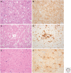
Neuropathology of sporadic Creutzfeldt–Jakob disease (sCJD) subtypes: (A,B) MM1/MV1; (C,D) MM2; and (E,F) sporadic fatal insomnia. (A) The frontal cortex in the MM1 and MV1 subtypes shows microvacuolar spongiform change. Hematoxylin and eosin, ×40. (B) PrP accumulates in the cerebral cortex in the MM1/MV1 subtypes in a diffuse granular/synaptic pattern. 12F10 antibody, ×40. (C) The frontal cortex in the MM2 cortical subtype shows confluent spongiform change. Hematoxylin and eosin, ×20. (D) PrP accumulates in the cerebral cortex in the MM2 cortical subtype in a dense perivacuolar pattern. 12F10 antibody, ×20. (E) The thalamus in sporadic fatal insomnia (MM2 thalamic subtype) exhibits severe neuronal loss without significant vacuolation. Hematoxylin and eosin, ×40. (F) Marked thalamic gliosis in sporadic fatal insomnia is demonstrated with an antibody to glial fibrillary acidic protein, ×40.
Figure 5.
Figure 5.
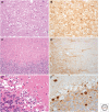
Neuropathology of sporadic Creutzfeldt–Jakob disease (sCJD) subtypes: (A,B) VV1; (C,D) VV2; (E,F) MV2. (A) Spongiform change in the temporal cortex in the VV1 subtype has vacuoles intermediate in size between the MM1 and MM2 subtypes. Hematoxylin and eosin, ×40. (B) Prion protein (PrP) accumulates in the temporal cortex in a patchy coarse granular pattern in the VV1 subtype. 12F10 antibody, ×40. (C) Spongiform change in the VV2 subtype is most marked in the molecular layer of the cerebellum. Hematoxylin and eosin, ×20. (D) PrP accumulates in the frontal cortex in a perineuronal/granular pattern in the VV2 subtype. 12F10 antibody, ×20. (E). The pathological hallmark of the MV2 subtype is the kuru-type plaque in the cerebellum, composed of a dense amyloid core with paler peripheral fibrils. Hematoxylin and eosin, ×40. (F) PrP accumulates in the kuru-type plaques and in a granular/synaptic pattern in the cerebellum of the MV2 subtype. 12F10 antibody, ×40.
Figure 6.
Figure 6.
Western blot for protease-resistant prion protein (PrPres) in sporadic, iatrogenic, and variant Creutzfeldt–Jakob disease (CJD) as well as in variably protease-sensitive prionopathy (VPSPr). (A) Schematic representation of the PrPres types recognized to characterize sporadic CJD (sCJD), iatrogenic CJD (iCJD), variant CJD (vCJD), and VPSPr brain tissue. The typing scheme has two component parts, the mobility of the bottom (nonglycosylated) fragment (type 1 at ∼21 kDa and/or type 2 at ∼19 kDa) and glycosylation site occupancy (A, in which the middle, monoglycosylated band predominates, or B, in which the top, diglycosylated band predominates). (B) Western blot analysis of brain tissue from the two most common subtypes of sCJD (MM1 and VV2), vCJD, and VPSPr. The VPSPr lane was loaded with 10 times the amount of brain homogenate loaded in the CJD lanes. (C) Western blot analysis of brain tissue from a case of human growth hormone (hGH) iCJD (VV), flanked by samples of sCJD of the MM1 and VV2 subtypes. Typing of the sample is designated below each lane.
Figure 7.
Figure 7.
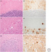
Neuropathology: (A,B) variably protease-sensitive prionopathy (VPSPr) (C,D) kuru; and (E,F) iatrogenic Creutzfeldt–Jakob disease (iCJD). (A) The cerebellar molecular layer in VPSPr shows patchy spongiform change. Hematoxylin and eosin, ×20. (B) Prion protein (PrP) accumulates in the cerebellar cortex in numerous microplaques, one of the pathological hallmarks of VPSPr. 12F10 antibody, ×20. (C) The granular layer of the cerebellum in kuru often contains characteristic plaques, with a dense amyloid core and pale peripheral fibrils. Hematoxylin and eosin, ×40. (D) PrP accumulates in the cerebellar plaques in kuru and in a patchy distribution in the granular layer. KG9 antibody, ×20. (E) The cerebellum in iCJD in hGH recipients is atrophic, with marked neuronal loss and gliosis. Hematoxylin and eosin, ×20. (F) PrP accumulates in the frontal cortex in iCJD in an hGH recipient in a plaque-like pattern with widespread granular/synaptic positivity. 12F10 antibody, ×40.
Figure 8.
Figure 8.
Epidemiologic curve of variant Creutzfeldt–Jakob disease (vCJD) in the United Kingdom.
Figure 9.
Figure 9.
Magnetic resonance imaging (MRI) brain scan in variant Creutzfeldt–Jakob disease (vCJD) showing bilateral pulvinar high signal.
Figure 10.
Figure 10.
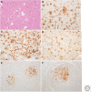
Neuropathology of variant Creutzfeldt–Jakob disease (vCJD) and subclinical vCJD infection. (A) The pathological hallmark of vCJD in the frontal cortex is the florid plaque, composed of a dense amyloid core with pale radiating fibrils surrounded by spongiform change. Hematoxylin and eosin, ×40. (B) Prion protein (PrP) accumulates in the florid plaques in vCJD and in numerous smaller clusters of plaques and amorphous deposits. 12F10 antibody, ×20. (C) In the basal ganglia in vCJD, PrP accumulates in a linear periaxonal pattern. 12F10 antibody, ×40. (D) The pulvinar in vCJD exhibits severe neuronal loss and gliosis, with numerous large reactive astrocytes. Glial fibrillary acidic protein antibody, ×40. (E) PrP accumulates in the follicular dendritic cells within the lymphoid follicles of the tonsil in vCJD. 12F10 antibody, ×20. (F) PrP accumulates in the follicular dendritic cells of a cervical lymph node in a patient (PRNP codon 129 MV genotype) who died with an asymptomatic vCJD infection following a contaminated red blood cell transfusion. 12F10 antibody, ×20.

References

    1. Alcalde-Cabero E, Almazán-Isla J, Brandel JP, Breithaupt M, Catarino J, Collins S, Haybäck J, Höftberger R, Kahana E, Kovacs GG, et al. 2012. Health professionals and risk of sporadic Creutzfeldt–Jakob disease, 1965 to 2010. Eurosurveillance 17: 1–10. - PubMed
    1. Barbot C, Castro L, Oliveira C, Carpenter C. 2010. Variant Creutzfeldt–Jakob disease: The first confirmed cases from Portugal shows early onset, long duration and unusual pathology. J Neurol Neurosurg Psychiatry 81: 112–115. - PubMed
    1. Binelli S, Agazzi P, Giaccone G, Will RG, Bugiani O, Franceschetti S, Tagliavini F. 2006. Periodic electroencephalogram complexes in a patient with variant Creutzfeldt–Jakob disease. Ann Neurol 59: 423–427. - PubMed
    1. Bishop MT, Ritchie DL, Will RG, Ironside JW, Head MW, Thomson V, Bruce M, Manson JC. 2008. No major change in vCJD agent strain after secondary transmission via blood transfusion. PLoS ONE 3: 1–6. - PMC - PubMed
    1. Bishop MT, Will RG, Manson JC. 2010. Defining sporadic Creutzfeldt–Jakob disease strains and their transmission properties. Proc Natl Acad Sci 107: 12005–12010. - PMC - PubMed

LinkOut - more resources