. 2016 Dec 9;291(50):26216-26225.
doi: 10.1074/jbc.M116.756502. Epub 2016 Oct 28.

The Role of Sialylated Glycans in Human Platelet Endothelial Cell Adhesion Molecule 1 (PECAM-1)-mediated Trans Homophilic Interactions and Endothelial Cell Barrier Function

Affiliations

The Role of Sialylated Glycans in Human Platelet Endothelial Cell Adhesion Molecule 1 (PECAM-1)-mediated Trans Homophilic Interactions and Endothelial Cell Barrier Function

Panida Lertkiatmongkol et al. J Biol Chem. .

Abstract

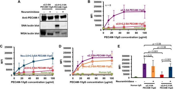
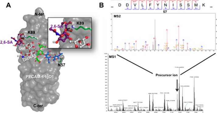
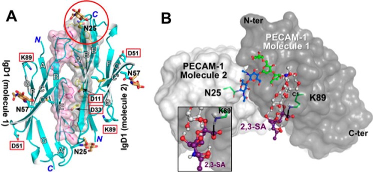
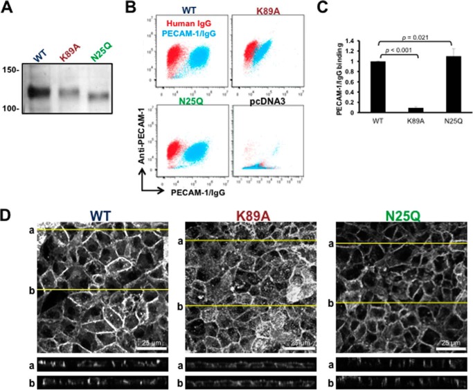
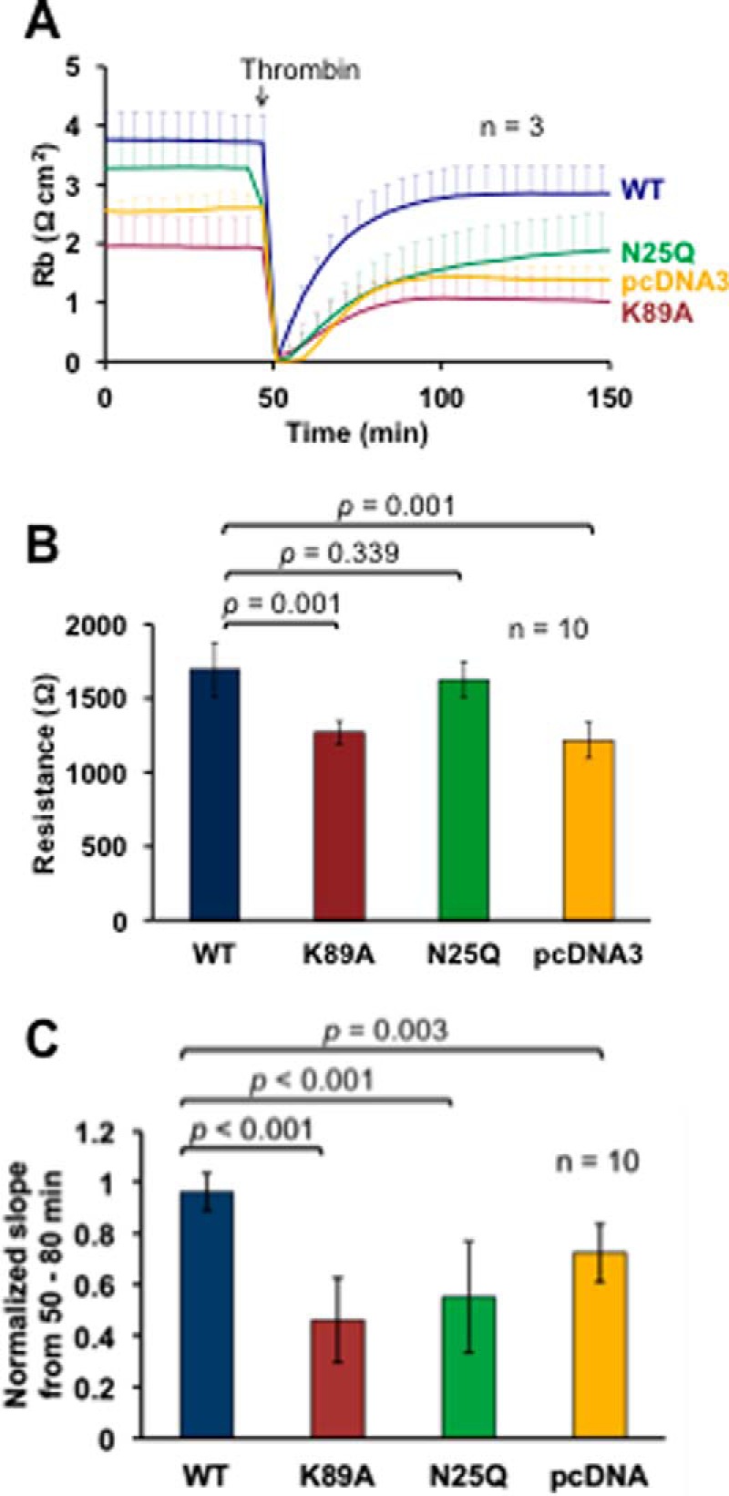
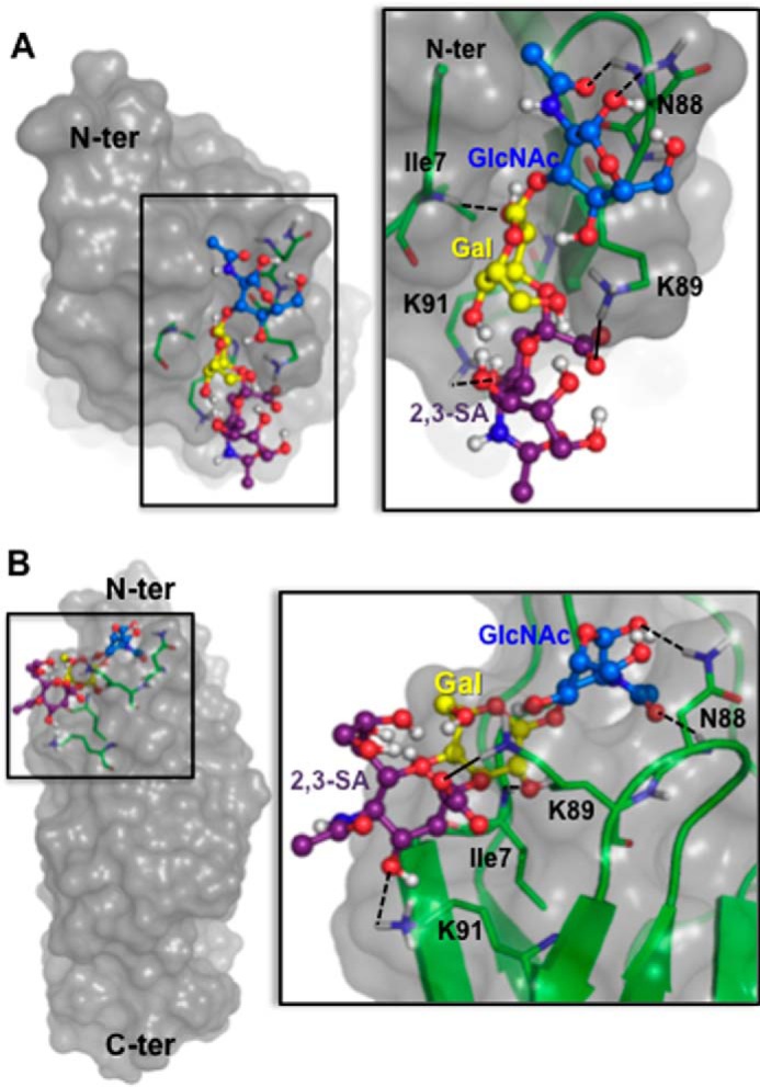
Platelet Endothelial Cell Adhesion Molecule 1 (PECAM-1) is a major component of the endothelial cell intercellular junction. Previous studies have shown that PECAM-1 homophilic interactions, mediated by amino-terminal immunoglobulin homology domain 1, contribute to maintenance of the vascular permeability barrier and to its re-establishment following inflammatory or thrombotic insult. PECAM-1 glycans account for ∼30% of its molecular mass, and the newly solved crystal structure of human PECAM-1 immunoglobulin homology domain 1 reveals that a glycan emanating from the asparagine residue at position 25 (Asn-25) is located within the trans homophilic-binding interface, suggesting a role for an Asn-25-associated glycan in PECAM-1 homophilic interactions. In support of this possibility, unbiased molecular docking studies revealed that negatively charged α2,3 sialic acid moieties bind tightly to a groove within the PECAM-1 homophilic interface in an orientation that favors the formation of an electrostatic bridge with positively charged Lys-89, mutation of which has been shown previously to disrupt PECAM-1-mediated homophilic binding. To verify the contribution of the Asn-25 glycan to endothelial barrier function, we generated an N25Q mutant form of PECAM-1 that is not glycosylated at this position and examined its ability to contribute to vascular integrity in endothelial cell-like REN cells. Confocal microscopy showed that although N25Q PECAM-1 concentrates normally at cell-cell junctions, the ability of this mutant form of PECAM-1 to support re-establishment of a permeability barrier following disruption with thrombin was significantly compromised. Taken together, these data suggest that a sialic acid-containing glycan emanating from Asn-25 reinforces dynamic endothelial cell-cell interactions by stabilizing the PECAM-1 homophilic binding interface.

Keywords: adhesion; endothelial cell; glycosylation; permeability; platelet endothelial cell adhesion molecule (PECAM); sialic acid; vascular biology.


Figures

FIGURE 1.
FIGURE 1.
α2,6-linked sialic acid residues inhibit the homophilic interactions of human PECAM-1. A, α2,3-sialylated PECAM-1/IgG was purified from the culture supernatant of PECAM-1/IgG-transfected wild-type CHO cells, which express only α2,3-sialyltransferases. ST6Gal-1-transfected CHO cells were employed to express α2,6+2,3-sialylated PECAM-1/IgG. Both types of PECAM-1/IgG were also desialylated with neuraminidase and subjected to immunoblotting and lectin blotting analyses. Note that only PECAM-1/IgG purified from ST6Gal-1-transfected CHO cells is SNA-positive. Also note that neuraminidase treatment results in the generation of species of lower apparent molecular weight and reduced reactivity with wheat germ agglutinin, which binds to sialic acid residues and, to a lesser extent, the GlcNAc residues that are exposed following neuraminidase treatment. B, concentration-dependent binding of PECAM-1/IgG to PECAM-1-transfected REN cells. Note that the presence of α2,6 sialic acid on PECAM-1/IgG inhibits its trans homophilic binding down to background levels, as defined by the binding of normal human IgG. C, removal of sialic acid residues restores the binding of α2,6-linked sialylated PECAM-1/IgG while having little effect on the binding of α2,3 sialylated PECAM-1/IgG (D). E, quantification of PECAM-1/IgG binding from eight independent FACS binding assays of PECAM-1/IgG at a concentration of 100 μg/ml. The p values were derived from a Student's t test. MFI, median fluorescence intensity.
FIGURE 2.
FIGURE 2.
The Asn-57-linked glycan of PECAM-1/IgG from ST6Gal-1-transfected CHO cells contains an α2,6-linked sialic acid that blocks a key residue involved in human PECAM-1-mediated homophilic binding. A, unbiased molecular docking of α2,6-sialylated lactosamine to the X-ray crystal structure of human PECAM-1 IgD1 predicts that the carboxyl moiety of α2,6-linked sialic acid (violet ball and stick) binds across the face of the domain in such a way as to form an intrachain hydrogen bond (black line in the inset) with the ϵ amino group of amino acid Lys-89, thus blocking a residue shown previously to be important for PECAM-1-mediated homophilic binding. The GlcNAc-Gal disaccharide (white rings) of the bound α2,6-sialylated lactosamine are adjacent to the GlcNAc-GlcNAc-Man3 antennae (two blue rings followed by three green rings) that emanate from Asn-57. Side chains of Lys-89 and Asn-57 are shown as green sticks. N, O, and H atoms are colored blue, red, and white, respectively. C-ter, C terminus; N-ter, N terminus. B, the MS1 spectrum of deglycosylated α2,6-sialylated glycopeptides derived from α2,6+α2,3-sialylated PECAM-1/IgG shows a 715.8296 m/z ion (z = 2, 1431.6592 Da) that was subsequently fragmented by collision-induced dissociation. The amino acid sequence shown in the MS2 spectrum of the 715.8296 m/z ion includes Asn-57.
FIGURE 3.
FIGURE 3.
The Asn-25 glycosylation site, present in human but not murine PECAM-1, is located within the IgD1/IgD1 trans homophilic-binding interface. A, side view (adapted from Ref. 8) of IgD1/D1 trans homophilic interactions, with the homophilic interface shown as a pink space-filling model. Asn-25 is circled in red. The truncated GlcNAc-GlcNAc-Man3 glycan emanating from Asn-25 in PECAM-1 molecule 2 is represented by orange-tipped yellow sticks. The amino acid residues in red boxes have been shown previously by Newton et al. (7) to be implicated in PECAM-1 homophilic interactions. Note that Lys-89 is not located on the binding interface. B, en face view of the likely full-length complex carbohydrate emanating from Asn-25 molecule 2. The first two GlcNAc residues emanating from this residue are shown in blue, with the Man3 antenna shown in green. A trisaccharide comprised of N-acetylglucosamine, galactose, and α2,3-linked sialic acid (α2,3-sialylated lactosamine) was subjected to unbiased molecular docking and found to bind with high affinity to PECAM-1 molecule 1 in such a way as to hydrogen-bond (black line) with the ϵ-amino group of Lys-89. Thus, this glycan forms an intermolecular bridge between the two opposing PECAM-1 IgD1 domains interacting in trans. C-ter, C terminus; N-ter, N terminus.
FIGURE 4.
FIGURE 4.
Elimination of the N-glycan attached to Asn-25 has little effect on steady-state PECAM-1-mediated homophilic binding. A, detergent lysates from WT-, K89A-, and N25Q-PECAM-1-transfected REN cells were immunoprecipitated with mouse anti-PECAM-1 mAb 1.3 and analyzed on silver-stained SDS gels. Removal of the Asn-25-linked glycan decreased the molecular mass of N25Q PECAM-1 as expected. B, FACS analysis of α2,3-sialylated PECAM-1/IgG homophilic binding to WT-, K89A-, and N25Q-PECAM-1-transfected REN cells. REN cells transfected with vector pcDNA3 did not bind to α2,3-sialylated PECAM-1/IgG or express PECAM-1. Thus, they were not further quantitated. C, quantitation of PECAM-1/IgG binding from 12 independent experiments, normalized for the level of PECAM-1 expression in each cell line. Student's t test revealed that absence of the Asn-25 glycan has a negligible difference in trans homophilic binding of PECAM-1/IgG. D, REN cells transfected with the indicated PECAM-1 isoforms were stained with the mouse anti-human PECAM-1 mAb 1.3. Projection of the entire Z stack is shown in the top panel. Two cross-sections of each transfected REN cells (yellow lines) are displayed in the bottom panel. Note that although the homophilically crippled K89A mutant form of PECAM-1 is uniformly distributed along the entire surface of the cell, both WT PECAM-1 and the N25Q mutant form of PECAM-1 are able to concentrate at cell-cell junctions.
FIGURE 5.
FIGURE 5.
The Asn-25 glycan emanating from PECAM-1 IgD1 supports dynamic PECAM-1 homophilic interactions. A, multifrequency ECIS analysis of baseline barrier function (Rb) and the rate of recovery following thrombin challenge. Note that absence of the Asn-25 glycan has little effect on the baseline steady state barrier function but a dramatic effect on the dynamic rate and extent of barrier function following thrombin challenge. The data shown are from three independent experiments. B, baseline resistance of REN cells expressing the indicated mutant forms of PECAM-1 from 10 independent experiments. Resistance was measured at 4000 Hz. C, linear regression analysis of the rate of recovery of barrier function after thrombin challenge in 10 separate experiments, measuring the slope from the 50- to 80-min time points, as illustrated in A. Data reflect the standard deviation in duplicate wells normalized to the rate of recovery of cells expressing WT PECAM-1.
FIGURE 6.
FIGURE 6.
Molecular details of the trans homophilic interaction between the α2,3-sialylated glycan emanating from Asn-25 of PECAM-1/IgG and four highly conserved amino acids in IgD1 of cellular PECAM-1 to which it binds. A, in addition to the molecular interaction of the carboxylate moiety of α2,3 sialic acid (purple ball and stick) with the ϵ amino group of Lys-89 (black line, detailed in Fig. 3B), the hydroxyl group of the bound sialic acid also forms a hydrogen bond (black dashed line) with Lys-91. Glycan interactions are additionally stabilized by a hydrogen bond between the α amino group of Ile-7 and the hydroxyl group of galactose (yellow ring) and two hydrogen bonds between Asn-88 and GlcNAc (blue ring). All four of these glycan-binding amino acids are completely conserved in human, pig, cow, dog, and mouse PECAM-1. B, 90° rotation view of A. C-ter, C terminus; N-ter, N terminus.

Similar articles

Cited by

References

    1. Newman P. J. (1997) The biology of PECAM-1. J. Clin. Invest. 99, 3–8 - PMC - PubMed
    1. Newman P. J. (1999) Switched at birth: a new family for PECAM-1. J. Clin. Invest. 103, 5–9 - PMC - PubMed
    1. Newman P. J., Berndt M. C., Gorski J., White G. C. 2nd, Lyman S., Paddock C., and Muller W. A. (1990) PECAM-1 (CD31) cloning and relation to adhesion molecules of the immunoglobulin gene superfamily. Science 247, 1219–1222 - PubMed
    1. Newman P. J., and Newman D. K. (2003) Signal transduction pathways mediated by PECAM-1: new roles for an old molecule in platelet and vascular cell biology. Arterioscler. Thromb. Vasc. Biol. 23, 953–964 - PubMed
    1. Sun Q.-H., DeLisser H. M., Zukowski M. M., Paddock C., Albelda S. M., and Newman P. J. (1996) Individually distinct Ig homology domains in PECAM-1 regulate homophilic binding and modulate receptor affinity. J. Biol. Chem. 271, 11090–11098 - PubMed

MeSH terms

LinkOut - more resources