. 2016 Dec 1;25(23):5059-5068.
doi: 10.1093/hmg/ddw310.

PINK1 and Parkin are genetic modifiers for FUS-induced neurodegeneration

Affiliations

PINK1 and Parkin are genetic modifiers for FUS-induced neurodegeneration

Yanbo Chen et al. Hum Mol Genet. .

Abstract

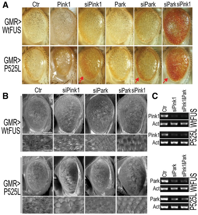
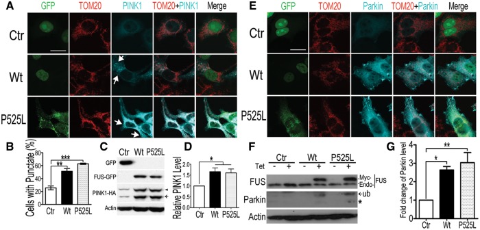
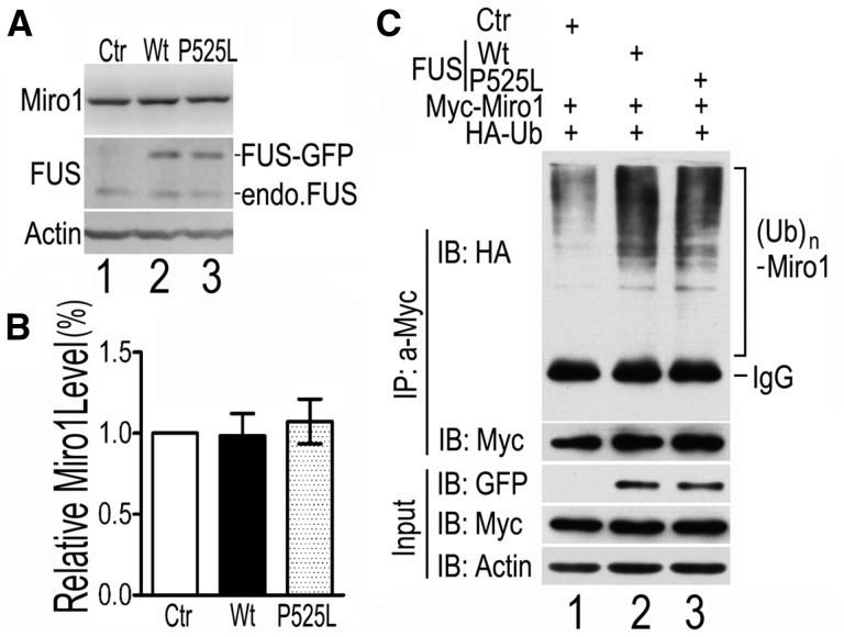
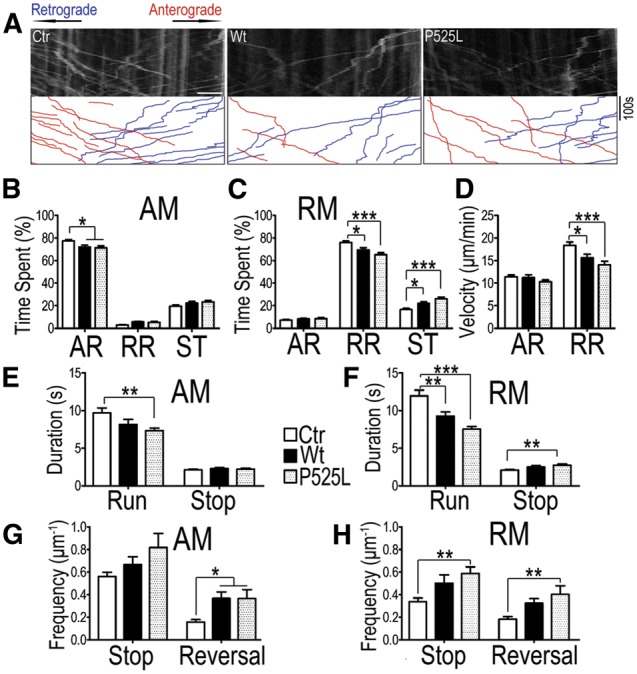
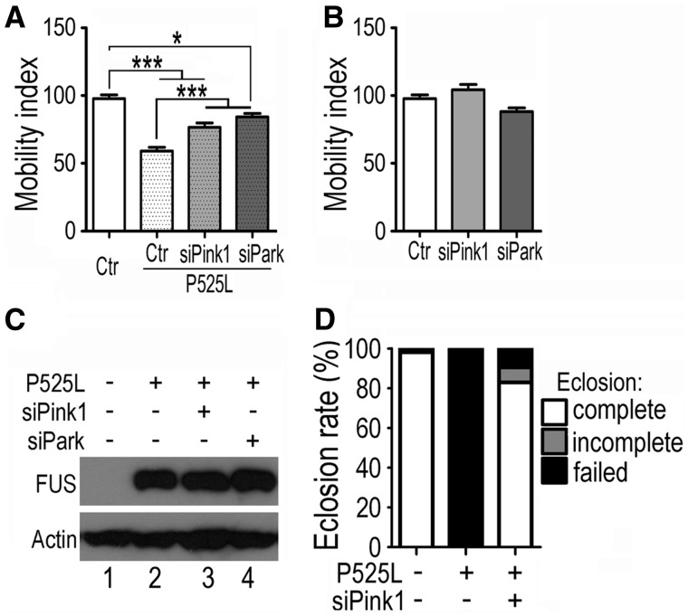
Dysregulation of Fused in Sarcoma (FUS) gene expression is associated with fronto-temporal lobar degeneration (FTLD), and missense mutations in the FUS gene have been identified in patients affected by amyotrophic lateral sclerosis (ALS). However, molecular and cellular defects underlying FUS proteinopathy remain to be elucidated. Here, we examined whether genes important for mitochondrial quality control play a role in FUS proteinopathy. In our genetic screening, Pink1 and Park genes were identified as modifiers of neurodegeneration phenotypes induced by wild type (Wt) or ALS-associated P525L-mutant human FUS. Down-regulating expression of either Pink1 or Parkin genes ameliorated FUS-induced neurodegeneration phenotypes. The protein levels of PINK1 and Parkin were elevated in cells overexpressing FUS. Remarkably, ubiquitinylation of Miro1 protein, a downstream target of the E3 ligase activity of Parkin, was also increased in cells overexpressing FUS protein. In fly motor neurons expressing FUS, both motility and processivity of mitochondrial axonal transport were reduced by expression of either Wt- or P525L-mutant FUS. Finally, down-regulating PINK1 or Parkin partially rescued the locomotive defects and enhanced the survival rate in transgenic flies expressing FUS. Our data indicate that PINK1 and Parkin play an important role in FUS-induced neurodegeneration. This study has uncovered a previously unknown link between FUS proteinopathy and PINK1/Parkin genes, providing new insights into the pathogenesis of FUS proteinopathy.


Figures

Figure 1.
Figure 1.
Down-regulating Pink1 or Parkin in Drosophila partially rescues retinal degeneration in flies expressing Wt- or P525L-mutant FUS. (A) Bright field microscopy (A) of fly eyes show that RNAi against Pink1 or Parkin (siPink1 or siPark, respectively) mitigates retinal degeneration caused by Wt- or P525L-mutant FUS, whereas overexpression of Pink1 exacerbates the eye phenotype in flies expressing the P525L-mutant FUS. (B) Scanning EM (SEM) images showing areas of the fly eyes with restored ommatidial organization. Scale bars in panel B: 100 μm (upper panels) or 20 μm (lower panels at higher magnification of SEM). (C) The down-regulation of Pink1 and Park gene expression was verified by RT-PCR.
Figure 2.
Figure 2.
Expression of Wt- or P525L-mutant FUS in mammalian cells led to accumulation of PINK1 and Parkin proteins. (A) Immunofluorescent staining reveals the punctate distribution of PINK1 in cells expressing either Wt- or P525L-mutant FUS. Following co-transfection of HEK293 cells with different plasmids [PINK1-HA together with the nuclear localized GFP control (NLS-GFP), or Wt-FUS or P525L-mutant FUS tagged with GFP], immunostaining was performed using anti-TOM20 and anti-HA antibodies 20 h post-transfection. (B) Less than 25% of the control cells exhibited punctate PINK1 staining, whereas cells expressing either Wt- or P525L-mutant FUS show a significant increase in the percentages of cells with the punctate pattern of PINK1 distribution (puncta are marked by the white arrows). Data represent three independent experiments with at least 50 cells in each group in each experiment. (C, D) PINK1 accumulated in Wt-or P525L-FUS expressing cells. (E) Immunostaining images of cells co-transfected with different plasmid combinations (Parkin-Flag together with NLS-GFP, or with Wt- or P525L-mutant FUS tagged with GFP). Cells were stained with anti-TOM20 and anti-Flag antibodies 20h after transfection. (F, G) The level of Parkin protein was determined by Western blotting analyses of cell lysates prepared from inducible HEK293 cells expressing either Wt- or P525L-mutant FUS upon induction using tetracycline (0.5 μg/mL; 20 h). Data represent independent experiments. Scale bars: 20 μm. Data were analyzed using one-way ANOVA (*: P < 0.05; **: P < 0.01; ***: P < 0.001).
Figure 3.
Figure 3.
Expression of Wt- or P525L-mutant FUS enhances MIRO1 ubiquitination. (A, B) Endogenous MIRO1 protein level is not altered in Wt- or P525L-mutant FUS expressing cells. Cells were co-transfected with different plasmids (NLS-GFP control or Wt- or P525L-mutant FUS tagged with GFP). Protein levels were determined using Western blotting with corresponding antibodies. No significant changes were detected in the MIRO1 protein level among cells expressing either GFP control or Wt-FUS or P525L-mutant FUS. Data represent three independent experiments and are analyzed using one-way ANOVA. (C) Expression of Wt-FUS or P525L-mutant FUS promotes MIRO1 ubiquitination. Cells were co-transfected with different combinations of plasmids as shown above the gel: HA-ubiquitin (HA-Ub) together with myc-MIRO1 and GFP, or Wt- or P525L-mutant FUS. Following immunoprecipitation using anti-myc, the ubiquitinyted MIRO1 was detected by HA antibody. Poly-ubiquitinated MIRO1 was increased in cells expressing Wt- or P525L-mutant FUS as compared with the control cells. Data represent three independent experiments.
Figure 4.
Figure 4.
Axonal mitochondrial transport defects in MNs of FUS-transgenic flies as detected by the mitochondrial imaging in MN axons in the ventral nerve cord (VNC). (A) Representative kymographs of axonal mitochondria labeled by mito-GFP in MNs of control (Ctr) or transgenic flies expressing either Wt- or P525L-mutant FUS. A 40-μm axonal region was photo-bleached before image acquisition and video recorded for 6 min with VNC placed on the left of the acquired images. The anterograde (plus end-directed) and retrograde (minus end-directed) trafficking of mitochondria are marked in red and blue respectively. Scale bar: 5 μm. (B–H) Quantification of mitochondrial transport in MN axons of flies expressing Wt- or P525L-mutant FUS as compared with the control flies. Images were processed and quantified using ImageJ. (B, C) show duty cycles of the mitochondria that exhibit net anterograde movement (AM) and retrograde movement (RM). Duty cycles of individual mitochondria are divided into anterograde run (AR), retrograde run (RR), and stop (ST). A significant decrease in the time spent on moving (AR in B and RR in C, respectively) together with a significant increase in time spent pausing is detected in flies expressing either Wt- or P525L-mutant FUS as compared with the control flies. (D) The velocity of moving mitochondria in the anterograde and retrograde runs (AR and RR respectively). Flies expressing either Wt- or P525L-mutant FUS showed significantly reduced velocity of RR mitochondria, as compared with the control flies. (E, F) Duration of individual mitochondrial running or pausing events in different fly groups. Duration of mitochondrial anterograde movement (AM) in flies expressing the P525L-mutant FUS is reduced (E); whereas the duration of retrograde movement (RM) was decreased in flies expressing either Wt- or P525L-mutant FUS as compared with the control group. (G, H). The frequency of reversal of mitochondrial movement in AM or RM. The reversal of AM mitochondria was increased in flies expressing either Wt- or P525L-mutant FUS; whereas the P525L-mutant FUS group showed a significant increase in the frequencies of both stop and reversal of mitochondrial RM. At least 120 mitochondria from > 10 fly larvae were analyzed for each group. Data were analyzed using one-way ANOVA (*, P < 0.05; ***, P < 0.001). Fly genotypes, Ctr: D42-Gal4/UAS-mitoGFP/UAS-RFP; Wt: D42-Gal4/UAS-mitoGFP/UAS-Wt-hFUS-RFP; P525L: D42-Gal4/UAS-mitoGFP/UAS-P525L-hFUS-RFP.
Figure 5.
Figure 5.
Knockdown of Pink1 or Parkin in fly MNs ameliorates functional defects in larval movement and eclosion induced by FUS expression. (A, B) The impaired locomotion of larvae expressing P525L-mutant FUS was partially restored by knockdown of Pink1 or Parkin in MNs (A), although down-regulation of Pink1 or Parkin did not affect locomotive function in the control flies (B). At least 30 larvae from each group were analyzed, and data were analyzed using one-way ANOVA (*: P < 0.05; ***: P < 0.001). (C) The FUS protein level was not affected by knockdown of Pink1 or Parkin, as determined by Western blotting. (D) Knockdown of Pink1 largely restored the eclosion ability of pupae expressing the P525L-mutant FUS. At least 250 pupae were examined for each group.

References

    1. Vance C., Rogelj B., Hortobagyi T., De Vos K.J., Nishimura A.L., Sreedharan J., Hu X., Smith B., Ruddy D., Wright P. et al. (2009) Mutations in FUS, an RNA processing protein, cause familial amyotrophic lateral sclerosis type 6. Science, 323, 1208–1211. - PMC - PubMed
    1. Kwiatkowski T.J., Jr., Bosco D.A., Leclerc A.L., Tamrazian E., Vanderburg C.R., Russ C., Davis A., Gilchrist J., Kasarskis E.J., Munsat T. et al. (2009) Mutations in the FUS/TLS gene on chromosome 16 cause familial amyotrophic lateral sclerosis. Science, 323, 1205–1208. - PubMed
    1. Lattante S., Rouleau G.A., Kabashi E. (2013) TARDBP and FUS mutations associated with amyotrophic lateral sclerosis: summary and update. Hum. Mutat., 34, 812–826. - PubMed
    1. Ling S.C., Polymenidou M., Cleveland D.W. (2013) Converging mechanisms in ALS and FTD: disrupted RNA and protein homeostasis. Neuron, 79, 416–438. - PMC - PubMed
    1. Da Cruz S., Cleveland D.W. (2011) Understanding the role of TDP-43 and FUS/TLS in ALS and beyond. Curr. Opin. Neurobiol., 21, 904–919. - PMC - PubMed

MeSH terms

Substances