. 2016 Sep 16;8(10):2308-2323.
doi: 10.18632/aging.101038.

SIRT6 delays cellular senescence by promoting p27Kip1 ubiquitin-proteasome degradation

Affiliations

SIRT6 delays cellular senescence by promoting p27Kip1 ubiquitin-proteasome degradation

Ganye Zhao et al. Aging (Albany NY). .

Abstract

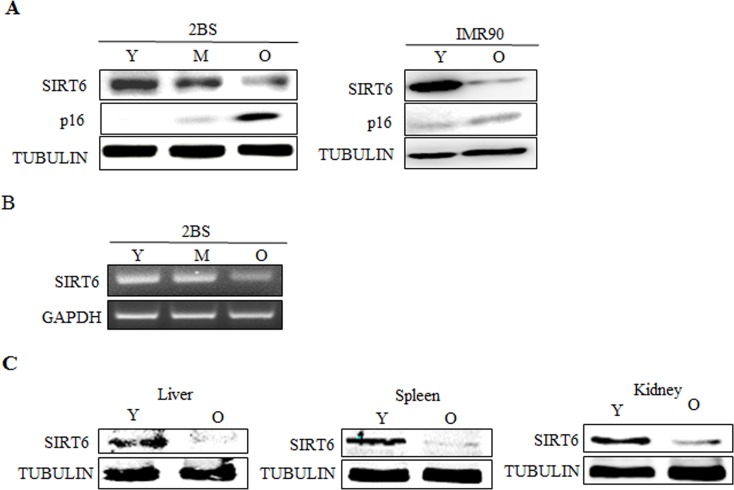
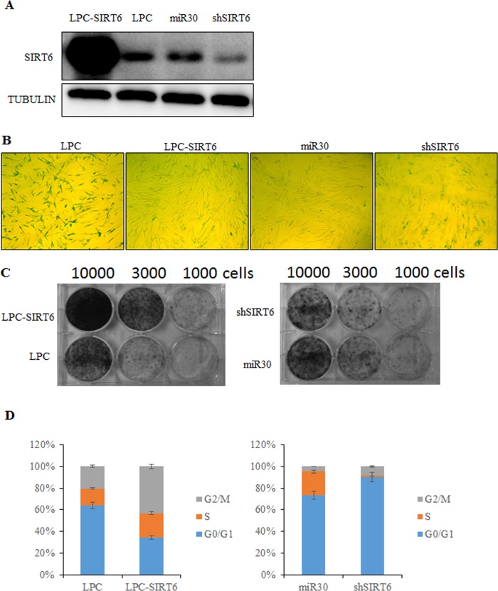
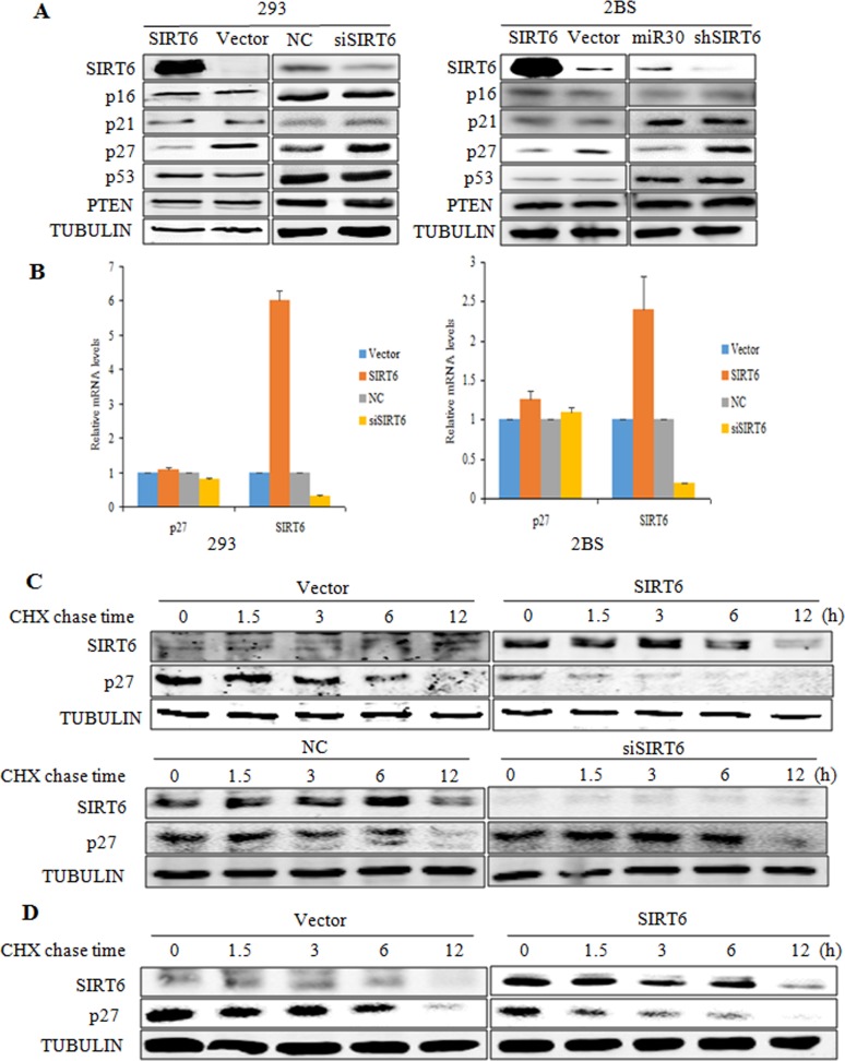
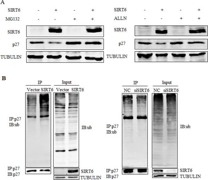
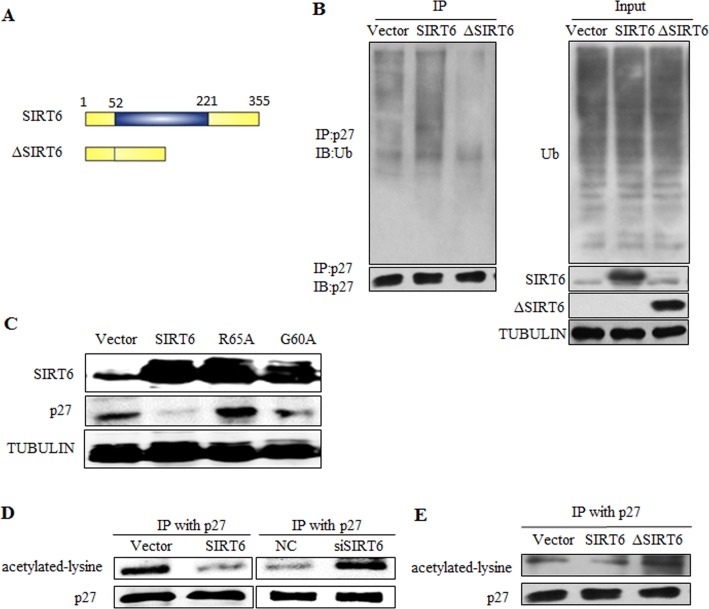
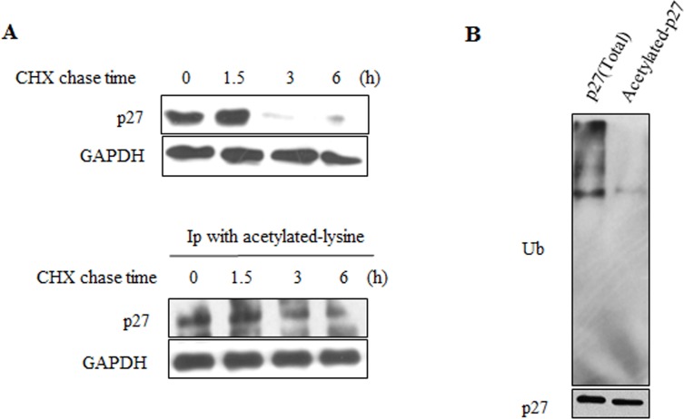
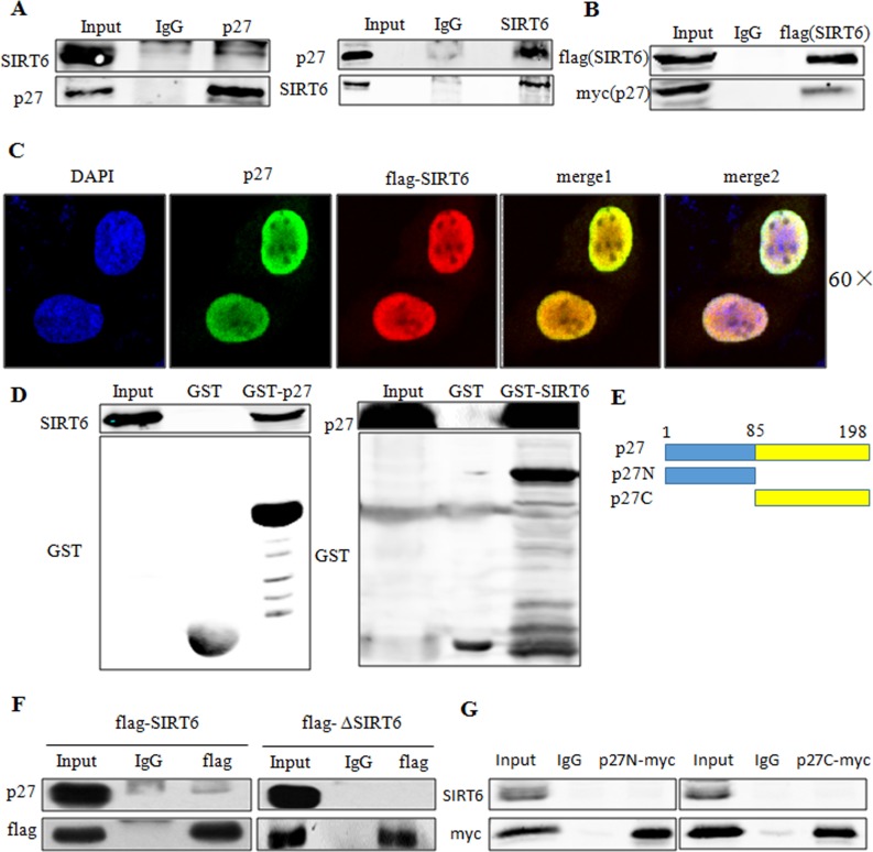
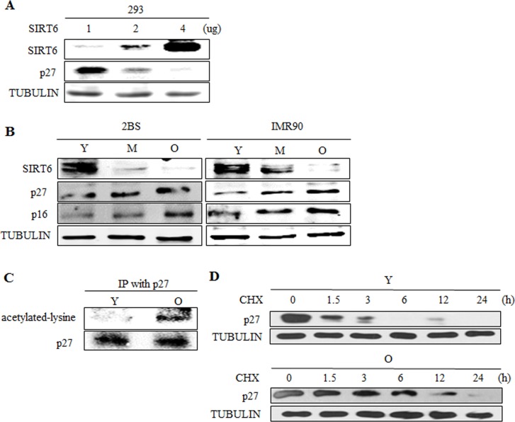
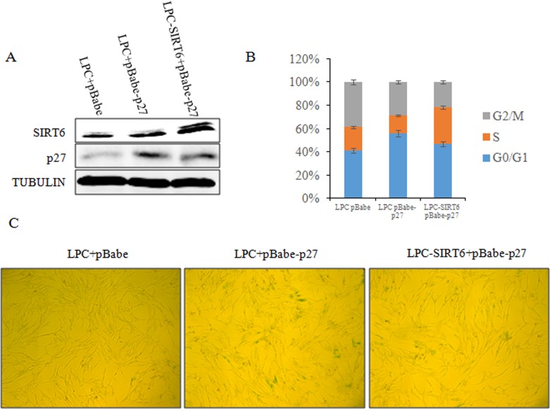
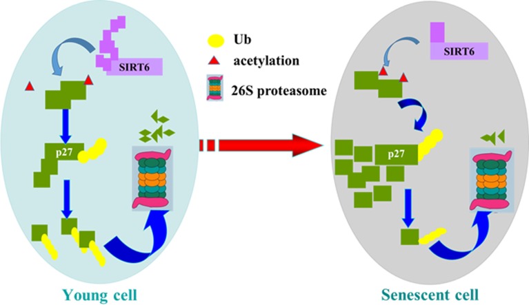
Sirtuin6(SIRT6) has been implicated as a key factor in aging and aging-related diseases. However, the role of SIRT6 in cellular senescence has not been fully understood. Here, we show that SIRT6 repressed the expression of p27Kip1 (p27) in cellular senescence. The expression of SIRT6 was reduced during cellular senescence, whereas enforced SIRT6 expression promoted cell proliferation and antagonized cellular senescence. In addition, we demonstrated that SIRT6 promoted p27 degradation by proteasome and SIRT6 decreased the acetylation level and protein half-life of p27. p27 acetylation increased its protein stability. Furthermore, SIRT6 directly interacted with p27. Importantly, p27 was strongly acetylated and had a prolonged protein half-life with the reduction of SIRT6 when cells were senescent, compared with those young cells. Finally, SIRT6 markedly rescued senescence induced by p27. Our findings indicate that SIRT6 decreases p27 acetylation, leading to its degradation via ubiquitin-proteasome pathway and then delays cellular senescence.

Keywords: SIRT6; acetylation; cellular senescence; p27kip1; ubiquitination.


Conflict of interest statement

The authors have no conflict of interests to declare.

Figures

Figure 1
Figure 1. Expression patterns of SIRT6 in young and senescent cells
(A) Left, Western blot analysis of SIRT6 expression in young (Y, ≈PD 30), middle-aged (M, ≈PD 40) and senescent (O, ≈PD 55) 2BS cells. Total protein was extracted, and immunoblotting was performed using specific antibodies against SIRT6, p16 as indicated. Tubulin served as a loading control. Right, the levels of SIRT6, p16 and TUBULIN in young (Y) and senescent (O) IMR90 cells were analysed by western blot analysis. (B) RT-PCR analysis of SIRT6 in young, middle-aged and senescent 2BS cells. Total mRNA was extracted and assessed by RT-PCR using specific primers. GAPDH was used as a loading control. (C) Protein levels of SIRT6 in indicated tissues of young (3 months) and old (18 months) BALB/c mice were determined with western blotting. Tubulin serves as loading control.
Figure 2
Figure 2. SIRT6 represses senescent-associated features in 2BS cells
(A) Western blot analysis of SIRT6 expression levels in SIRT6-overexpression (LPC-SIRT6) and SIRT6-knockdown (shSIRT6) 2BS cells. (B) SIRT6-overexpression and SIRT6-knockdown 2BS cells were stained for SA-β-gal activity. (C) Colony formation assay was performed using SIRT6-overexpression and SIRT6-knockdown 2BS cells. (D) Flow cytometry analysis of SIRT6-overexpression and SIRT6-knockown 2BS cells. Values represent the means ± S.E. of triplicate points from a representative experiment (n=3), which was repeated three times.
Figure 3
Figure 3. SIRT6 decreases p27 protein levels
(A) Western blot analysis of SIRT6, p16, p21, p27, p53 and PTEN expression levels was carried out in SIRT6 overexpressing or knockdown 293 (left) and 2BS (right) cells. (B) Real-time PCR analysis of SIRT6, p27 was performed in cells as in A. Specific real-time PCR primers were used. (C) The protein half-life of p27 was evaluated in 293 cells with altered SIRT6 expression. (D) The protein half-life of p27 was evaluated in 2BS cells with SIRT6 overexpression.
Figure 4
Figure 4. SIRT6 promotes the ubiquitination of p27
(A) 293 cells were transfected with SIRT6 and vector, twenty four hours after transfection, cells were treated with MG132 or acetyl-leu-leu-norleucinal (ALLN) 5 h before harvesting. Proteins were analyzed by western blot. (B) An in vivo ubiquitination assay was performed. 293 cells were transiently transfected with SIRT6 and vector, siRNA-SIRT6 and NC (negative control). Forty two hours later, cells were treated with MG132 for 5 h. Cells were lysed and proceeded for co-immunoprecipitation using the p27 antibody.
Figure 5
Figure 5. SIRT6 regulates p27 via its deacetylation activity
(A) Schematic representation of full-length SIRT6 and its central core domain deleted mutant ΔSIRT6. (B) In vivo ubiquitination assay of p27 influenced by SIRT6 deleted mutant ΔSIRT6. (C) Overexpress SIRT6 and its mutants R65A (lack deacetylase activity) and G60A (lack mono-ADP-ribosyltransferase activity) in 293 cells, then protein levels of p27 was detected. (D) In vivo acetylation assay of p27 was carried out after overexpressing and knocking down SIRT6 in 293 cells. (E) The acetylation level of p27 was detected after overexpressing wild-type SIRT6 and its deletion ΔSIRT6.
Figure 6
Figure 6. Acetylated-p27 is more stable
(A) 293 cells were treated with CHX for different time periods, cells were then lysed and IP was performed with acetylated-lysine antibody (lower). The protein half-life of acetylated-p27 (lower) was analyzed by western blot. The protein half-life of p27 was detected as control (upper). (B) The ubiquitination of acetylated-p27 was detected. After treatment with MG132, 293 cells were harvested and IP was performed with acetylated-lysine antibody, then IP with p27 antibody was performed using the elution products. The second elution products were loaded on the SDS-PAGE gel for western blot to detect the ubiquitination level of acetylated-p27.
Figure 7
Figure 7. SIRT6 interacts with p27 in vivo and in vitro
(A) Co-IP of endogenous SIRT6 and p27 was performed in 293 cells. (B) Co-IP of exogenous SIRT6 (flag-SIRT6) and p27 (myc-p27) was performed using flag antibody in 293 cells. (C) 293 cells were transfected with flag-SIRT6 and p27, then immunofluorescence assay was carried out using flag and p27 antibodies to investigate the co-localization of these two proteins. (D) GST-pulldown assay using in vitro transcribed and translated SIRT6 or p27 and purified GST-p27 or GST-SIRT6 from E.coli BL21 cells. Blots were evaluated with SIRT6, p27 and GST antibodies. (E) Schematic representation of full-length p27 and its deletions. (F) 293 cells were transfected with flag-SIRT6 and flag-ΔSIRT6. Cell lysates were then used for co-IP with the flag antibody. Blots were evaluated with flag and p27 antibody. (G). 293 cells were transfected with flag-SIRT6 and myc-p27N or myc-p27C, co-IP was then performed using anti-myc antibody. Blots were evaluated with myc and SIRT6 antibodies.
Figure 8
Figure 8. SIRT6 expression correlates with p27 acetylation level and protein half-life during cellular senescence
(A) Different amounts of SIRT6 were expressed in 293 cells. 48 h after transfection, SIRT6 and p27 were analysed by western blot. (B) Western blot analysis of SIRT6, p16 and p27 expression in young (Y, ≈PD 30), middle-aged (M, ≈PD 40) and senescent (O, ≈PD 55) 2BS and IMR90 cells. (C) The acetylation level of p27 was detected in young (Y, ≈PD 30) and senescent (O, ≈PD 55) 2BS cells. (D) The protein half-life of p27 during cellular senescence in 2BS was detected.
Figure 9
Figure 9. SIRT6 represses cellular senescence induced by p27
(A) 2BS cells were stably co-transfected with LPC vector and pBabe vector, LPC vector and pBabe-p27 or LPC-SIRT6 and pBabe-p27 plasmids. The expression of SIRT6 and p27 protein levels in the stable transformants were analyzed by western blot. (B) Cell cycle of cells from A was detected. Values represent the means ± S.E. of triplicate points from a representative experiments (n=3), which was repeated three times. (C) Cells from A were stained for SA-β-gal activity.
Figure 10
Figure 10. Model of SIRT6 regulating p27 during cellular senescence
SIRT6 deacetylates p27, which promotes the ubiquitination of p27 and increases the degradation of p27 by proteasome. Then SIRT6 can repress cellular senescence.

Similar articles

Cited by

References

    1. Gertler AA, Cohen HY. SIRT6, a protein with many faces. Biogerontology. 2013;14:629–39. doi: 10.1007/s10522-013-9478-8. - DOI - PubMed
    1. Imai S, Armstrong CM, Kaeberlein M, Guarente L. Transcriptional silencing and longevity protein Sir2 is an NAD-dependent histone deacetylase. Nature. 2000;403:795–800. doi: 10.1038/35001622. - DOI - PubMed
    1. Kaeberlein M, McVey M, Guarente L. The SIR2/3/4 complex and SIR2 alone promote longevity in Saccharomyces cerevisiae by two different mechanisms. Genes Dev. 1999;13:2570–80. doi: 10.1101/gad.13.19.2570. - DOI - PMC - PubMed
    1. Rogina B, Helfand SL. Sir2 mediates longevity in the fly through a pathway related to calorie restriction. Proc Natl Acad Sci USA. 2004;101:15998–6003. doi: 10.1073/pnas.0404184101. - DOI - PMC - PubMed
    1. Tissenbaum HA, Guarente L. Increased dosage of a sir-2 gene extends lifespan in Caenorhabditis elegans. Nature. 2001;410:227–30. doi: 10.1038/35065638. - DOI - PubMed

Publication types

MeSH terms

LinkOut - more resources