. 2017 Nov;35(15):3354-3369.
doi: 10.1080/07391102.2016.1254682. Epub 2016 Nov 29.

Molecular dynamics simulations of aptamer-binding reveal generalized allostery in thrombin

Affiliations

Molecular dynamics simulations of aptamer-binding reveal generalized allostery in thrombin

Jiajie Xiao et al. J Biomol Struct Dyn. 2017 Nov.

Abstract

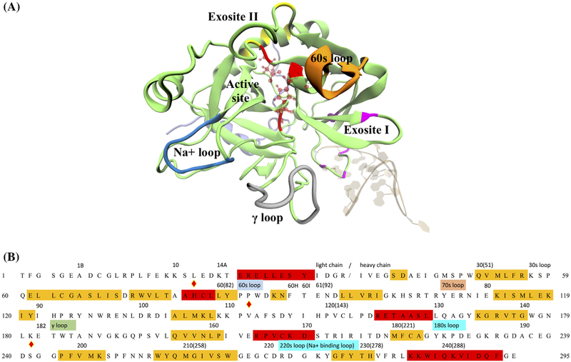
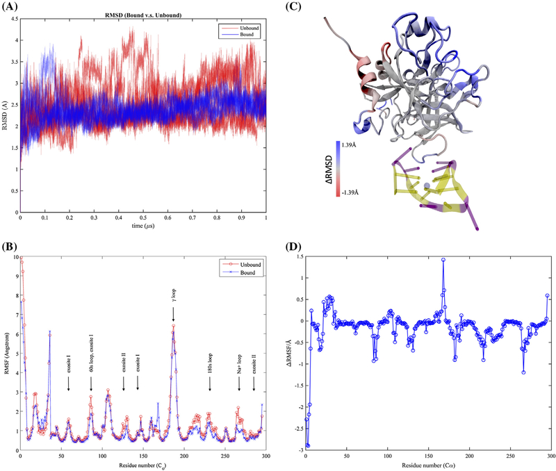
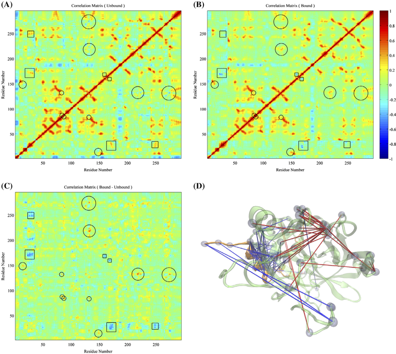
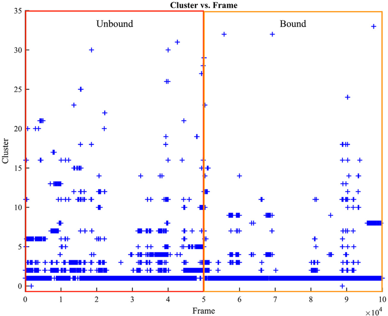
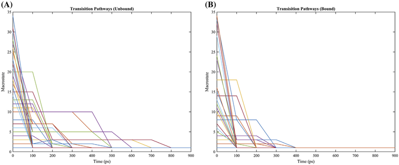
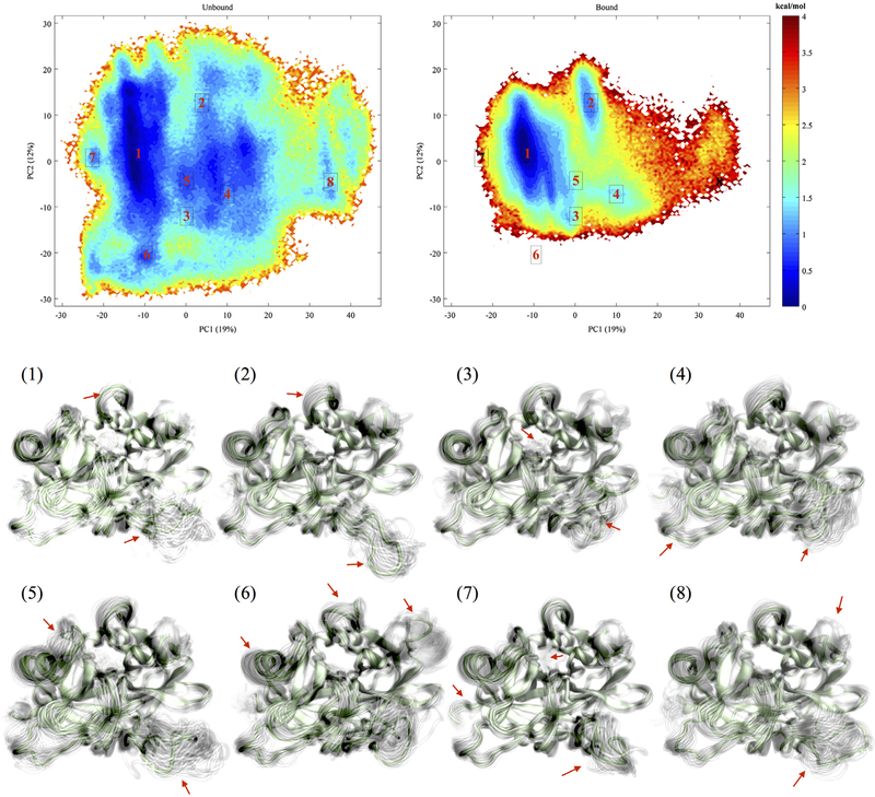
Thrombin is an attractive target for antithrombotic therapy due to its central role in thrombosis and hemostasis as well as its role in inducing tumor growth, metastasis, and tumor invasion. The thrombin-binding DNA aptamer (TBA), is under investigation for anticoagulant drugs. Although aptamer binding experiments have been revealed various effects on thrombin's enzymatic activities, the detailed picture of the thrombin's allostery from TBA binding is still unclear. To investigate thrombin's response to the aptamer-binding at the molecular level, we compare the mechanical properties and free energy landscapes of the free and aptamer-bound thrombin using microsecond-scale all-atom GPU-based molecular dynamics simulations. Our calculations on residue fluctuations and coupling illustrate the allosteric effects of aptamer-binding at the atomic level, highlighting the exosite II, 60s, γ and the sodium loops, and the alpha helix region in the light chains involved in the allosteric changes. This level of details clarifies the mechanisms of previous experimentally demonstrated phenomena, and provides a prediction of the reduced autolysis rate after aptamer-binding. The shifts in thrombin's ensemble of conformations and free energy surfaces after aptamer-binding demonstrate that the presence of bound-aptamer restricts the conformational freedom of thrombin suggesting that conformational selection, i.e. generalized allostery, is the dominant mechanism of thrombin-aptamer binding. The profound perturbation on thrombin's mechanical and thermodynamic properties due to the aptamer-binding, which was revealed comprehensively as a generalized allostery in this work, may be exploited in further drug discovery and development.

Keywords: aptamer; generalized allostery; molecular dynamics; molecular recognition; thrombin.


Conflict of interest statement

Disclosure statement

The authors of this manuscript declare no conflicts of interest.

Figures

Figure 1.
Figure 1.
Human α-thrombin and its functional sites. (A) Tertiary structure of thrombin in PDB 4DII was showed in cartoon representation. The light and heavy chains were respectively colored in light violet and lime. Several known function sites were indicated by the colors and nearby labels. The thrombin-binding aptamer in the same PDB was shown in NewRibbons representation. (B) Sequence of human α-thrombin was listed in one-letter amino acid code. The residue indices in the original PDB file and positions of several function sites were labeled above the sequence. Residues under the red and yellow stand for alpha helix and beta sheet regions. The catalytic triad was marked by the diamond signs above the letter. For the convenience of counting, a new residue number was assigned to each residue as labeled in the beginnings and ends of each row of sequence and the numbers in the parentheses.
Figure 2.
Figure 2.
Thermodynamic fluctuations of thrombin. Free thrombin has heavier fluctuations in comparison with aptamer-bound thrombin with respect to (A) the root-mean-square distances (RMSD) to the same reference of the initial structure along simulation trajectories and (B) the root-mean-squared fluctuations (RMSF) of each alpha carbon atom. The regions with distinct fluctuations on free and aptamer-bound thrombin were also indicated by the colored regions in plot (C). The aptamer-bound thrombin has larger fluctuations in red regions and smaller fluctuations in blue regions. These fluctuation differences were further indicated via the plot of RMSF differences between aptamer-bound and aptamer-unbound thrombin in (D).
Figure 3.
Figure 3.
Comparisons on overall motions between the aptamer-unbound and aptamer-bound thrombin. (A) Correlations of each alpha carbon’s displacement from average position of the free thrombin. (B) Correlations of motions of each alpha carbon’s displacement from average position of the aptamer-bound thrombin. (C) Subtraction of correlation matrix of free thrombin from the one of aptamer-bound thrombin. The circles and squares highlight the regions with significant positive and negative increments of correlations, respectively. (D) Pairs of residues with significantly distinct correlated motions in both systems were highlighted via lines and beads. The blue and red lines indicate residue pairs with negative and positive subtractions of correlation matrices of aptamer-bound and aptamer-unbound thrombin respectively.
Figure 4.
Figure 4.
Structure clustering evolution of thrombin in unbound and bound simulations. The red and orange regions include five 1-microsecond-long trajectories from the free thrombin and aptamer-bound thrombin simulations respectively.
Figure 5.
Figure 5.
Transition pathways of clusters in the simulations of aptamer-unbound and aptamer-bound simulations. Only sampled clusters for each system were plotted. Every 100 ps stands for one iteration step in the transition pathway calculation.
Figure 6.
Figure 6.
Free energy landscapes of aptamer-unbound and aptamer-bound thrombins and representative structures corresponding each labeled well.

Similar articles

Cited by

References

    1. Abdi H, & Williams LJ (2010). Principal component analysis. Wiley Interdisciplinary Reviews: Computational Statistics, 2, 433–459. doi:10.1002/wics.101 - DOI
    1. Adams TE, & Huntington JA (2006). Thrombin-cofactor interactions: Structural insights into regulatory mechanisms. Arteriosclerosis, Thrombosis, and Vascular Biology, 26, 1738–1745. doi:10.1161/01.ATV.0000228844.65168.d1 - DOI - PubMed
    1. Akhavan H, Mannucci PM, Lak H, Mancuso H, Mazzucconi MG, Rocino H, … Perkins SJ (2000). Identification and three-dimensional structural analysis of nine novel mutations in patients with prothrombin deficiency. Thrombosis and Haemostasis, 84, 989–997. - PubMed
    1. Article H, Steering H, For H, & Thrombosis H. (2014). Thrombosis: A major contributor to the global disease burden. Journal of Thrombosis and Haemostasis, 12, 1580–1590. doi: 10.1111/jth.12698 - DOI - PubMed
    1. Baglin TP, Carrell RW, Church FC, Esmon CT, & Huntington JA (2002). Crystal structures of native and thrombin-complexed heparin cofactor II reveal a multistep allosteric mechanism. Proceedings of the National Academy of Sciences, 99, 11079–11084. doi:10.1073/pnas.162232399 - DOI - PMC - PubMed